. 2014 Feb;27(1):133-44.
doi: 10.1007/s10278-013-9640-5.

Computer-aided segmentation system for breast MRI tumour using modified automatic seeded region growing (BMRI-MASRG)

Affiliations

Computer-aided segmentation system for breast MRI tumour using modified automatic seeded region growing (BMRI-MASRG)

Ali Qusay Al-Faris et al. J Digit Imaging. 2014 Feb.

Abstract

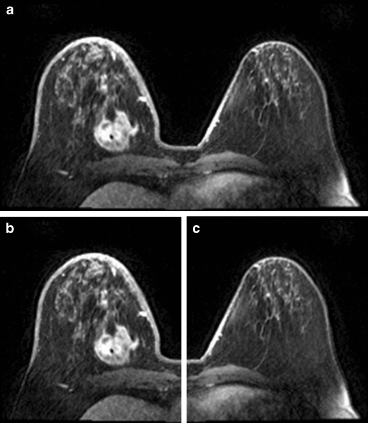
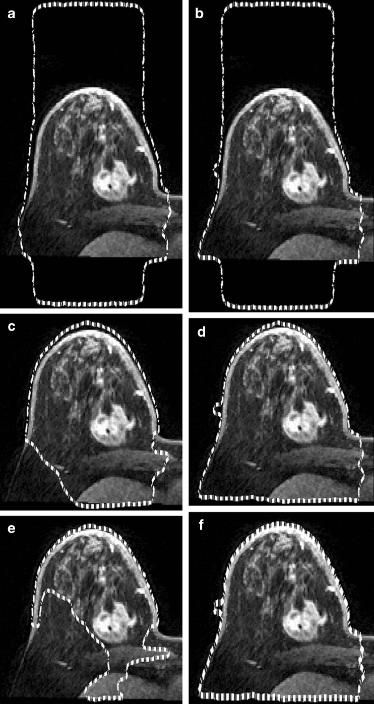
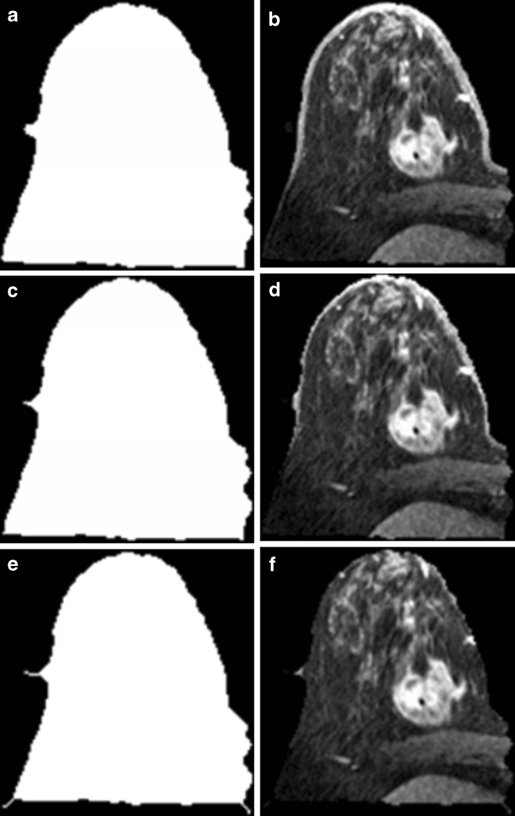
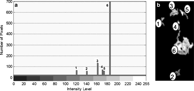
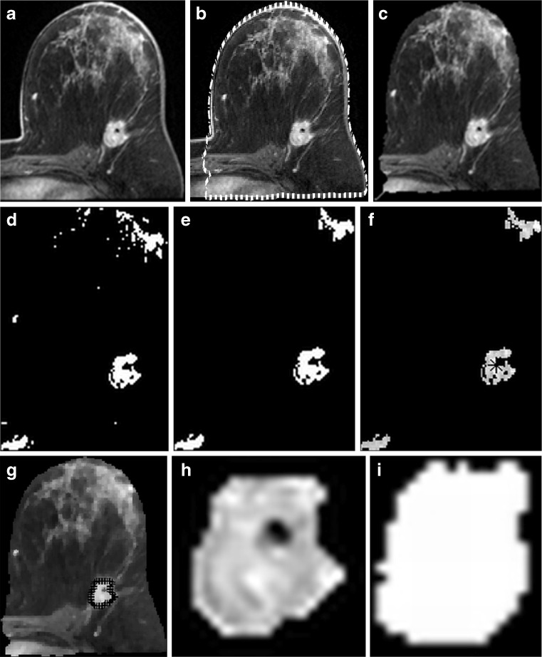
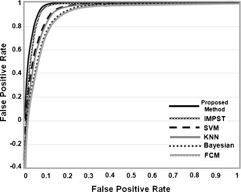
In this paper, an automatic computer-aided detection system for breast magnetic resonance imaging (MRI) tumour segmentation will be presented. The study is focused on tumour segmentation using the modified automatic seeded region growing algorithm with a variation of the automated initial seed and threshold selection methodologies. Prior to that, some pre-processing methodologies are involved. Breast skin is detected and deleted using the integration of two algorithms, namely the level set active contour and morphological thinning. The system is applied and tested on 40 test images from the RIDER breast MRI dataset, the results are evaluated and presented in comparison to the ground truths of the dataset. The analysis of variance (ANOVA) test shows that there is a statistically significance in the performance compared to the previous segmentation approaches that have been tested on the same dataset where ANOVA p values for the evaluation measures' results are less than 0.05, such as: relative overlap (p = 0.0002), misclassification rate (p = 0.045), true negative fraction (p = 0.0001) and sum of true volume fraction (p = 0.0001).


Figures

Fig. 1
Fig. 1
Methodology flowchart
Fig. 2
Fig. 2
Results of image splitting; a Original image. b Left side. c Right side
Fig. 3
Fig. 3
Different results after applied Chunming’s algorithm with different values of σ and N LS. a σ = 3, N LS = 100; b σ = 1.5, N LS = 100; c σ = 3, N LS = 300; d σ = 1.5, N LS = 300; e σ = 3, N LS = 700; f σ = 1.5, N = 700
Fig. 4
Fig. 4
Results after applying morphological thinning algorithm with three different iteration numbers on the resultant image of Chunming’s algorithm; a after applying thinning (N Th = 1) on binary image; b after reconverting thinning image (N Th = 1) to its original grey scale; c after applying thinning (N Th = 3) on binary image; d after reconverting thinning image (N Th = v3) to its original grey scale; e after applying thinning (N Th = 7) on binary image; f after converting thinning image (N Th = 7) to its original grey scale
Fig. 5
Fig. 5
Region ranking according to their pixels’ density values. a Image regions flags. b Bar chart for the ranked regions according to the highest mean intensity values
Fig. 6
Fig. 6
The initial seed pixel and its eight neighbouring pixels
Fig. 7
Fig. 7
The proposed system approach processes on one of the RIDER images: a original image; b breast skin detection after applying level set algorithm; c breast skin deletion after applying the thinning algorithm; d after applying the thresholding process using MMRT algorithm; e after applying morphological open operation; f initial seed selected using the proposed method (star sign (*)); g after the SRG method is applied using the proposed method of SRG threshold value; h The segmented tumour regions; i GT of the image
Fig. 8
Fig. 8
The ROC curves for the proposed method and the previous methods

References

    1. Gardiner I: CAD improves breast MRI workflow: increasing throughput while maintaining accuracy in breast MRI reads requires powerful workflow tools, 2010
    1. Lehman CD, Peacock S, DeMartini WB, Chen X. A new automated software system to evaluate breast MR examinations: improved specificity without decreased sensitivity. Am J Roentgenol. 2006;187:51–56. doi: 10.2214/AJR.05.0269. - DOI - PubMed
    1. Li L, Clark RA, Thomas JA. Computer-aided diagnosis of masses with full-field digital mammography. Acad Radiol. 2002;9:4–12. doi: 10.1016/S1076-6332(03)80290-8. - DOI - PubMed
    1. Verma B, Zakos J. A computer-aided diagnosis system for digital mammograms based on fuzzy-neural and feature extraction techniques. IEEE Trans Inf Technol Biomed. 2001;5:46–54. doi: 10.1109/4233.908389. - DOI - PubMed
    1. Ibrahim S, Khalid NEA, Manaf M. Empirical study of brain segmentation using particle swarm optimization. Selangor: Shah Alam; 2010.

LinkOut - more resources