. 2013 Oct 21;210(11):2371-85.
doi: 10.1084/jem.20122348. Epub 2013 Oct 7.

RhoA GTPase controls cytokinesis and programmed necrosis of hematopoietic progenitors

Affiliations

RhoA GTPase controls cytokinesis and programmed necrosis of hematopoietic progenitors

Xuan Zhou et al. J Exp Med. .

Abstract

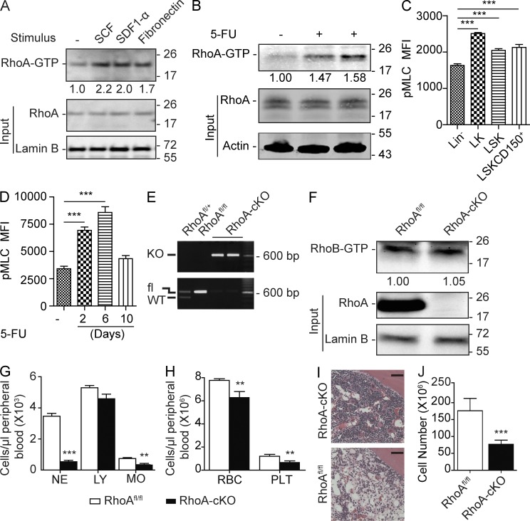
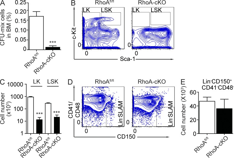
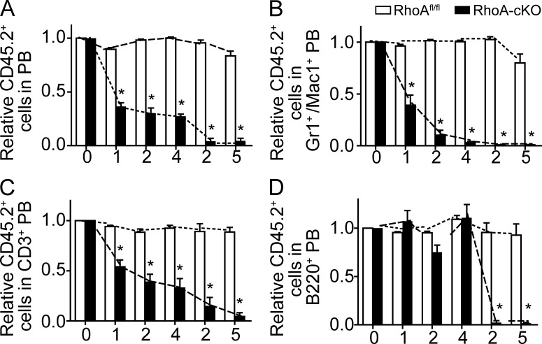
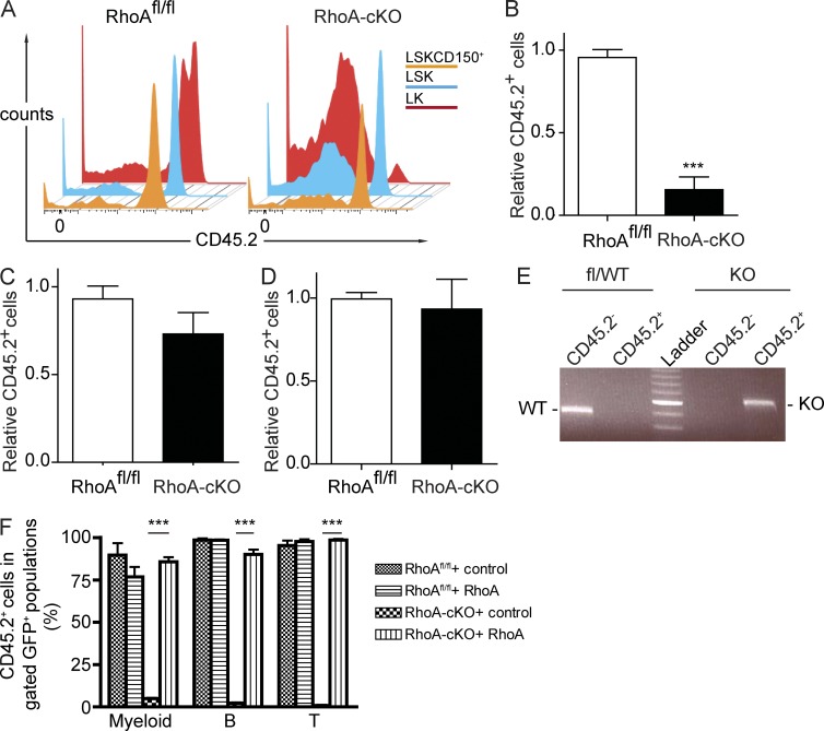
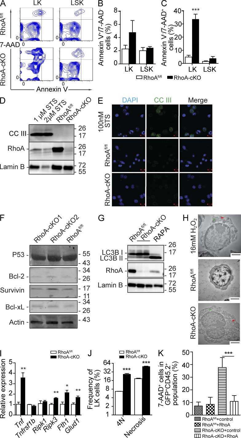
Hematopoietic progenitor cells (HPCs) are central to hematopoiesis as they provide large numbers of lineage-defined blood cells necessary to sustain blood homeostasis. They are one of the most actively cycling somatic cells, and their precise control is critical for hematopoietic homeostasis. The small GTPase RhoA is an intracellular molecular switch that integrates cytokine, chemokine, and adhesion signals to coordinate multiple context-dependent cellular processes. By using a RhoA conditional knockout mouse model, we show that RhoA deficiency causes a multilineage hematopoietic failure that is associated with defective multipotent HPCs. Interestingly, RhoA(-/-) hematopoietic stem cells retained long-term engraftment potential but failed to produce multipotent HPCs and lineage-defined blood cells. This multilineage hematopoietic failure was rescued by reconstituting wild-type RhoA into the RhoA(-/-) Lin(-)Sca-1(+)c-Kit(+) compartment. Mechanistically, RhoA regulates actomyosin signaling, cytokinesis, and programmed necrosis of the HPCs, and loss of RhoA results in a cytokinesis failure of HPCs manifested by an accumulation of multinucleated cells caused by failed abscission of the cleavage furrow after telophase. Concomitantly, the HPCs show a drastically increased death associated with increased TNF-RIP-mediated necrosis. These results show that RhoA is a critical and specific regulator of multipotent HPCs during cytokinesis and thus essential for multilineage hematopoiesis.


Figures

Figure 1.
Figure 1.
RhoA deficiency causes acute hematopoietic failure. (A) Isolated Lin cells were stimulated with SCF, SDF-1α, and fibronectin, and relative RhoA activity was determined by normalizing to total RhoA input. (B) Low-density monocytes were isolated 6 d after 5-FU injection, and relative RhoA activity was normalized to total RhoA input. (C) Phosphorylation of MLC (Ser19) in the WT primitive HSPC populations was determined by flow cytometry. MFI, mean fluorescence intensity. (D) Kinetics of MLC phosphorylation (Ser19) in LinSLAM population after 5-FU treatment was determined by flow cytometry. (E) RhoA deletion efficiency of the Mx-cre+RhoAfl/fl mice was determined by genotyping PCR using Lin cells 3 d after induction. (F) RhoB activity was assessed using Lin cells isolated 3 d after poly I:C induction. RhoB activity was determined and normalized against Lamin B. (G and H) PB counts of the congenic transplantation recipients. CD45.2+ RhoAfl/fl; Mx-cre+ or Mx-cre cells were transplanted into lethally irradiated CD45.1+ WT recipients. Three poly I:C injections were administrated 2 mo after transplantation. Recipients were sacrificed for analysis at 5 d after the last poly I:C injections. NE, neutrophils; LY, lymphocytes; MO, monocytes; RBC, red blood cells; PLT, platelets. (I) Representative H&E staining of femur sections. Bars, 40 µm. (J) Absolute number of BM white cells in the tibia, femur, and iliac crest 5 d after three poly I:C injections. Numbers of samples analyzed: four (C and D) or five (G, H, and J) per group. (A–D, F–H, and J) The results from a representative experiment of two independent experiments are shown. (A, B, and F) Molecular masses (kilodaltons) are indicated to the right of the blots. Error bars indicate SEM. **, P < 0.01; ***, P < 0.001.
Figure 2.
Figure 2.
RhoA deficiency reduces hematopoietic progenitors but not HSCs. (A) Frequency of CFCs in BM 5 d after poly I:C treatments. (B and C) Number of LK and LSK cells in BM 5 d after poly I:C treatments. (B) Representative flow cytometry plots of BM LK and LSK populations. (C) Quantification of LK and LSK cell numbers in tibia, femur, and iliac crest. (D and E) The number of LinSLAM cells in BM. (D) Representative flow cytometry plots of the LinSLAM population. (E) Quantification of the number of LinSLAM cells in tibia, femur, and iliac crest. All mice used in this figure were congenic transplantation recipients as described in Fig. 1 G. Numbers of samples analyzed: five per group (A and C) and four (RhoAfl/fl) and five (RhoA-cKO; E). (A–E) The results from a representative experiment of two independent experiments are shown. Error bars indicate SEM. ***, P < 0.001.
Figure 3.
Figure 3.
RhoA deficiency impairs multilineage hematopoiesis in a competitive transplantation experiment. (A–D) Lethally irradiated CD45.1+ recipient mice were reconstituted with equal numbers of CD45.2+ donor BM cells of the RhoAfl/flMx-cre+ or Mx-cre genotype with CD45.1+ WT competitors. Three poly I:C injections were administrated into the recipients 8 wk after transplantation, and the chimerism of PB was analyzed monthly thereafter. 4 mo after poly I:C inductions, BM cells from the primary recipients were pooled and transplanted into secondary recipients. The kinetics of relative chimerism of donor-derived (CD45.2+) cells in total PB (A), myeloid (B), T (C), and B (D) cells in the peripheral circulation of recipients are shown. The x axes show units in months. Months 0–4: primary recipients; months 2–5: secondary recipients. Seven mice per genotype were examined. The results from a representative experiment of three independent experiments are shown. Error bars indicate SEM. *, P < 0.001.
Figure 4.
Figure 4.
RhoA deficiency depletes HPCs but not HSCs in competitive setting. (A) CD45.2+ donor BM cells of the RhoAfl/flMx-cre+ or Mx-cre genotype were cotransplanted into lethally irradiated CD45.1+ recipients together with equal number of CD45.1+ WT competitor cells. Three poly I:C injections were administrated 8 wk after the transplantation, and BM cells from the primary recipients were transplanted into secondary recipients 4 mo after the poly I:C injections. Data were analyzed 5 mo after secondary transplantation. Representative flow cytometry plots of CD45.2 expression within the LK, LSK, and LSKCD150+ population in the competitive transplant recipients are shown. (B–D) Relative chimerism of donor-derived LK (B), LSK (C), and LSKCD150+ (D) in BM of recipients. (E) DNA from sorted CD45.2+ LSK cells was used as PCR template to determine the deletion efficiency of RhoA. Data were analyzed 5 mo after secondary transplantation. Brightest band of ladder: 600 bp. (F) LSK cells from poly I:C–injected competitive transplantation recipients were transduced with RhoA-expressing or control virus and transplanted into secondary recipients. PB samples were examined 16 wk after transplantation. Percentage of donor-derived (CD45.2+) cells in gated GFP+ mature lineages of PB was determined by flow cytometry. Numbers of mice analyzed: six (A–D) and four (F) mice per group. The results from a representative experiment of three (A–D) or two (F) independent experiments are shown. Error bars indicate SEM. ***, P < 0.001.
Figure 5.
Figure 5.
RhoA regulates HSPC actomyosin signaling, migration, and adhesion. (A) MLC phosphorylation (Ser19) in Lin BM cells 3 d after poly I:C induction. Relative MLC phosphorylation was determined by normalizing to total MLC expression. (B) Cofilin (Ser3) phosphorylation in isolated Lin BM cells 3 d after poly I:C induction. Relative cofilin phosphorylation was determined by normalizing to total cofilin. (A and B) Molecular masses (kilodaltons) are indicated to the right of the blots. (C and D) Cortical F-actin formation in response to 100 ng/ml SDF-1α stimulation in Lin cells. Lin cells were isolated from mice 3 d after induction and stained with Alexa Fluor 488 phalloidin. (C) Representative ImageStream images of F-actin staining in Lin cells. (D) Quantification of cortical F-actin signaling. MFI, mean fluorescence intensity. (E) Lin BM cells were cultured in the top chamber of Transwells. 100 ng/ml SDF-1α was added into the bottom chamber as a chemoattractant. The percentage of cells migrated into the bottom chamber 4 h later is shown. (F) Lin BM cells were cultured at 37°C for 2 h in CH-296–coated 96-well plates containing IMDM supplemented with 10% FBS. Nonadherent cells were removed, and the percentage of adherent cells is shown. Cells used in this figure were isolated from poly I:C–injected RhoAfl/fl; Mx-Cre+ or Mx-Cre mice. Numbers of samples analyzed: three (D and F) or four (E) mice per group. The results from a representative experiment of two (A–E) or four (F) independent experiments are shown. Error bars indicate SEM. *, P < 0.05.
Figure 6.
Figure 6.
RhoA–ROCK and RhoA–mDia interactions are required for HPC cytokinesis. (A) Noncompetitive transplantation experiments were performed similarly as described in Fig. 1 G. A BrdU chase at 0.5 mg/recipient was performed 30 min before the analysis. Cell cycle distribution of LK and LSK cells in the noncompetitive transplanted recipients was examined by BrdU incorporation assay. (top) Representative flow cytometry plot. The red rectangle indicates G0/G1 phase, green rectangle S phase, and purple rectangle G/M phase. (bottom) Quantification of cell cycle distribution. (B) Competitive transplantations were performed similarly as described in Fig. 3. Samples were analyzed 5 mo after secondary transplantation. BrdU injection at 0.5 mg/recipient was performed 30 min before experiment. G1-S transition of LK and LSK cells in the competitive transplanted recipients was examined by BrdU incorporation assay. (top) Representative flow cytometry plot. (bottom) Quantification of BrdU+ cells. (C) Nuclei contents of LK cells. LK cells were FACS isolated 3 d after two poly I:C injections, and Giemsa staining was performed to determine nuclei contents. (top) Representative Giemsa staining. (bottom) Quantification of multinucleated cell frequency. Insets show enlarged representative cells. Red arrowheads indicate multinuclear cells. (D) LK cells were isolated from RhoAfl/fl or RhoA-cKO mice 3 d after two consecutive poly I:C injections. More than 30 mitotic cells per group were analyzed by morphology and DNA staining. Data were statistically analyzed by the χ2 test. (E) Activation and distribution of Aurora A/B/C within LK cells. Cells were isolated 3 d after induction. The representative metaphase and telophase cells are shown. BF, brightfield. (F) Control or RhoA-cKO LK cells isolated 3 d after induction were transduced with mock retrovirus (REW13) or retrovirus expressing WT or effector binding–deficient mutant forms (E40L, R68A, and Y42C) of RhoA. Exogenous expression of RhoA was detected by both RhoA (top) and HA (middle) antibodies. Exogenous HA-RhoA mobilizes more slowly than endogenous RhoA because of the N-terminal 3x HA tag. Molecular masses (kilodaltons) are indicated to the right of the blots. (G and H) Percentage of 4N cells after expressing WT or mutant forms of RhoA. DNA contents were analyzed by Hoechst 33342 staining. (G) Representative flow cytometry plot. (H) Quantification of 4N cell frequency. Numbers of samples analyzed: four (RhoAfl/fl) and five (RhoA-cKO; A), seven per group (B), four (RhoAfl/fl) and six (RhoA-cKO; C), and three per group (D and H). (A–H) The results from a representative experiment of two independent experiments are shown. Error bars indicate SEM. *, P < 0.05; **, P < 0.01. Bars, 5 µm.
Figure 7.
Figure 7.
RhoA deficiency causes HPC necrosis but not apoptosis. (A–C) Congenic transplantation recipients described in Fig. 1 G were used, and BM cells were isolated 3 d after poly I:C induction. Cells were gated on the LK and LSK populations and analyzed by flow cytometry for Annexin V/7-AAD staining (A), apoptotic cells (B), and dead cells (C). (D) Cleavage of caspase 3 (CCIII) in Lin cells of RhoAfl/fl; Mx-cre+ or Mx-cre mice. Lin cells were isolated 3 d after poly I:C injections. Staurosporine (STS)-treated WT Lin cells were used as a positive apoptosis control. (E) CCIII staining of isolated LK cells. (F) p53, Bcl-xL, Bcl-2, and Survivin expression in Lin cells of RhoAfl/fl; Mx-cre+ or Mx-cre mice. Protein levels of apoptotic-related proteins were determined at 3 d after induction. (G) Levels of LC3B isoforms in Lin cells of RhoAfl/fl; Mx-cre+ or Mx-cre mice. Lin cells were isolated 3 d after poly I:C injections. 100 nM Rapamycin (RAPA)–treated WT Lin cells were used a positive autophagy control. (D, F, and G) Molecular masses (kilodaltons) are indicated to the right of the blots. (H) Representative electron micrographs of Lin cells in native mice. 16 mM H2O2–treated Lin cells were used as a positive necrosis control. Red arrowheads indicate features of membrane integrity loss. Green arrowheads indicate enlarged organelles. (I) Expression of TNF–RIP-related genes in LK cells 3 d after induction. (J) Association between cytokinesis failure (4N) and increased necrosis (7-AAD+) in RhoA-deficient HPCs. BM Lin cells were isolated at 3 d after induction. (K) Necrosis analysis of LK cells from recipients transplanted with LSK cells reconstituted with exogenous RhoA as described in Fig. 4 F. Numbers of samples analyzed: three (B, C, and J) or four (I and K) per group. (A–K) The results from a representative experiment of two independent experiments are shown. Error bars indicate SEM. *, P < 0.05; **, P < 0.01; ***, P < 0.001. Bars: (E) 10 µm; (H) 2 µm.

Similar articles

Cited by

References

    1. Andrews P.D., Knatko E., Moore W.J., Swedlow J.R. 2003. Mitotic mechanics: the auroras come into view. Curr. Opin. Cell Biol. 15:672–683 10.1016/j.ceb.2003.10.013 - DOI - PubMed
    1. Arai F., Hirao A., Ohmura M., Sato H., Matsuoka S., Takubo K., Ito K., Koh G.Y., Suda T. 2004. Tie2/angiopoietin-1 signaling regulates hematopoietic stem cell quiescence in the bone marrow niche. Cell. 118:149–161 10.1016/j.cell.2004.07.004 - DOI - PubMed
    1. Boggs D.R., Boggs S.S. 1976. Editorial: The pathogenesis of aplastic anemia: a defective pluripotent hematopoietic stem cell with inappropriate balance of differentiation and self-replication. Blood. 48:71–76 - PubMed
    1. Bonnet D., Dick J.E. 1997. Human acute myeloid leukemia is organized as a hierarchy that originates from a primitive hematopoietic cell. Nat. Med. 3:730–737 10.1038/nm0797-730 - DOI - PubMed
    1. Broudy V.C. 1997. Stem cell factor and hematopoiesis. Blood. 90:1345–1364 - PubMed

Publication types

MeSH terms