

Optogenetic-mediated increases in in vivo spontaneous activity disrupt pool-specific but not dorsal-ventral motoneuron pathfinding
- PMID: 24101487
- PMCID: PMC3808638
- DOI: 10.1073/pnas.1316457110
Optogenetic-mediated increases in in vivo spontaneous activity disrupt pool-specific but not dorsal-ventral motoneuron pathfinding
Abstract
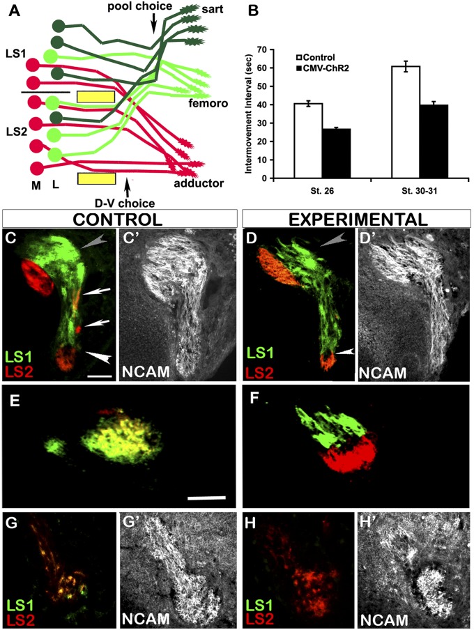
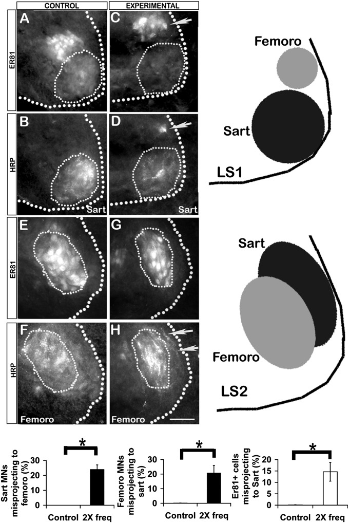
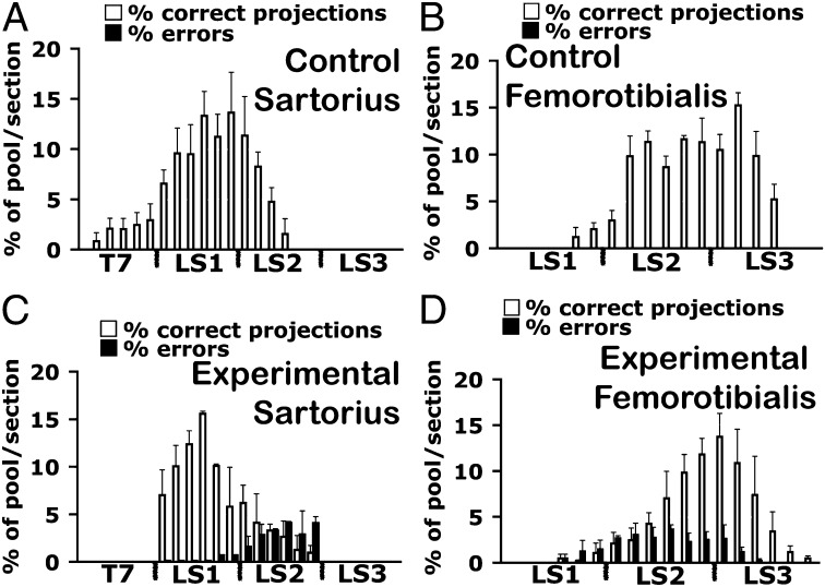
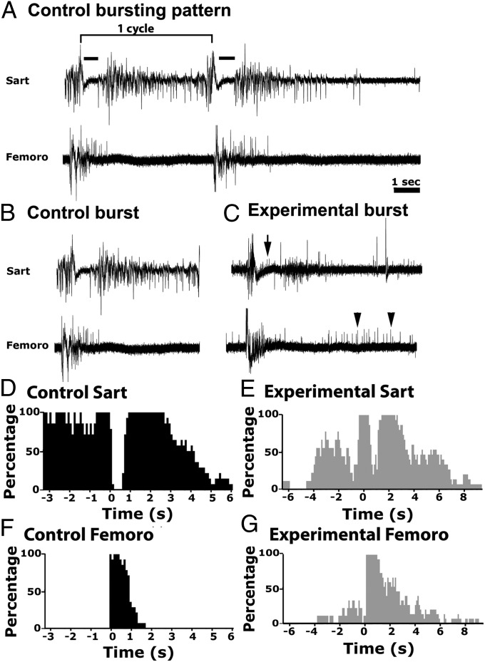
Rhythmic waves of spontaneous electrical activity are widespread in the developing nervous systems of birds and mammals, and although many aspects of neural development are activity-dependent, it has been unclear if rhythmic waves are required for in vivo motor circuit development, including the proper targeting of motoneurons to muscles. We show here that electroporated channelrhodopsin-2 can be activated in ovo with light flashes to drive waves at precise intervals of approximately twice the control frequency in intact chicken embryos. Optical monitoring of associated axial movements ensured that the altered frequency was maintained. In embryos thus stimulated, motor axons correctly executed the binary dorsal-ventral pathfinding decision but failed to make the subsequent pool-specific decision to target to appropriate muscles. This observation, together with the previous demonstration that slowing the frequency by half perturbed dorsal-ventral but not pool-specific pathfinding, shows that modest changes in frequency differentially disrupt these two major pathfinding decisions. Thus, many drugs known to alter early rhythmic activity have the potential to impair normal motor circuit development, and given the conservation between mouse and avian spinal cords, our observations are likely relevant to mammals, where such studies would be difficult to carry out.
Keywords: axonal guidance; motoneuron development; spinal cord development; spontaneous neural activity.
Conflict of interest statement
The authors declare no conflict of interest.
Figures
Similar articles
-
Spontaneous rhythmic activity in early chick spinal cord influences distinct motor axon pathfinding decisions.Brain Res Rev. 2008 Jan;57(1):77-85. doi: 10.1016/j.brainresrev.2007.06.021. Epub 2007 Aug 1. Brain Res Rev. 2008. PMID: 17920131 Free PMC article. Review.
-
In vivo activation of channelrhodopsin-2 reveals that normal patterns of spontaneous activity are required for motoneuron guidance and maintenance of guidance molecules.J Neurosci. 2010 Aug 4;30(31):10575-85. doi: 10.1523/JNEUROSCI.2773-10.2010. J Neurosci. 2010. PMID: 20686000 Free PMC article.
-
Characterization of rhythmic Ca2+ transients in early embryonic chick motoneurons: Ca2+ sources and effects of altered activation of transmitter receptors.J Neurosci. 2009 Dec 2;29(48):15232-44. doi: 10.1523/JNEUROSCI.3809-09.2009. J Neurosci. 2009. PMID: 19955376 Free PMC article.
-
Increasing the frequency of spontaneous rhythmic activity disrupts pool-specific axon fasciculation and pathfinding of embryonic spinal motoneurons.J Neurosci. 2006 Dec 6;26(49):12769-80. doi: 10.1523/JNEUROSCI.4170-06.2006. J Neurosci. 2006. PMID: 17151280 Free PMC article.
-
General principles of spinal motor circuit development: early contributions from research on avian embryos.Int J Dev Biol. 2018;62(1-2-3):235-243. doi: 10.1387/ijdb.170305LL. Int J Dev Biol. 2018. PMID: 29616732 Review.
Cited by
-
Fragile X Mental Retardation Protein Requirements in Activity-Dependent Critical Period Neural Circuit Refinement.Curr Biol. 2017 Aug 7;27(15):2318-2330.e3. doi: 10.1016/j.cub.2017.06.046. Epub 2017 Jul 27. Curr Biol. 2017. PMID: 28756946 Free PMC article.
-
Optogenetic Restoration of Disrupted Slow Oscillations Halts Amyloid Deposition and Restores Calcium Homeostasis in an Animal Model of Alzheimer's Disease.PLoS One. 2017 Jan 23;12(1):e0170275. doi: 10.1371/journal.pone.0170275. eCollection 2017. PLoS One. 2017. PMID: 28114405 Free PMC article.
-
Spatiotemporal integration of developmental cues in neural development.Dev Neurobiol. 2015 Apr;75(4):349-59. doi: 10.1002/dneu.22254. Epub 2014 Dec 10. Dev Neurobiol. 2015. PMID: 25484201 Free PMC article. Review.
-
Activity-dependent FMRP requirements in development of the neural circuitry of learning and memory.Development. 2015 Apr 1;142(7):1346-56. doi: 10.1242/dev.117127. Development. 2015. PMID: 25804740 Free PMC article.
-
Connectivity of pacemaker neurons in the neonatal rat superficial dorsal horn.J Comp Neurol. 2015 May 1;523(7):1038-1053. doi: 10.1002/cne.23706. Epub 2015 Feb 17. J Comp Neurol. 2015. PMID: 25380417 Free PMC article.
References
Publication types
MeSH terms
Substances
Grants and funding
LinkOut - more resources
Full Text Sources
Other Literature Sources
