. 2013 Nov 12;110(46):18380-5.
doi: 10.1073/pnas.1309729110. Epub 2013 Oct 7.

3D printing of microscopic bacterial communities

Affiliations

3D printing of microscopic bacterial communities

Jodi L Connell et al. Proc Natl Acad Sci U S A. .

Abstract

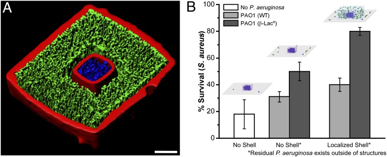
Bacteria communicate via short-range physical and chemical signals, interactions known to mediate quorum sensing, sporulation, and other adaptive phenotypes. Although most in vitro studies examine bacterial properties averaged over large populations, the levels of key molecular determinants of bacterial fitness and pathogenicity (e.g., oxygen, quorum-sensing signals) may vary over micrometer scales within small, dense cellular aggregates believed to play key roles in disease transmission. A detailed understanding of how cell-cell interactions contribute to pathogenicity in natural, complex environments will require a new level of control in constructing more relevant cellular models for assessing bacterial phenotypes. Here, we describe a microscopic three-dimensional (3D) printing strategy that enables multiple populations of bacteria to be organized within essentially any 3D geometry, including adjacent, nested, and free-floating colonies. In this laser-based lithographic technique, microscopic containers are formed around selected bacteria suspended in gelatin via focal cross-linking of polypeptide molecules. After excess reagent is removed, trapped bacteria are localized within sealed cavities formed by the cross-linked gelatin, a highly porous material that supports rapid growth of fully enclosed cellular populations and readily transmits numerous biologically active species, including polypeptides, antibiotics, and quorum-sensing signals. Using this approach, we show that a picoliter-volume aggregate of Staphylococcus aureus can display substantial resistance to β-lactam antibiotics by enclosure within a shell composed of Pseudomonas aeruginosa.

Keywords: antibiotic resistance; microfabrication; multiphoton lithography; polymicrobial.


Conflict of interest statement

The authors declare no conflict of interest.

Figures

Fig. 1.
Fig. 1.
Gelatin-based micro-3D printing in the presence of bacteria. (A) Schematic depicting in situ microfabrication around cells encapsulated in thermally set gelatin. The red and blue arrows indicate steps performed at 37 °C or 18–22 °C, respectively. (B) Confocal fluorescence isosurfaces show isolated Pseudomonas aeruginosa microcolonies within a surface-anchored 2-pL pyramid (Top; two cells initially) and an untethered 3-pL torus (Bottom; partially transparent on the right views; one cell initially; see also Fig. S1). (C) Partially transparent and cut-out views of confocal fluorescence isosurfaces illustrate six physically segregated P. aeruginosa populations organized in three dimensions within a series of spheroid cavities (2–15 pL in volume) tethered to the glass surface by two cylindrical posts, where the right-most spheroid is vacant and the others initially contained either one or two cells. A side view of these clusters is shown in the lower image in which the upper portion of the red channel is digitally removed to reveal bacterial clusters. The upper image provides a top-down view of the same community, with the transparency of the red channel adjusted to reveal bacterial clusters. (D) A bright-field image acquired 10 min postfabrication (Left), and side-on and partially transparent top-down views of a confocal fluorescence isosurface (Center and Right, respectively) showing colony growth at 18 h. The spheroidal chambers are physically connected by channels such that motile P. aeruginosa cells can distribute throughout the 5-pL structure according to preference. Cylindrical posts extend from the base of each spheroid to tether the structure to the glass surface. See also Fig. S2. Cells are false-colored green in B–D for visualization. (Scale bars, 20 μm.)
Fig. 2.
Fig. 2.
Nesting of a mobile bacterial microcluster within a physically isolated population. The 70-s sequence shows a time series acquired ∼6 h post-fabrication in which a tightly packed, 1-pL spheroidal population (initially one cell) of P. aeruginosa is buffeted by freely swimming P. aeruginosa contained within an exterior 55-pL “shell” (initially two cells; see also Movie S1). The 5-μm-thick roof on the exterior shell is optically transparent and not visible in the images. The green, pink, and yellow reference markers show the rotation of the sphere in three dimensions. (Scale bar, 10 μm.)
Fig. 3.
Fig. 3.
Engineering polymicrobial communities. (A) Staphylococcus aureus and P. aeruginosa cells are embedded in one precursor gel to form a physically mixed polymicrobial community. Bright-field images acquired at 37 °C over 10 h show growth of the mixed colony, with the enclosure (initially 8 pL) roof and walls distending dramatically over the last several hours of growth (Movie S2). Eventually, the structure’s 2-μm-thick roof ruptured, releasing cells into the surrounding medium (not shown). S. aureus and P. aeruginosa cells are false-colored red and green, respectively, for visualization in the “0 min” and “80 min” images. (B) Cut-away 3D mask reconstructions (Upper) and bright-field images (Lower) depict examples of nested polymicrobial communities of varying geometries and cell densities (see also Fig. S3). Low-density (Left) and high-density (Right) S. aureus microclusters are confined in rectangular and hemispherical cavities, respectively, surrounded by high-density P. aeruginosa populations. Inner cavities are 1 pL and outer chambers are 30 pL. The 5-μm-thick roofs used to seal in inner cavities and the outer chambers are not visible in the bright-field images. (Scale bars, 10 μm.)
Fig. 4.
Fig. 4.
Sharing of antibiotic resistance within a polymicrobial community containing P. aeruginosa and S. aureus. (A) Cut-away view of a confocal fluorescence isosurface revealing a nested S. aureus microcolony surrounded by P. aeruginosa on all sides except for the coverglass surface (see also Fig. S3; scale bar, 10 μm). (B) Confined microcolonies of S. aureus display resistance to ampicillin via the action of cocultured P. aeruginosa. Monocultures of S. aureus experience approximately an 80% kill rate when exposed to ampicillin at the MIC for 2 h (white bar, Left). Confined S. aureus with residual P. aeruginosa in the surrounding growth medium exhibits some resistance to the β-lactam antibiotic (Center). Survival of S. aureus is significantly enhanced when confined microclusters are nested within high-density populations of P. aeruginosa (Right). Additionally, PAO1 that overproduces β-lactamase [PAO1 (β-LacR); dark gray bars] provides enhanced protection from ampicillin relative to PAO1 wild-type [PAO1 (WT); light gray bars]. (Inset) Schematics depicting the three different dosing conditions. S. aureus and P. aeruginosa cells appear blue and green, respectively. S. aureus microcolonies initially contained two to eight cells within a 1- to 4-pL cavity. Nested populations are surrounded by 10- to 20-μm-thick layer of P. aeruginosa in the x–y dimensions that extends 15–50 μm in the z dimension (shell volumes ranged between 20 and 150 pL). Error bars represent 1 SD between individual microstructures containing S. aureus (n ≥ 4) pooled from multiple biological replicates (NBio ≥ 2).

References

    1. Parsek MR, Greenberg EP. Sociomicrobiology: The connections between quorum sensing and biofilms. Trends Microbiol. 2005;13(1):27–33. - PubMed
    1. Horswill AR, Stoodley P, Stewart PS, Parsek MR. The effect of the chemical, biological, and physical environment on quorum sensing in structured microbial communities. Anal Bioanal Chem. 2007;387(2):371–380. - PMC - PubMed
    1. Balaban NQ, Merrin J, Chait R, Kowalik L, Leibler S. Bacterial persistence as a phenotypic switch. Science. 2004;305(5690):1622–1625. - PubMed
    1. Hall-Stoodley L, Stoodley P. Biofilm formation and dispersal and the transmission of human pathogens. Trends Microbiol. 2005;13(1):7–10. - PubMed
    1. Boedicker JQ, Vincent ME, Ismagilov RF. Microfluidic confinement of single cells of bacteria in small volumes initiates high-density behavior of quorum sensing and growth and reveals its variability. Angew Chem Int Ed Engl. 2009;48(32):5908–5911. - PMC - PubMed

Publication types

MeSH terms