. 2013 Oct 22;110(43):17582-7.
doi: 10.1073/pnas.1308987110. Epub 2013 Oct 7.

Arabidopsis CRY2 and ZTL mediate blue-light regulation of the transcription factor CIB1 by distinct mechanisms

Affiliations

Arabidopsis CRY2 and ZTL mediate blue-light regulation of the transcription factor CIB1 by distinct mechanisms

Hongtao Liu et al. Proc Natl Acad Sci U S A. .

Abstract

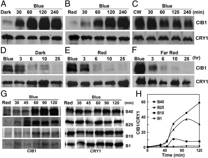
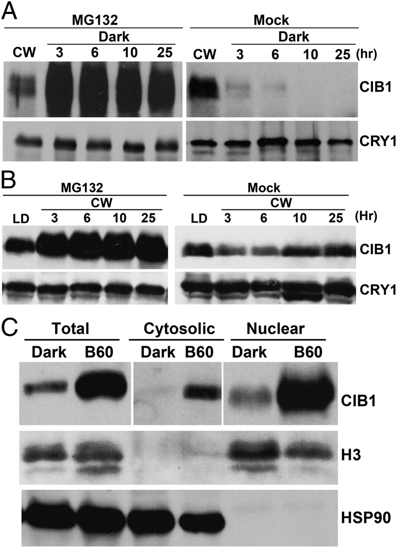
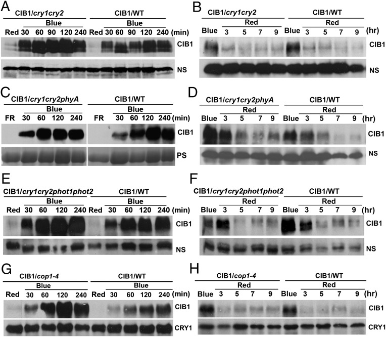
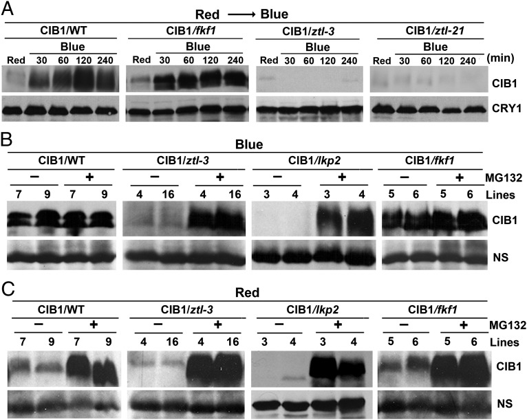
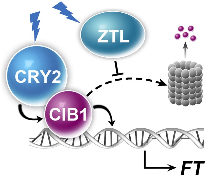
Plants possess multiple photoreceptors to mediate light regulation of growth and development, but it is not well understood how different photoreceptors coordinate their actions to jointly regulate developmental responses, such as flowering time. In Arabidopsis, the photoexcited cryptochrome 2 interacts with the transcription factor CRYPTOCHROME-INTERACTING basic helix-loop-helix 1 (CIB1) to activate transcription and floral initiation. We show that the CIB1 protein expression is regulated by blue light; CIB1 is highly expressed in plants exposed to blue light, but levels of the CIB1 protein decreases in the absence of blue light. We demonstrate that CIB1 is degraded by the 26S proteasome and that blue light suppresses CIB1 degradation. Surprisingly, although cryptochrome 2 physically interacts with CIB1 in response to blue light, it is not the photoreceptor mediating blue-light suppression of CIB1 degradation. Instead, two of the three light-oxygen-voltage (LOV)-domain photoreceptors, ZEITLUPE and LOV KELCH PROTEIN 2, but not FLAVIN-BINDING KELCH REPEAT 1, are required for the function and blue-light suppression of degradation of CIB1. These results support the hypothesis that the evolutionarily unrelated blue-light receptors, cryptochrome and LOV-domain F-box proteins, mediate blue-light regulation of the same transcription factor by distinct mechanisms.

Keywords: gene expression; photomorphogenesis; protein degradation.


Conflict of interest statement

The authors declare no conflict of interest.

Figures

Fig. 1.
Fig. 1.
The CRY2-interacting bHLH protein CIB1 is degraded in the absence of blue light. Immunoblots show the expression of the CIB1 protein in transgenic plants expressing the 35S::MycCIB1 transgene. Samples were fractionated by 10% SDS/PAGE, blotted, probed with the anti-Myc antibody, stripped, and reprobed with the anti-CRY1 antibody as the loading controls. (AC) Plants were grown in CW for 3 wk, transferred to dark (A) or red light (20 μmol⋅m−2⋅s−1) (B) or remained in CW (C) for 16 h, and then transferred to blue light (35 μmol⋅m−2⋅s−1) for the indicated time before sample collection. (DF) Three-week-old plants were grown in CW, transferred to continuous blue light (blue, 35 μmol m−2 s−1) for 16 h, and then transferred to dark (D), red light (20 μmol⋅m−2⋅s−1) (E), or far-red light (5 μmol⋅m−2⋅s−1) (F), respectively, for the indicated time before sample collection. (G and H) Results of a fluence rate response showing the CIB1 protein expression changes in response to blue light. Plants were grown in continuous red light for 3 wk and then transferred to blue light of indicated fluence rate (1–40 μmol⋅m−2⋅s−1) and time indicated before sample collection. (H) The immunoblots shown in G were analyzed by the quantitative Odyssey analysis.
Fig. 2.
Fig. 2.
The CIB1 protein is degraded in the absence of blue light by the 26S proteasome. (A and B) Immunoblot showing the inhibition of CIB1 degradation by the proteasome inhibitor MG132. Plants were grown in CW for 3 wk, and leaves were excised and incubated with MG132 (50 µmol/L) or mock solution (0.1% DMSO) in darkness (A) or white light (B) for the indicated time before sample collection. (C) Immunoblot showing the CIB1 protein in cytosolic and nuclear fractions. LD [16-h light/8-h dark (16hL/8hD)]-grown plants were transferred to dark for 16 h and then transferred to blue light (35 μmol⋅m−2⋅s−1) for 60 min. Total protein, cytosolic protein, and nuclear proteins were extracted, fractionated by 10% SDS/PAGE, blotted, and probed by the anti-Myc (CIB1), anti-histone H3 (nuclear marker), and anti-HSP90 (cytosol marker) antibodies.
Fig. 3.
Fig. 3.
Lack of effect of CRY, phyA, and COP1 on CIB1 protein expression. (A, C, E, and G) Transgenic plants expressing the 35S::MycCIB1 transgene in wild-type (WT) or the indicated mutant backgrounds (cry1cry2, cry1cry2phyA, cry1cry2phot1phot2, or cop1) were grown in LD (16hL/8hD) for 3 wk and exposed to red light (20 μmol⋅m−2⋅s−1) for 16 h (A, E, and G) or exposed to far-red light (5 μmol⋅m−2⋅s−1) for 16 h (C) and then transferred to blue light (35 μmol⋅m−2⋅s−1) for the indicated time before sample harvest. (B, D, F, and H) Alternatively, the 3-wk-old plants were exposed to blue light (35 μmol⋅m−2⋅s−1) for 16 h and then transferred to red light (20 μmol⋅m−2⋅s−1) for the indicated time. Samples were fractionated by 10% SDS/PAGE, blotted, and probed by the anti-Myc antibody (CIB1). CRY1 or nonspecific bands (NS) are shown as the loading controls. Because of uncontrolled exposure times of ECL of different immunoblots, results of different blots are not directly comparable.
Fig. 4.
Fig. 4.
ZTL and LKP2, but not FKF1, are required for the accumulation of CIB1 protein in response to blue light. (A) Immunoblot showing the lack of blue-light–dependent CIB1 accumulation in two different ztl mutant alleles. The transgenic plants expressing the 35S::MycCIB1 transgene in the wild-type (CIB1/WT) and ztl-3 (CIB1/ztl-3) and ztl-21 (CIB1/ztl-21) mutant backgrounds were grown in LD (16hL/8hD) for 3 wk, transferred to continuous red light (20 µmol⋅m−2⋅s−1) for 16 h, and then transferred to blue light (35 µmol⋅m−2⋅s−1) for the indicated time before sample collection. Immunoblot was probed with the anti-Myc antibody, stripped, and reprobed with the anti-CRY1 antibody as the loading control. (B) Immunoblots showing levels of the CIB1 protein in different genetic backgrounds treated with blue light in the absence or presence of the proteasome inhibitor MG132. The transgenic plants expressing the 35S::MycCIB1 transgene in the wild-type (CIB1/WT) or ztl (CIB1/ztl-3), lkp2 (CIB1/lkp2), or fkf1 (CIB1/fkf1) mutants were grown in LD (16hL/8hD) for 3 wk and transferred to blue light (35 µmol⋅m−2⋅s−1) for 16 h. Leaves were excised, incubated in MG132 (50 µmol/L) or mock solution (0.1% DMSO) in blue light for 3 h, and the samples were analyzed by immunoblot probed with the anti-Myc antibody. A nonspecific band (NS) is included as the loading control. Two independent transgenic lines of each genotype were tested and shown (Lines). (C) Immunoblots showing levels of the CIB1 protein in different genetic backgrounds treated with red light in the absence or presence of the proteasome inhibitor MG132. The transgenic plants expressing the 35S::MycCIB1 transgene in wild-type (WT) or ztl-3, lkp2, or fkf1 mutants were grown in LD for 3 wk and transferred to red light (20 µmol⋅m−2⋅s−1) for 16 h. Leaves were excised, incubated in MG132 (50 µmol/L) or mock solution (0.1% DMSO) under red light for 3 h, and analyzed by immunoblot probed with the anti-Myc antibody. A nonspecific band (NS) is included as the loading control. Two independent transgenic lines of each genotype were tested and shown (Lines).
Fig. 5.
Fig. 5.
The CIB1 activity promoting floral initiation is dependent on cryptochrome, ZTL, and LKP2, but independent from FKF1. (A, C, E, and G) Images of transgenic plants overexpressing CIB1 in the indicated genetic backgrounds and the respective parents. Plants were grown in LD photoperiods. (B, D, F, and H) The time to flowering, the number of rosette leaves at the time of flowering of the respective genotypes, and the SDs (n > 20) are shown.
Fig. 6.
Fig. 6.
A hypothetical model depicting CRY2- and ZTL-mediated blue-light regulation of CIB1. The model hypothesizes that in response to blue light, CRY2 interacts with CIB1 to activate the activity of CIB1 promoting transcription of the FT gene, whereas ZTL suppresses CIB1 degradation by the 26S proteasome (barrel).

References

    1. Cashmore AR. Cryptochromes: Enabling plants and animals to determine circadian time. Cell. 2003;114(5):537–543. - PubMed
    1. Sancar A. Structure and function of DNA photolyase and cryptochrome blue-light photoreceptors. Chem Rev. 2003;103(6):2203–2237. - PubMed
    1. Chaves I, et al. The cryptochromes: Blue light photoreceptors in plants and animals. Annu Rev Plant Biol. 2011;62:335–364. - PubMed
    1. Liu H, Liu B, Zhao C, Pepper M, Lin C. The action mechanisms of plant cryptochromes. Trends Plant Sci. 2011;16(12):684–691. - PMC - PubMed
    1. Ahmad M, Cashmore AR. HY4 gene of A. thaliana encodes a protein with characteristics of a blue-light photoreceptor. Nature. 1993;366(6451):162–166. - PubMed

Publication types

MeSH terms

Substances