. 2013 Oct 22;110(43):17368-73.
doi: 10.1073/pnas.1308236110. Epub 2013 Oct 7.

Serum deprivation inhibits the transcriptional co-activator YAP and cell growth via phosphorylation of the 130-kDa isoform of Angiomotin by the LATS1/2 protein kinases

Affiliations

Serum deprivation inhibits the transcriptional co-activator YAP and cell growth via phosphorylation of the 130-kDa isoform of Angiomotin by the LATS1/2 protein kinases

Jacob J Adler et al. Proc Natl Acad Sci U S A. .

Abstract

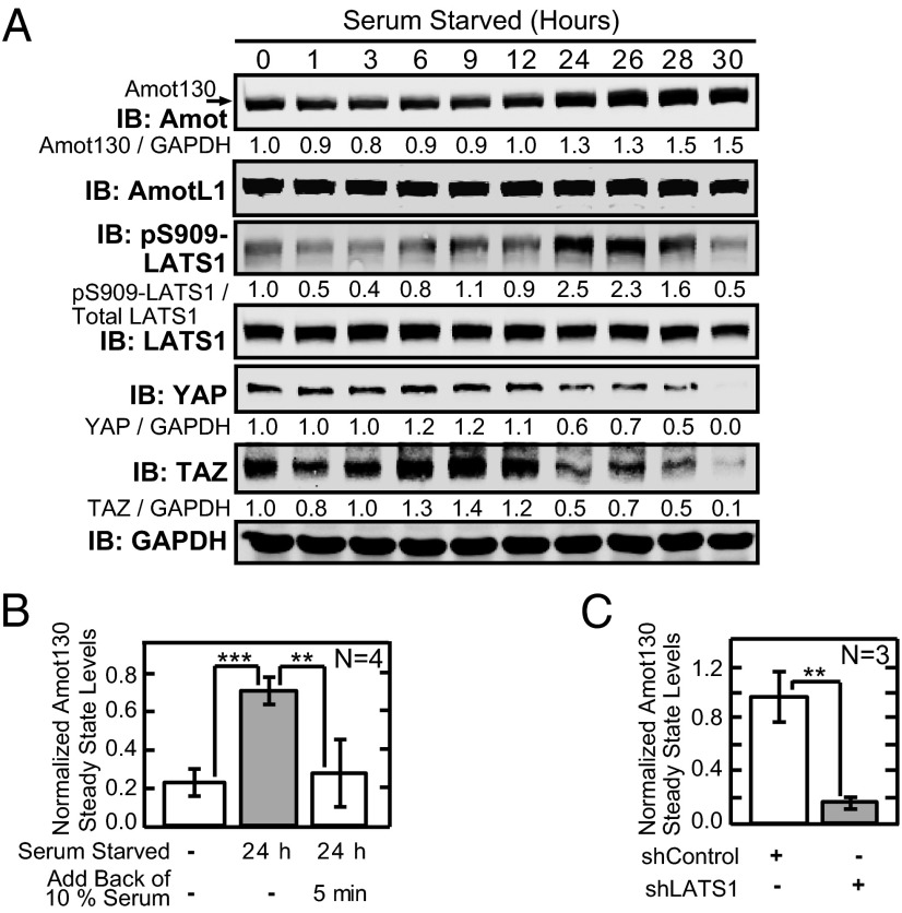
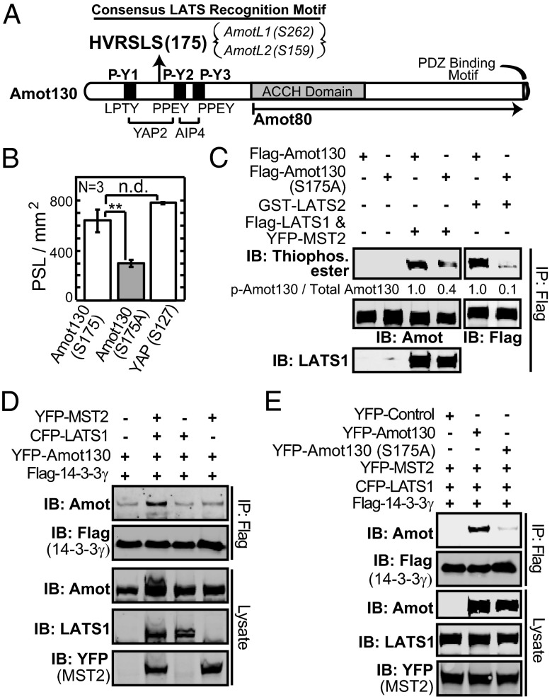
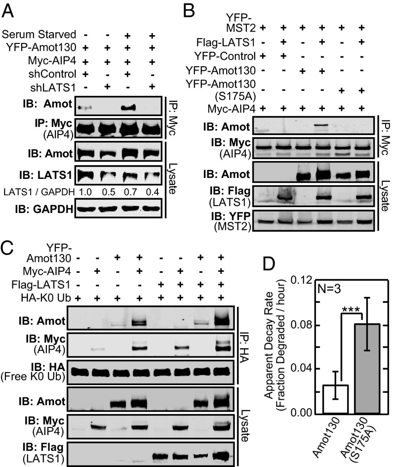
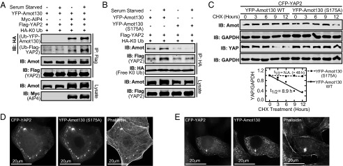
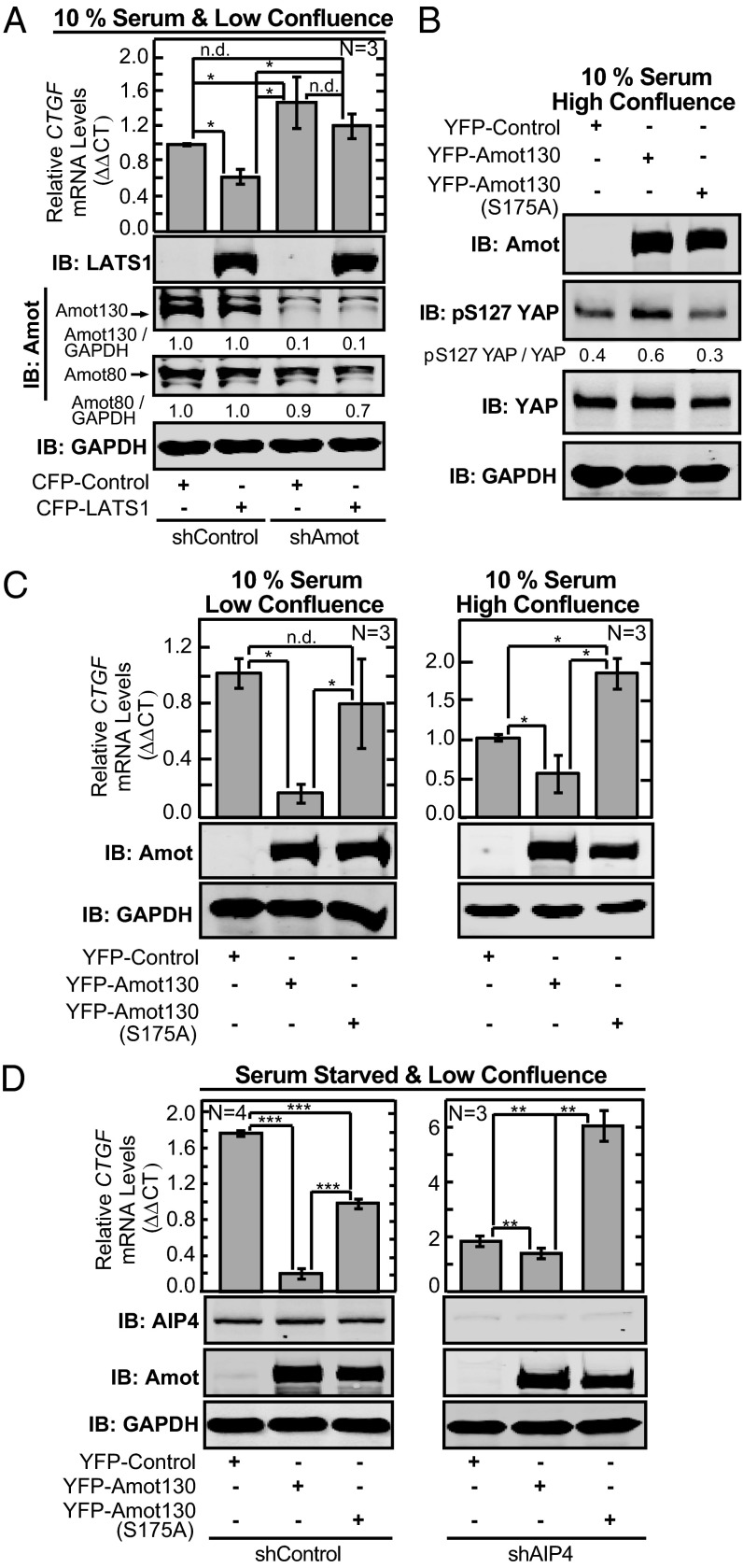
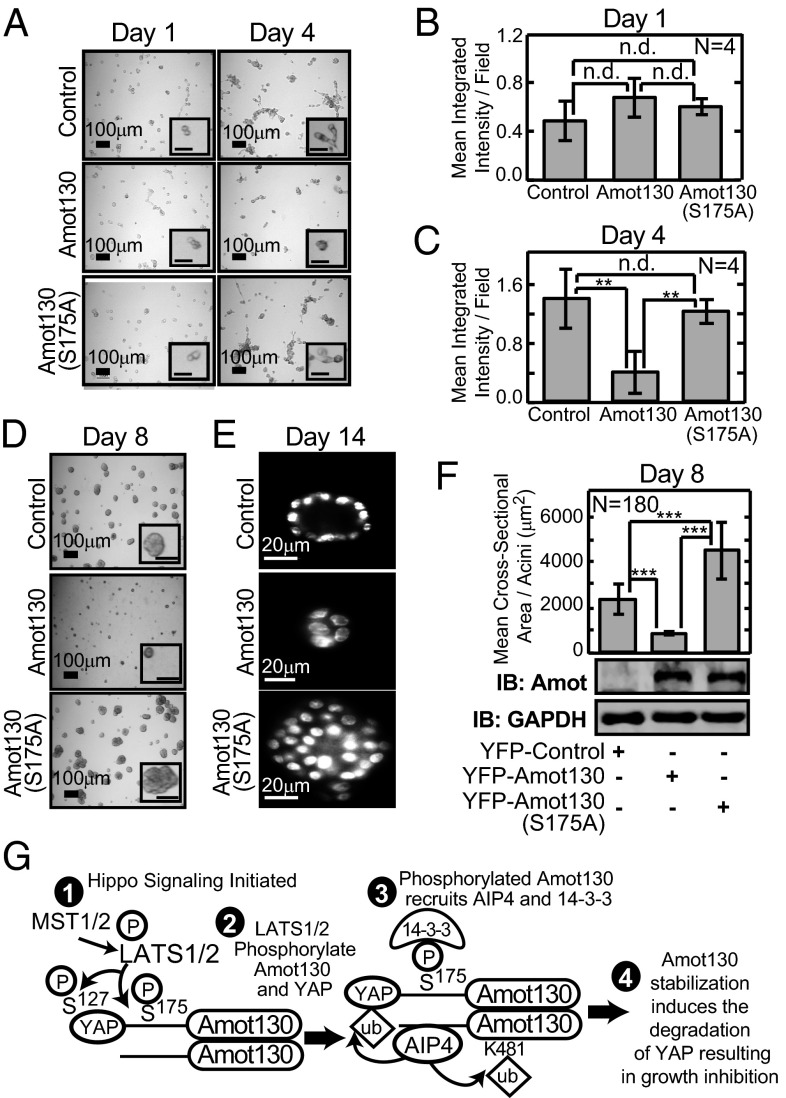
Large tumor suppressor (LATS)1/2 protein kinases transmit Hippo signaling in response to intercellular contacts and serum levels to limit cell growth via the inhibition of Yes-associated protein (YAP). Here low serum and high LATS1 activity are found to enhance the levels of the 130-kDa isoform of angiomotin (Amot130) through phosphorylation by LATS1/2 at serine 175, which then forms a binding site for 14-3-3. Such phosphorylation, in turn, enables the ubiquitin ligase atrophin-1 interacting protein (AIP)4 to bind, ubiquitinate, and stabilize Amot130. Consistently, the Amot130 (S175A) mutant, which lacks LATS phosphorylation, bound AIP4 poorly under all conditions and showed reduced stability. Amot130 and AIP4 also promoted the ubiquitination and degradation of YAP in response to serum starvation, unlike Amot130 (S175A). Moreover, silencing Amot130 expression blocked LATS1 from inhibiting the expression of connective tissue growth factor, a YAP-regulated gene. Concordant with phosphorylated Amot130 specifically mediating these effects, wild-type Amot130 selectively induced YAP phosphorylation and reduced transcription of connective tissue growth factor in an AIP4-dependent manner versus Amot130 (S175A). Further, Amot130 but not Amot130 (S175A) strongly inhibited the growth of MDA-MB-468 breast cancer cells. The dominant-negative effects of Amot130 (S175A) on YAP signaling also support that phosphorylated Amot130 transduces Hippo signaling. Likewise, Amot130 expression provoked premature growth arrest during mammary cell acini formation, whereas Amot130 (S175A)-expressing cells formed enlarged and poorly differentiated acini. Taken together, the phosphorylation of Amot130 by LATS is found to be a key feature that enables it to inhibit YAP-dependent signaling and cell growth.

Keywords: Itch; breast cancer; growth control.


Conflict of interest statement

The authors declare no conflict of interest.

Figures

Fig. 1.
Fig. 1.
Serum starvation and LATS1 increase Amot130 protein levels. (A) The levels of endogenous proteins at the indicated times after initiation of serum starvation were measured by immunoblot (IB) from lysates of MDA-MB-468 cells. Pixel intensities of endogenous proteins normalized to GAPDH and the ratios of phosphorylated Ser-909 (pS909) LATS1 to total LATS1 are provided. (B) A graph of the mean ratios of endogenous Amot130 to GAPDH from four experiments (n = 4) from HEK 293T cells grown in DMEM with 10% serum or following 24 h of no serum followed by add back of DMEM containing 0% or 10% serum for 5 min. (C) A graph showing the mean ratio of endogenous Amot130 to GAPDH from three experiments (n = 3) detected by immunoblot from lysates prepared from HEK 293T cells stably expressing control or LATS1 shRNA. Error bars represent ± SD. ***P < 0.0001; **P < 0.005.
Fig. 2.
Fig. 2.
LATS1 and LATS2 phosphorylate Amot130 at serine 175. (A) Schematic of the predicted LATS phosphorylation motifs in Amot130, AmotL1, and AmotL2. (B) Graph of the mean incorporation of [32P]phosphate from three experiments (n = 3) into the indicated immobilized 15-residue peptides by purified active LATS2. Values represent photostimulated luminescence density (PSL per mm2). (C) Immunoblot detecting the ATP-γ-S incorporated into immunoprecipitated (IP) Flag-tagged Amot130 or Amot130 (S175A) by purified LATS2 or immunoprecipitated Flag-tagged LATS1 using an antibody against thiophosphate esters (Thiophos. ester) (Top) and the ratio of phosphorylated Amot130 to total Amot130, Flag-tagged Amot130 with antibodies against Amot or Flag (Middle), or LATS1 (Bottom). (D) Immunoblot of the levels of YFP-tagged Amot130 in anti-Flag (14-3-3γ) immunoprecipitations and the indicated proteins in lysates from HEK 293T cells transfected as indicated. (E) Immunoblot of the levels of YFP-tagged Amot130 and Amot130 (S175A) in an anti-Flag (14-3-3γ) immunoprecipitation and the indicated proteins in lysates from HEK 293T cells transfected as indicated. Error bars represent ± SD. **P < 0.005; n.d., no statistical difference.
Fig. 3.
Fig. 3.
Active LATS1 drives the association of Amot130 with AIP4 resulting in the ubiquitination and increased stability of Amot130. (A) An immunoblot of YFP-tagged Amot130 in immunoprecipitates of Myc-tagged AIP4 as well as the total levels of indicated proteins in lysates from HEK 293T cells that were coinfected with the indicated combinations of shRNA and tagged proteins and then grown with or without 10% serum for 24 h. Pixel intensities of LATS1 over GAPDH (below the fourth panel) are indicated. (B) Immunoblot of YFP-tagged Amot130 or Amot130 (S175A) in lysates and immunoprecipitations of Myc-tagged AIP4 (anti-Myc) prepared from HEK 293T cells expressing the indicated combinations of proteins. (C) Immunoblot of ubiquitinated Amot130 and AIP4 in immunoprecipitates of HA-Lys-0 ubiquitin (HA-K0 Ub) along with the indicated proteins in lysates from HEK 293T cells expressing the indicated combinations of proteins. (D) A graph of the mean regression slopes from three independent experiments (n = 3) of immunoblots of the levels of YFP-tagged Amot130 or Amot130 (S175A) in lysates of MDA-MB-468 cells treated with vehicle (DMSO) or cycloheximide (CHX) for 0, 4, or 8 h. Error bars represent ± SD. ***P < 0.05.
Fig. 4.
Fig. 4.
Amot130 and AIP4, but not Amot130 (S175A), cooperatively induce the ubiquitination and degradation of YAP. (A) Immunoblot with an anti-HA antibody used to detect ubiquitinated Flag-tagged YAP2 (anti-Flag) and Amot130 in immunoprecipitations with anti-Flag antibody from HEK 293T cells expressing HA-tagged Lys-0 ubiquitin (HA-K0 Ub) and the indicated combinations of proteins. Before lysis, cells were cultured in DMEM with 10% or 0% serum for 24 h. (B) Ubiquitinated proteins were immunoprecipitated with an anti-HA antibody from HEK 293T cells expressing the indicated combinations of recombinant proteins. Individual proteins were then detected by immunoblot as indicated. (C) The levels of YFP-tagged Amot130, YFP-tagged Amot130 (S175A), CFP-tagged YAP2, and endogenous GAPDH were detected by immunoblot of lysates from MCF7 cells stably expressing these proteins following treatment for the indicated times with vehicle (DMSO) or CHX. (Lower) A graph of the ratios of CFP-tagged YAP2 to GAPDH and the resulting half-lives (t1/2) in cells expressing (▪) YFP-tagged Amot130 (S175A) or (♦) YFP-tagged Amot130. (D and E) Confocal images of fixed MCF7 cells stained with phalloidin-594 (for actin) and expressing CFP-tagged YAP2 in combination with (D) YFP-tagged Amot130 (S175A) or (E) YFP-tagged Amot130.
Fig. 5.
Fig. 5.
Phosphorylation of Amot130 at Ser-175 underlies its role in mediating Hippo signaling to inhibit YAP. (A) Real-time quantitative PCR measurements of the levels of CTGF mRNA in MDA-MB-468 cells coinfected for 24 h with lentivirus encoding combinations of Amot shRNA, CFP-tagged LATS1, or the indicated controls. (Lower) Immunoblots from paired cells validating Amot130-selective silencing and CFP-tagged LATS1 expression with pixel intensity values of Amot130 and Amot80 levels normalized to GAPDH. (B) Immunoblot of phospho-Ser-127 (pS127) YAP, total YAP, and GAPDH from MDA-MB-468 cells expressing YFP-tagged Amot130, Amot130 (S175A), or control vector. (C) Real-time quantitative PCR measurements of CTGF mRNA levels in MDA-MB-468 cells described in B and harvested at low (Left) or high confluence (Right). (D) CTGF mRNA levels from MDA-MB-468 cells coinfected with lentivirus expressing YFP-tagged Amot130, Amot130 (S175A), or control vector in combination with control shRNA (n = 4) or shRNA targeting AIP4 (n = 3), and cultured at low confluence in 0% serum for 24 h. Error bars represent ± SD. ***P < 0.0001; **P < 0.01; *P < 0.05; n.d., no statistical difference.
Fig. 6.
Fig. 6.
Phosphorylation of Amot130 at Ser-175 is essential for it to inhibit cell growth. (AC) MDA-MB-468 cells stably expressing YFP-tagged Amot130, Amot130 (S175A), or control vector were seeded onto Matrigel and imaged after 1 and 4 d. (AC) Representative bright-field stereo images (A) and plots of the mean pixel integrated intensity values per field from four experiments (n = 4) (B and C) at (B) day 1 and (C) day 4 are presented. (DF) MCF10A cells stably expressing proteins as in A were seeded onto Matrigel and grown for 14 d. (D) Bright-field stereo images of colonies at day 8 and (E) confocal fluorescence images of representative acini stained with Hoechst for nuclei at day 14 of growth. (F) Plots of the mean cross-sectional area per acini (μm2) from three experiments each with 60 acini (n = 180) at day 8 are graphed. (Lower) Immunoblot measuring YFP-tagged Amot130 or Amot130 (S175A) and GAPDH from cells extracted from Matrigel at day 8. (G) Model of the proposed mechanism for activation of Amot130 by serum deprivation and LATS resulting in the inhibition of YAP activity. Error bars represent ± SD. ***P < 1.0 × 10−60; **P < 0.01; n.d., no statistical difference. (Unlabeled scale bars, 50 μm.)

Similar articles

Cited by

References

    1. Zhao B, et al. Inactivation of YAP oncoprotein by the Hippo pathway is involved in cell contact inhibition and tissue growth control. Genes Dev. 2007;21(21):2747–2761. - PMC - PubMed
    1. Yu F-X, et al. Regulation of the Hippo-YAP pathway by G-protein-coupled receptor signaling. Cell. 2012;150(4):780–791. - PMC - PubMed
    1. Miller E, et al. Identification of serum-derived sphingosine-1-phosphate as a small molecule regulator of YAP. Chem Biol. 2012;19(8):955–962. - PubMed
    1. Oka T, Mazack V, Sudol M. Mst2 and Lats kinases regulate apoptotic function of Yes kinase-associated protein (YAP) J Biol Chem. 2008;283(41):27534–27546. - PubMed
    1. Pan D. Hippo signaling in organ size control. Genes Dev. 2007;21(8):886–897. - PubMed

Publication types

MeSH terms

Substances