. 2013 Oct 22;110(43):17456-61.
doi: 10.1073/pnas.1307309110. Epub 2013 Oct 7.

Identification and grafting of a unique peptide-binding site in the Fab framework of monoclonal antibodies

Affiliations

Identification and grafting of a unique peptide-binding site in the Fab framework of monoclonal antibodies

Joshua M Donaldson et al. Proc Natl Acad Sci U S A. .

Abstract

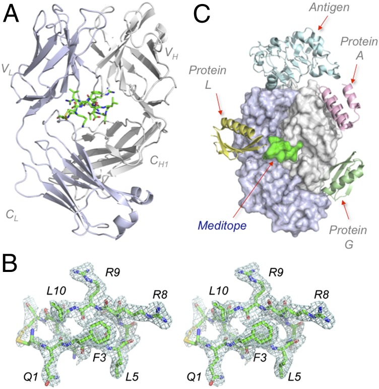
Capitalizing on their extraordinary specificity, monoclonal antibodies (mAbs) have become one of the most reengineered classes of biological molecules. A major goal in many of these engineering efforts is to add new functionality to the parental mAb, including the addition of cytotoxins and imaging agents for medical applications. Herein, we present a unique peptide-binding site within the central cavity of the fragment antigen binding framework region of the chimeric, anti-epidermal growth factor receptor mAb cetuximab. We demonstrate through diffraction methods, biophysical studies, and sequence analysis that this peptide, a meditope, has moderate affinity for the Fab, is specific to cetuximab (i.e., does not bind to human IgGs), and has no significant effect on antigen binding. We further demonstrate by diffraction studies and biophysical methods that the meditope binding site can be grafted onto the anti-human epidermal growth factor receptor 2 mAb trastuzumab, and that the antigen binding affinity of the grafted trastuzumab is indistinguishable from the parental mAb. Finally, we demonstrate a bivalent meditope variant binds specifically and stably to antigen-bearing cells only in the presence of the meditope-enabled mAbs. Collectively, this finding and the subsequent characterization and engineering efforts indicate that this unique interface could serve as a noncovalent "linker" for any meditope-enabled mAb with applications in multiple mAb-based technologies including diagnostics, imaging, and therapeutic delivery.

Keywords: cancer; molecular recognition; protein engineering.


Conflict of interest statement

Conflict of interest statement: D.A.H. and J.C.W. have founded Meditope Biosciences, a company based on some of the observations presented here.

Figures

Fig. 1.
Fig. 1.
Meditope binds to cetuximab Fab framework. (A) Ribbon representation of cetuximab Fab (light chain, blue-white; heavy chain, white) with a stick representation of the cyclic CQFDLSTRRLKC meditope (green). (B) Electron density maps of cQFD meditope, in stereo and contoured at 1σ. (C) Superposition of the cetuximab-EGFR (1YY9) structure to Fab framework ligands: protein L (1HEZ), protein A (1DEE), protein G (1QKZ), and cQFD meditope (4GW1).
Fig. 2.
Fig. 2.
Biophysical characterization of meditope–Fab interaction. (A) SPR measurements of immobilized cetuximab Fab with 20, 4, 0.8, 0.16, and 0.032 μM cQFD meditope and 100, 20, 4, 0.8, and 0.16 μM cQYN meditope passing over the chip. (B) SPR saturation experiments using immobilized cetuximab Fab and scFv and passing EGFRdIII (Upper) or cQFD meditope (Lower). Also, Inset is a schematic representation of the scFv and the Fab. (C) SPR measurements of immobilized EGFRdIII with 10, 5, 2.5, 1.3, 0.63, and 0.31 nM cetuximab Fab passed over in the absence (Upper) or presence of 10 μM cQFD meditope (Lower). (D) SEC of cetuximab Fab, EGFRdIII, MFC, Fab–EGFRdIII and Fab–MFC complexes and an admixture of all three. Each trace has been normalized to one. Shown on Left is a nonreducing SDS/PAGE of the new peak (shaded pink on SEC chromatogram) formed from the admixture.
Fig. 3.
Fig. 3.
Critical framework residues that confer meditope binding. (A) Alignment of cetuximab, CH14.18, trastuzumab, rituximab, and M425 light and heavy chain residues within 5 Å of the meditope-binding site. Magenta bars denote the combination of residues unique to cetuximab in Kabat notation. (B) A stereoview highlighting Arg9 of the cQFD meditope (green) and its occupation of a distinct pocket in cetuximab (yellow). Trastuzumab Fab (1N8Z; gray carbons) is superimposed onto cetuximab. Red dotted lines indicate salt bridge from Asp85 to the meditope Arg9 and Leu10. (C) Rotated by ∼90°, the hydrogen bond between Asn41 of cetuximab Fab to Thr7 of the meditope is shown. Also shown is the extended side chain of Arg8 of the meditope making a backbone hydrogen bond to Gln111 in the heavy chain.
Fig. 4.
Fig. 4.
Meditope-enabling of trastuzumab. (A) Sequence alignment of light and heavy chains of parental and memAb trastuzumab showing residues, in Kabat notation, mutated to confer meditope binding. (B) SPR sensogram showing the binding of 16, 8, 4, 2, 1, 0.5, 0.25, and 0.063 µM cQFD meditope to immobilized memAb trastuzumab ligand. (C) Superposition of memAb (yellow carbons) and parental trastuzumab (cyan carbons) with expanded view of critical mutations in meditope-enable trastuzumab. Cetuximab (white carbons) is included for comparison. (D). Stereoviews of the superposition of the meditope bound to memAb trastuzumab (yellow carbons) and cetuximab (white carbons). Phe3 and Leu10 are highlighted in Upper; Leu5, Arg8, and Arg-9 in Lower. The orientation of the disulfide bond between the terminal cysteines, however, is slightly different in the memAb trastuzumab.
Fig. 5.
Fig. 5.
Specific binding of meditope–Fc to memAb-labeled cells. (A) A schematic demonstrating the proposed enhanced affinity of the bivalent meditope to antigen overexpressing cells that are pretreated with a meditope-enabled mAb. (B) FACS analysis of AF488-labeled meditope–Fc (MFC) binding to untreated, AF647-labeled cetuximab-treated (C) or M425-treated (M) MDA-MB-468 cells. Monomeric meditope (cQFD) did not show appreciable binding. (C) Wide-field fluorescent microscopy showing meditope–Fc colocalization with cetuximab on MDA-MB-468 cells, but not to M425 pretreated cells (left two columns). Likewise, meditope–Fc colocalized with memAb trastuzumab, but not to parental trastuzumab pretreated SKBR3 cells. (Scale bars: 10 μm.)

Similar articles

Cited by

References

    1. Weiner LM, Murray JC, Shuptrine CW. Antibody-based immunotherapy of cancer. Cell. 2012;148(6):1081–1084. - PMC - PubMed
    1. Scott AM, Wolchok JD, Old LJ. Antibody therapy of cancer. Nat Rev Cancer. 2012;12(4):278–287. - PubMed
    1. Desjarlais JR, Lazar GA. Modulation of antibody effector function. Exp Cell Res. 2011;317(9):1278–1285. - PubMed
    1. Wolf E, Hofmeister R, Kufer P, Schlereth B, Baeuerle PA. BiTEs: Bispecific antibody constructs with unique anti-tumor activity. Drug Discov Today. 2005;10(18):1237–1244. - PubMed
    1. Goldenberg DM, et al. Pretargeted molecular imaging and radioimmunotherapy. Theranostics. 2012;2(5):523–540. - PMC - PubMed

Publication types

MeSH terms