. 2013 Oct 22;110(43):17380-5.
doi: 10.1073/pnas.1304266110. Epub 2013 Oct 7.

Cancer cell exosomes depend on cell-surface heparan sulfate proteoglycans for their internalization and functional activity

Affiliations

Cancer cell exosomes depend on cell-surface heparan sulfate proteoglycans for their internalization and functional activity

Helena C Christianson et al. Proc Natl Acad Sci U S A. .

Abstract

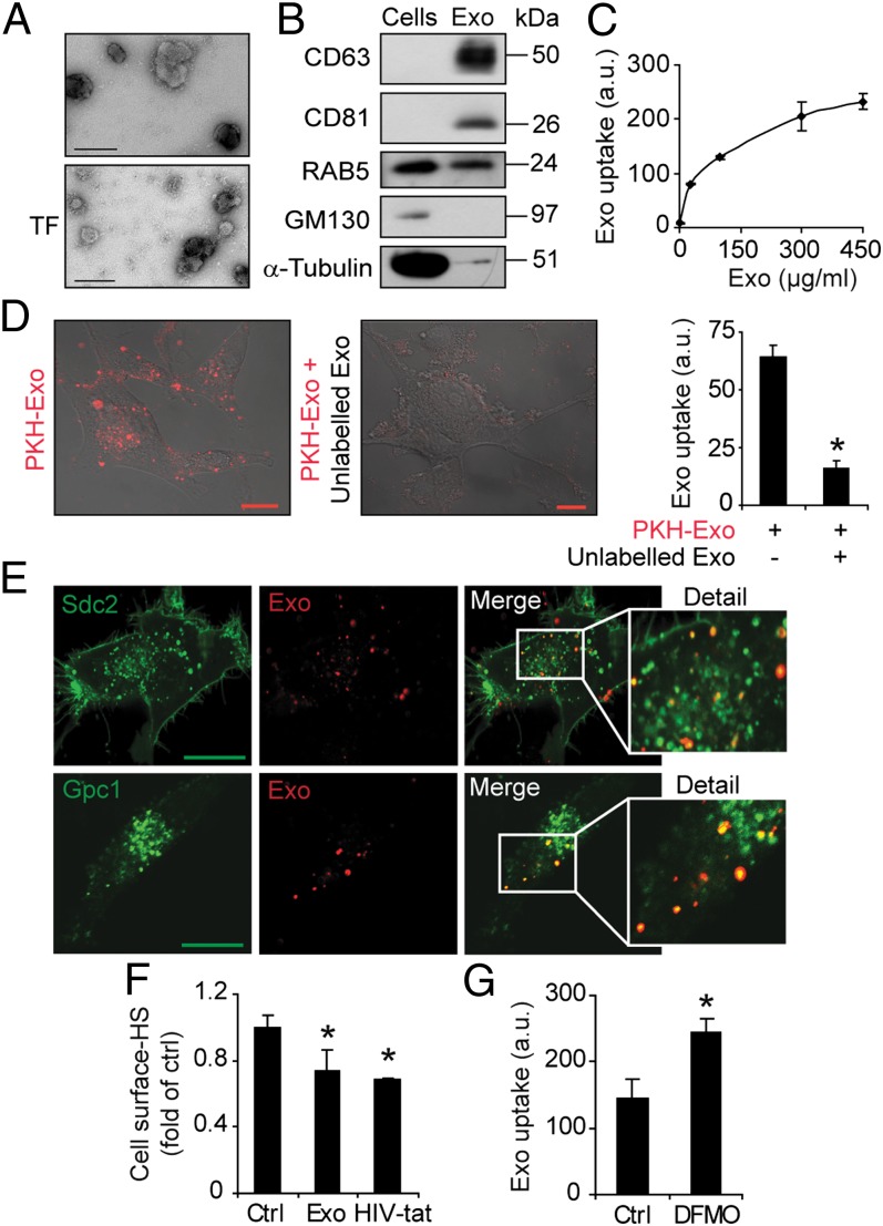
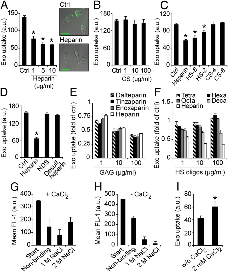
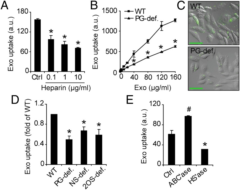
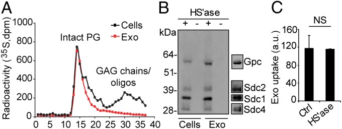
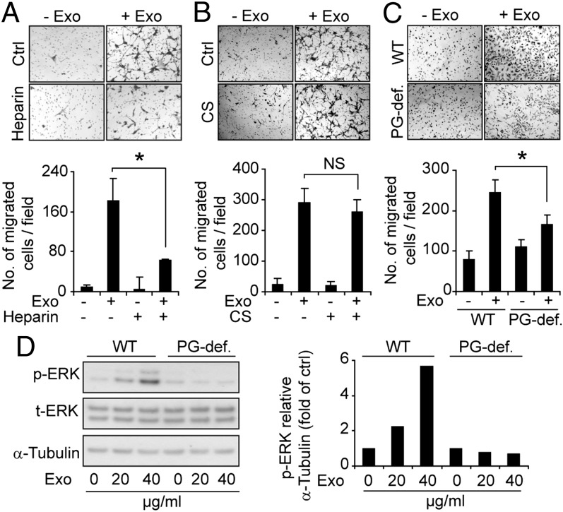
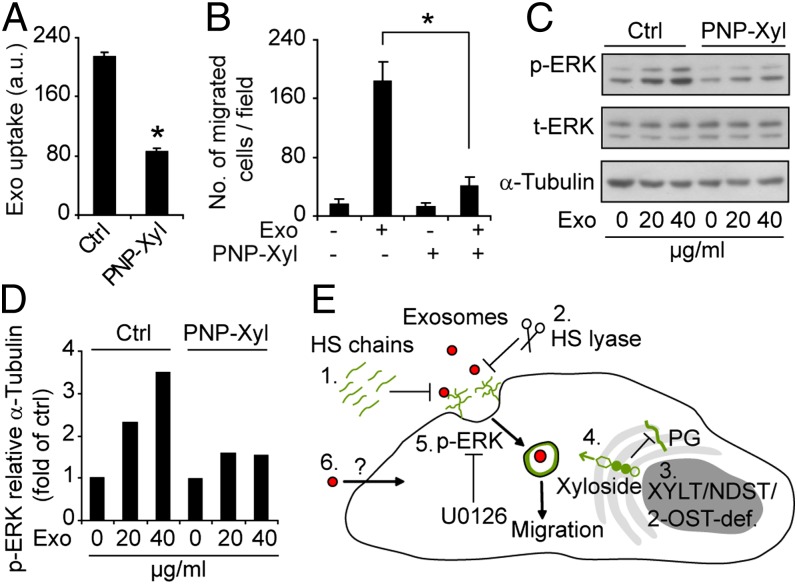
Extracellular vesicle (EV)-mediated intercellular transfer of signaling proteins and nucleic acids has recently been implicated in the development of cancer and other pathological conditions; however, the mechanism of EV uptake and how this may be targeted remain as important questions. Here, we provide evidence that heparan sulfate (HS) proteoglycans (PGs; HSPGs) function as internalizing receptors of cancer cell-derived EVs with exosome-like characteristics. Internalized exosomes colocalized with cell-surface HSPGs of the syndecan and glypican type, and exosome uptake was specifically inhibited by free HS chains, whereas closely related chondroitin sulfate had no effect. By using several cell mutants, we provide genetic evidence of a receptor function of HSPG in exosome uptake, which was dependent on intact HS, specifically on the 2-O and N-sulfation groups. Further, enzymatic depletion of cell-surface HSPG or pharmacological inhibition of endogenous PG biosynthesis by xyloside significantly attenuated exosome uptake. We provide biochemical evidence that HSPGs are sorted to and associate with exosomes; however, exosome-associated HSPGs appear to have no direct role in exosome internalization. On a functional level, exosome-induced ERK1/2 signaling activation was attenuated in PG-deficient mutant cells as well as in WT cells treated with xyloside. Importantly, exosome-mediated stimulation of cancer cell migration was significantly reduced in PG-deficient mutant cells, or by treatment of WT cells with heparin or xyloside. We conclude that cancer cell-derived exosomes use HSPGs for their internalization and functional activity, which significantly extends the emerging role of HSPGs as key receptors of macromolecular cargo.

Keywords: endocytosis; glioma; tumor.


Conflict of interest statement

The authors declare no conflict of interest.

Figures

Fig. 1.
Fig. 1.
Exosome-like EVs from GBM cells colocalize with cell-surface HSPGs. (A) Representative EM image shows EVs with a typical diameter of approximately 150 nm and positive staining for CD63 (Upper) and tissue factor (TF; Lower). (Scale bar, 100 nm.) (B) Equal amounts of total protein from EV and donor cell lysates were analyzed by immunoblotting for RAB5, CD63, CD81, α-tubulin, and GM130. (C) GBM cells were incubated with the indicated concentrations of PKH-labeled exosomes. Flow cytometry analysis shows dose-dependent and saturable uptake of exosomes. (D) Confocal microscopy analysis of GBM cells incubated with PKH26-labeled exosomes (10 µg/mL) in the absence (Left) or in the presence (Center) of 100 µg/mL unlabeled exosomes. (Right) Quantitative data from similar experiment (Left) analyzed by flow cytometry. (E) GBM cells transfected with syndecan-2–GFP (Sdc2-GFP; Upper) or glypican-1–GFP (Gpc1-GFP; Lower) encoding plasmid were incubated with PKH-labeled exosomes (10 µg/mL) for 30 min, washed with 1 M NaCl, and analyzed by confocal microscopy. Merged images of the indicated areas show colocalization of exosomes and Sdc2-GFP and Gpc1-GFP, respectively. (Scale bar, 20 µm.) (F) GBM cells were untreated (Ctrl) or incubated with exosomes or HIV-Tat peptide, followed by extensive washing to remove remaining cell surface-bound ligand, followed by staining with anti-HS antibody and flow cytometry analysis. (G) GBM cells were grown in the absence (Ctrl) or presence of 5 mM DFMO. Cells were then incubated with PKH-labeled exosomes (40 µg/mL) for 1 h and analyzed for uptake by flow cytometry. Data shown represent the mean ± SD from three independent experiments, each performed in triplicate (C) or duplicate (D and G). (F) Data are presented as fold of untreated control cells, and are the mean ± SD from three independent experiments, each performed in triplicate (*P < 0.05).
Fig. 2.
Fig. 2.
Effects of free heparin, HS, and CS chains on exosome uptake. (A) (Left) GBM cells were incubated with PKH-labeled exosomes (40 µg/mL) for 1 h in the absence (Ctrl) or in the presence of 1, 5, or 10 µg/mL heparin, and then analyzed for exosome uptake by flow cytometry. (Right) Representative confocal microscopy images of cells incubated with exosomes without (Ctrl; Upper) or with 10 µg/mL heparin (Lower). (B) Same experiments as in A without (Ctrl) or with 1, 10, or 100 µg/mL CS. (C) Same experiment as in A without (Ctrl) or with 10 µg/mL heparin, HS-6, HS-2, 4-O-sulfated CS (CS-4), or 6-O-sulfated CS (CS-6). (D) Same experiment as in A without (Ctrl) or with 10 µg/mL heparin, N-desulfated heparin (NDS), or completely desulfated heparin. (E) Same experiment as in A without (Ctrl) or with 1, 10, or 100 µg/mL full-length heparin, or either of LMHWs enoxaparin, dalteparin, or tinzaparin. (F) Same experiment as in A without (Ctrl) or with 1, 10, or 100 µg/mL heparin, or heparin oligosaccharides, as indicated. (G and H) Heparin-agarose and PKH-labeled exosomes were incubated with (G) or without (H) 2 mM CaCl2, and the fraction of nonbinding and binding (1 M and 2 M NaCl) exosomes was determined by fluorescence analyses. (I) Uptake of PKH-labeled exosomes with or without 2 mM CaCl2 was determined by flow cytometry analysis. Data shown represent the mean ± SD from three independent experiments, each performed in triplicate (AD) or duplicate (GI). (E and F) Data are presented as fold of untreated control cells, and are the mean ± SD from three independent experiments, each performed in triplicate (*P < 0.05).
Fig. 3.
Fig. 3.
Exosome uptake depends on cell-surface HSPGs. (A) WT CHO cells were incubated with PKH-labeled exosomes (40 µg/mL) for 1 h without (Ctrl) or with 0.1, 1, or 10 µg/mL heparin, and analyzed for exosome uptake by flow cytometry. (B) WT and PG-deficient (PG-def.) mutant CHO cells were incubated with the indicated concentrations of PKH-labeled exosomes, and analyzed for exosome uptake by flow cytometry. (C) WT (Upper) and PG-deficient (Lower) CHO cells were incubated with PKH-labeled exosomes (10 µg/mL) for 30 min and analyzed by confocal microscopy. (Scale bar, 50 µm.) (D) WT, PG-deficient, HS N-deacetylase sulfotransferase (NDST)–deficient (NS-def.), and HS 2-O-sulfotransferase–deficient (2OS-def.) mutant CHO cells were incubated with PKH-labeled exosomes (40 µg/mL) for 1 h and analyzed by flow cytometry. (E) GBM cells were untreated (Ctrl) or pretreated with chondroitinase ABC lyase (ABC’ase) or heparinase I and III lyases (HS’ase) to deplete cell-surface CSPG and HSPG, respectively. Uptake of PKH-labeled exosomes (40 µg/mL) for 1 h was determined by flow cytometry. Data shown represent the mean ± SD from three independent experiments, each performed in triplicate (A, B, and E). (D) Data are presented as fold of WT cells, and are the mean ± SD from three independent experiments, each performed in triplicate (*Significant decrease at P < 0.05 vs. control or WT cells; #Significant increase at P < 0.05 vs. control).
Fig. 4.
Fig. 4.
HSPGs are sorted to exosome vesicles but have no role in their uptake. (A) GBM cells were metabolically labeled with [35S]sulfate, and 35S-PGs and GAGs/oligosaccharides from cell and exosome lysates were size-fractionated on a Superose 6 column as described in SI Materials and Methods. (B) Isolated PGs from lysates of exosomes or GBM cells were untreated (−) or digested with heparinase III and ABC lyases (+). The digest products were separated by SDS page, and HSPG core proteins were visualized by immunoblotting with 3G10 anti-ΔHS antibody. Typical positions of members of the glypican and syndecan HSPG families are indicated (Right). (C) PKH-labeled exosomes (40 μg/mL) were untreated (Ctrl) or digested with heparinase I and III lyases (HS’ase), and then analyzed for uptake by GBM cells for 1 h by flow cytometry. Data shown represent the mean ± SD from three independent experiments, each performed in duplicate. NS, not significant (i.e., no statistically significant difference).
Fig. 5.
Fig. 5.
Exosome-mediated cell migration and signaling depend on HSPG. Transwell migration of GBM cells in the absence or presence of exosomes and with or without heparin (A) or CS (B) as indicated. (C) Migration of WT and mutant PG-deficient (PG-def.) CHO cells in the absence or presence of exosomes as indicated. (Upper) Representative images of migrated cells. (Lower) Quantitative data on number of migrated cells per field (n = 9). Data shown represent the mean ± SD from three independent experiments, each performed in triplicate [*P < 0.05; NS, not significant (i.e., no statistically significant difference)]. (D) WT and PG-deficient CHO cells were treated with the indicated concentrations of exosomes for 20 min, and cell lysates were analyzed by immunoblotting for p-ERK1/2, total ERK (t-ERK), and α-tubulin. (Left) Western blot from a representative experiment. (Right) Quantitative data of p-ERK/α-tubulin ratios in WT and PG-deficient cells.
Fig. 6.
Fig. 6.
Small-molecule inhibitor of PG biosynthesis reduces exosome uptake and attenuates exosome-mediated cell migration and signaling activation. (A) GBM cells were untreated (Ctrl) or pretreated with the PG biosynthesis inhibitor PNP-Xyl for 48 h, and then incubated with PKH-labeled exosomes (40 µg/mL) for 1 h, followed by flow cytometry analysis of exosome uptake. (B) Migration of untreated (Ctrl) or PNP-Xyl–pretreated GBM cells in the absence or presence of exosomes. Data shown represent the mean ± SD from three independent experiments, each performed in triplicate (*P < 0.05). (C and D) GBM cells were untreated or pretreated with PNP-Xyl and then incubated with exosomes for 20 min as indicated. Cell lysates were analyzed by immunoblotting for p-ERK1/2, total ERK (t-ERK), and α-tubulin. (C) Western blot from a representative experiment. (D) Quantitative data of p-ERK/α-tubulin ratios with the different treatments. (E) Schematic summary of major findings of the present work; 1, Exogenous HS inhibits exosome uptake in a size and charge dependent manner; 2, Enzymatic depletion of cell-surface HSPGs inhibits exosome uptake; 3, Genetic deficiency in XYLT, which catalyzes the initial step in PG formation, or NDST, which catalyses HS N-deacetylation/sulfation, or 2-O-sulfotransferase (2-OST), which catalyses HS 2-O-sulfation, all result in reduced exosome uptake; 4, Xyloside, i.e., small-molecule inhibitors of PG biosynthesis, inhibit exosome uptake; 5, Exosome-mediated ERK1/2 activation and cell migration are HSPG-dependent: and 6, Our data indicate the existence of alternative, HSPG-independent modes of internalization; however, we provide evidence that the HSPG-dependent pathway is essential for the functional activity of exosomes.

Similar articles

Cited by

References

    1. Raposo G, et al. B lymphocytes secrete antigen-presenting vesicles. J Exp Med. 1996;183(3):1161–1172. - PMC - PubMed
    1. Ratajczak J, Wysoczynski M, Hayek F, Janowska-Wieczorek A, Ratajczak MZ. Membrane-derived microvesicles: Important and underappreciated mediators of cell-to-cell communication. Leukemia. 2006;20(9):1487–1495. - PubMed
    1. Valadi H, et al. Exosome-mediated transfer of mRNAs and microRNAs is a novel mechanism of genetic exchange between cells. Nat Cell Biol. 2007;9(6):654–659. - PubMed
    1. Belting M, Wittrup A. Nanotubes, exosomes, and nucleic acid-binding peptides provide novel mechanisms of intercellular communication in eukaryotic cells: Implications in health and disease. J Cell Biol. 2008;183(7):1187–1191. - PMC - PubMed
    1. Al-Nedawi K, Meehan B, Rak J. Microvesicles: Messengers and mediators of tumor progression. Cell Cycle. 2009;8(13):2014–2018. - PubMed

Publication types

MeSH terms