. 2013 Nov 15;191(10):5124-38.
doi: 10.4049/jimmunol.1301415. Epub 2013 Oct 7.

Asymptomatic HLA-A*02:01-restricted epitopes from herpes simplex virus glycoprotein B preferentially recall polyfunctional CD8+ T cells from seropositive asymptomatic individuals and protect HLA transgenic mice against ocular herpes

Affiliations

Asymptomatic HLA-A*02:01-restricted epitopes from herpes simplex virus glycoprotein B preferentially recall polyfunctional CD8+ T cells from seropositive asymptomatic individuals and protect HLA transgenic mice against ocular herpes

Xavier Dervillez et al. J Immunol. .

Abstract

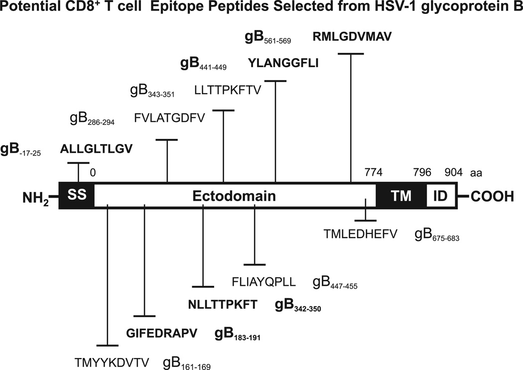
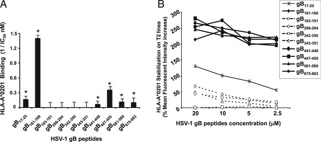
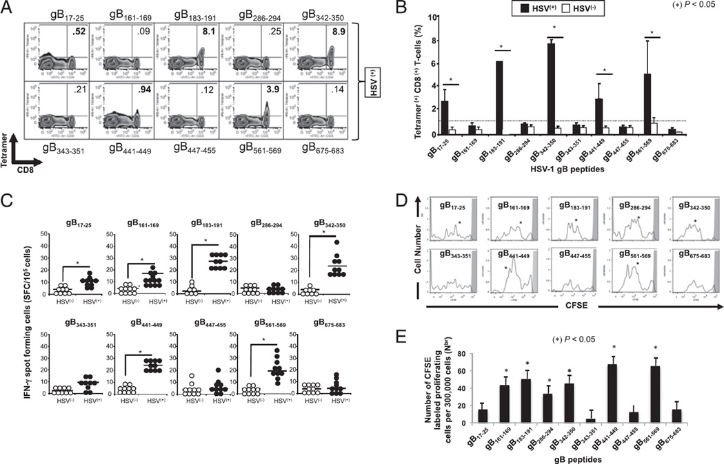
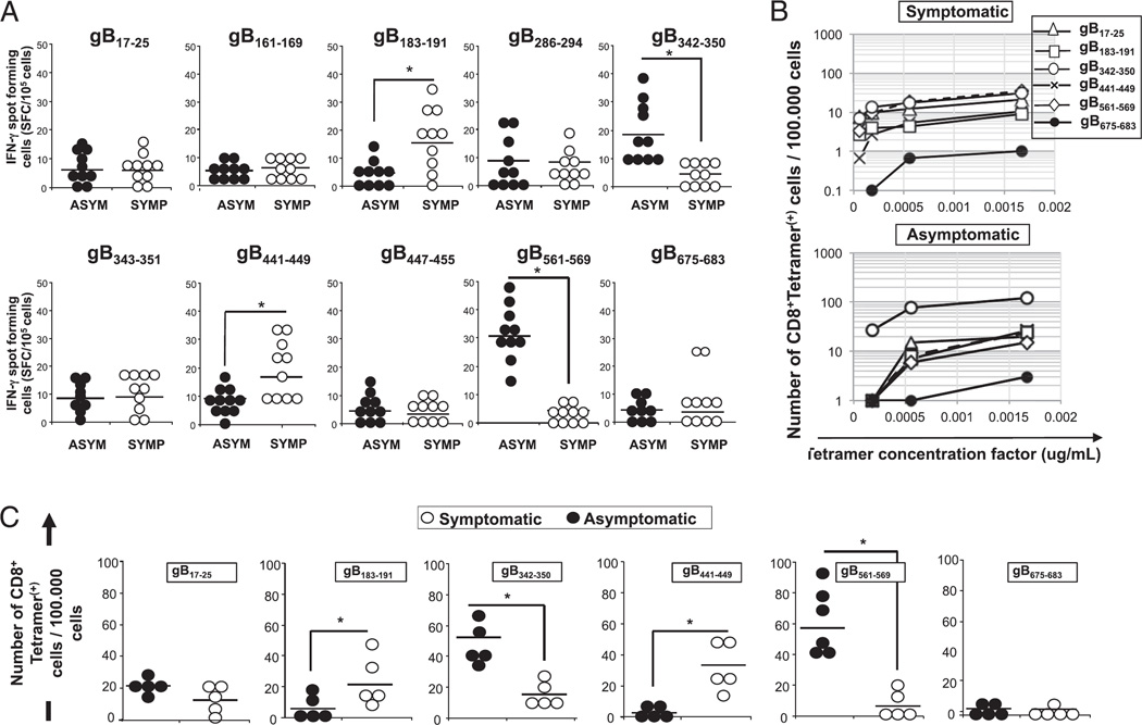
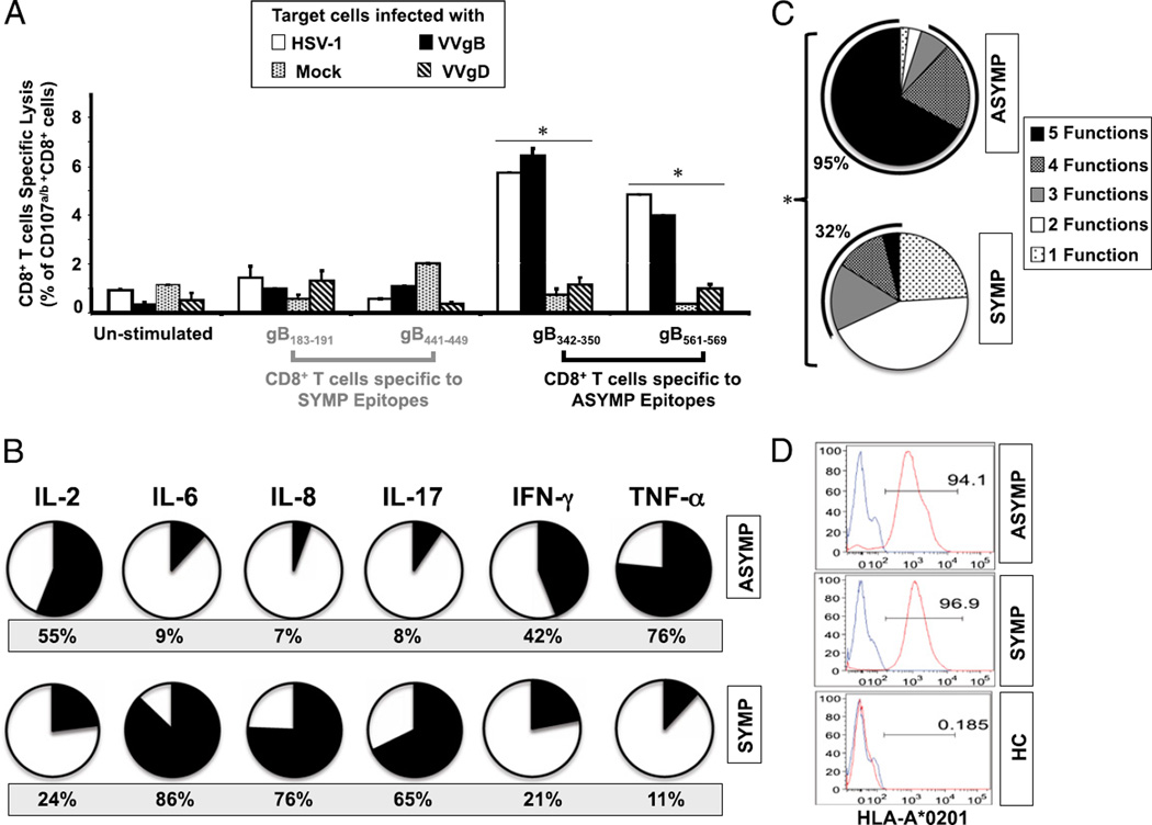
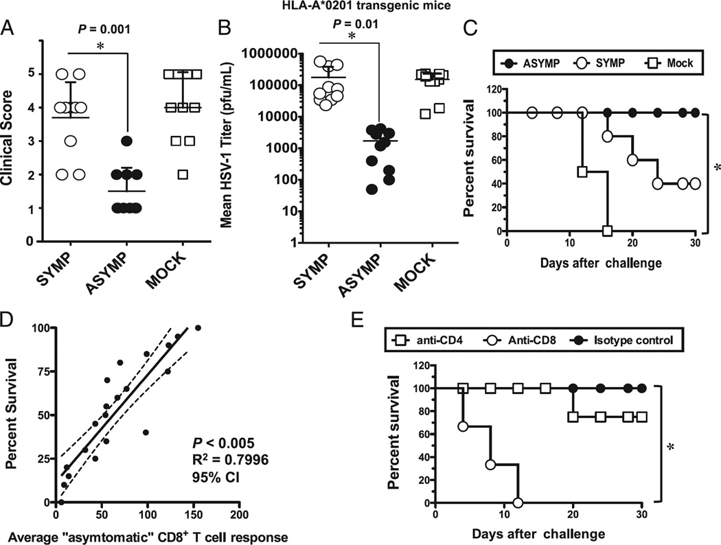
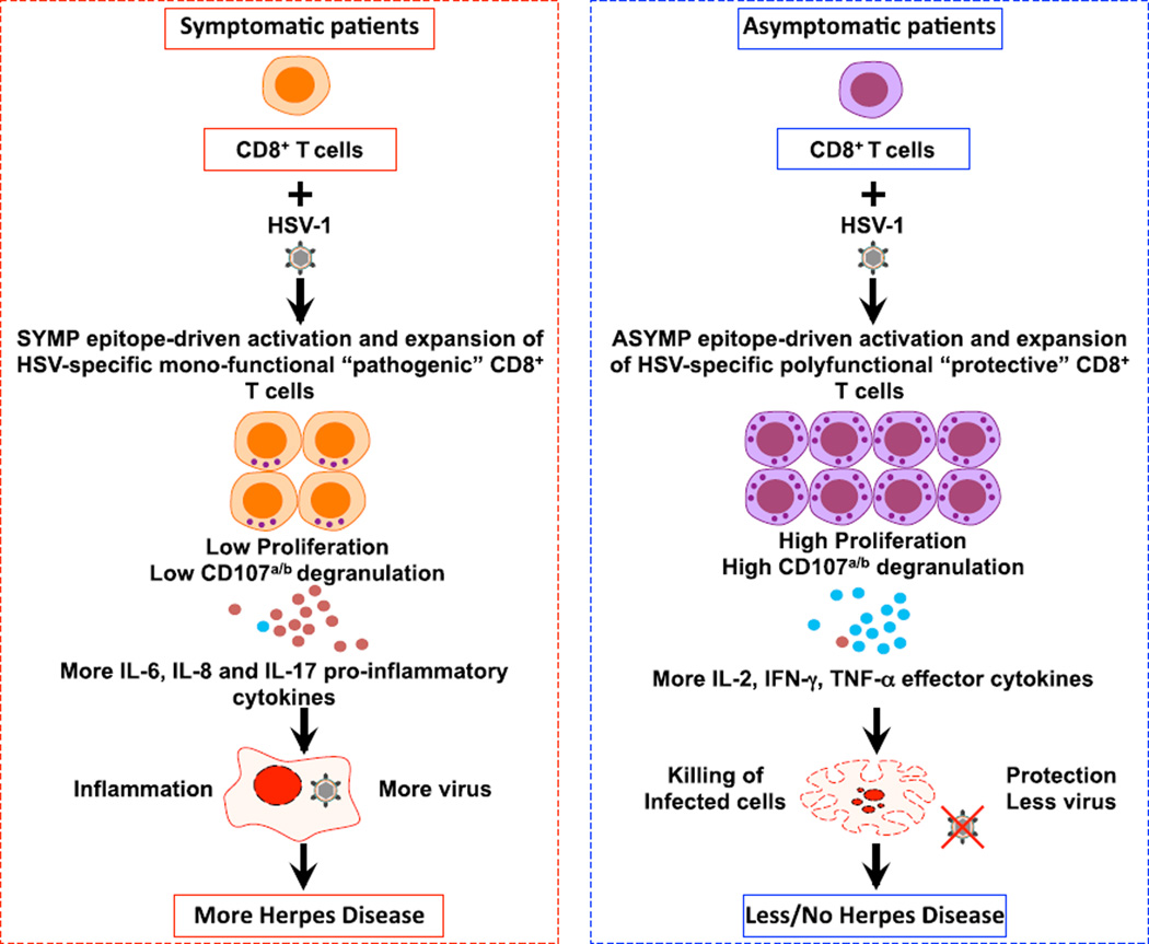
Evidence from C57BL/6 mice suggests that CD8(+) T cells, specific to the immunodominant HSV-1 glycoprotein B (gB) H-2(b)-restricted epitope (gB498-505), protect against ocular herpes infection and disease. However, the possible role of CD8(+) T cells, specific to HLA-restricted gB epitopes, in protective immunity seen in HSV-1-seropositive asymptomatic (ASYMP) healthy individuals (who have never had clinical herpes) remains to be determined. In this study, we used multiple prediction algorithms to identify 10 potential HLA-A*02:01-restricted CD8(+) T cell epitopes from the HSV-1 gB amino acid sequence. Six of these epitopes exhibited high-affinity binding to HLA-A*02:01 molecules. In 10 sequentially studied HLA-A*02:01-positive, HSV-1-seropositive ASYMP individuals, the most frequent, robust, and polyfunctional CD8(+) T cell responses, as assessed by a combination of tetramer, IFN-γ-ELISPOT, CFSE proliferation, CD107a/b cytotoxic degranulation, and multiplex cytokine assays, were directed mainly against epitopes gB342-350 and gB561-569. In contrast, in 10 HLA-A*02:01-positive, HSV-1-seropositive symptomatic (SYMP) individuals (with a history of numerous episodes of recurrent clinical herpes disease) frequent, but less robust, CD8(+) T cell responses were directed mainly against nonoverlapping epitopes (gB183-191 and gB441-449). ASYMP individuals had a significantly higher proportion of HSV-gB-specific CD8(+) T cells expressing CD107a/b degranulation marker and producing effector cytokines IL-2, IFN-γ, and TNF-α than did SYMP individuals. Moreover, immunization of a novel herpes-susceptible HLA-A*02:01 transgenic mouse model with ASYMP epitopes, but not with SYMP epitopes, induced strong CD8(+) T cell-dependent protective immunity against ocular herpes infection and disease. These findings should guide the development of a safe and effective T cell-based herpes vaccine.


Conflict of interest statement

Disclosures

The authors have no financial conflicts of interest.

Figures

FIGURE 1
FIGURE 1
Schematic representation showing the relative location within HSV-1 gB of the potential CD8+ T cell epitopes studied. HSV-1 (strain 17) gB regions carrying potential HLA A*0201 (HLA-A*0201)–restricted T cell epitopes were predicted using computer-assisted algorithms based on known HLA/peptide/TCR interactions, as described in Materials and Methods. The amino acid sequence, in a single letter code, and the peptide positions based on the 904-aa sequence of gB are shown. The high-affinity immunodominant epitopes identified in this study are shown in bold. ID, Intracellular domain; SS, signal sequence; TM, transmembrane domain.
FIGURE 2
FIGURE 2
Binding capacities of HSV-1 gB-derived epitope peptides to HLA-A*0201 molecules. (A) In vitro binding capacities of HSV-1 gB-derived epitope peptides to soluble HLA-A*0201 molecules. HSV-1 gB-derived peptides were tested by an ELISA binding assay specific for HLA-A*0201 molecules as described in Materials and Methods. A reference nonherpes peptide was used to validate each assay. Data are expressed as relative activity (ratio of the IC50 of the test peptide to the IC50 of the reference peptide) and are the means of two experiments. Six peptide epitopes with high-affinity binding to HLA-A*0201 molecules (IC50 < 100) are indicated by an asterisk. The columns show four peptide epitopes that failed to bind HLA-A*0201 molecules. (B) Stabilization of HLA-A*02:01 molecules on the surface of T2 cells by gB peptides. T2 cells (3 × 10⁵) were incubated with individual gB peptides at various concentrations (20, 10, 5, and 2.5 µM), as described in Materials and Methods. T2 cells were then stained with FITC-conjugated anti–HLA-A2 mAb (BB7.2). The graph represents the percentage of MFI of HLA-A*02:01 molecules on the surface of T2 cells following incubation with peptides. MFI was calculated as follows: percentage of MFI increase = [(MFI with the given peptide − MFI without peptide)/(MFI without peptide)] × 100. Error bars show SD for three independent experiments. Solid lines represent high levels of HLA-A*02:01 expression on the surface of T2 cells incubated with gB peptides. Broken lines represent low levels of stabilized HLA-A*02:01 expression.
FIGURE 3
FIGURE 3
High frequency of IFN-γ–producing CD8+ T cells specific to gB17–25, gB183–191, gB342–350, gB441–449, and gB561–569 epitopes detected in HLA-A*02:01–positive, HSV-seropositive individuals. PBMC-derived CD8+ T cells were analyzed ex vivo, after in vitro expansion, for the frequency of CD8+ T cells specific to 10 gB peptide/tetramer complexes representing each of the 10 potential gB epitopes shown in Fig. 1. (A) Representative FACS data of the frequencies of CD8+ T cells in an HSV-1–seropositive individual following a 5-d expansion with the indicated peptide. (B) Average frequency of PBMC-derived CD8+ T cells that recognize each of the indicated gB epitopes from 10 HLA-A*02:01–positive, HSV-1–seropositive individuals (HSV+) compared with 10 HLA-A*02:01–positive, HSV-seronegative individuals (HSV). (C) gB epitope-specific IFN-γ–producing CD8+ T cells detected in healthy HLA-A*02:01–positive, HSV-seropositive individuals. PBMC-derived CD8+ T cells, either from HSV+ or from HSV individuals, were stimulated with individual gB peptides. The number of gB epitope-specific, IFN-γ–producing T cells was determined by ELISPOT assay. Tests were performed in duplicates for each experiment. Spots were developed as described in Materials and Methods and calculated as spot-forming cells = mean number of spots in the presence of Ag − mean number of spots in the absence of stimulation. Open circles represent HSV individuals whereas filled circles represent HSV+ individuals. (D) Profile of T cell proliferation with individual gB peptides. The graphs represent the proliferation of CD8+ T cells in the presence of 10 µM of each individual gB peptide detected from HSV+ individuals using a CFSE assay. (E) Absolute numbers of dividing CD8+ T cells per 300,000 total cells after 5 d stimulation. The results are representative of two independent experiments. *p < 0.005, comparing HSV+ to HSV individuals using one-way ANOVA test.
FIGURE 4
FIGURE 4
Frequent and strong IFN-γ–producing CD8+ T cells preferentially induced by gB342–350 and gB561–569 epitopes in asymptomatic individuals. (A) PBMC-derived CD8+ T cells, either from 10 ASYMP or from 10 SYMP individuals, were stimulated with individual gB peptides. The number of gB epitope-specific, IFN-γ–producing T cells was determined by ELISPOT assay, as in Fig. 4. (B) Representative data from one SYMP and one ASYMP individual showing the number of gB epitope-specific CD8+ T cells per 100,000 cells detected ex vivo using several concentrations of tetramer. (C) The average number of gB epitope-specific, PBMC-derived CD8+ T cells per 100,000 cells from five HLA-A*02:01–positive, HSV-1–seropositive SYMP individuals versus five ASYMP individuals. The results are representative of two independent experiments. *p < 0.005, comparing ASYMP to SYMP individuals using one-way ANOVA test.
FIGURE 5
FIGURE 5
Asymptomatic epitopes preferentially induced polyfunctional CD8+ T cells. (A) ASYMP gB epitope-primed CD8+ T cells displayed lytic activity. CD8+ T cell lines specific to gB342–350 and gB561–569, gB183–191 and gB441–449 epitopes were derived from HLA-A*02:01–positive ASYMP (n = 5) and SYMP (n = 5) individuals. Each CD8+ T cell line was incubated with HSV-1– or VVgB-infected autologous target monocyte-derived dendritic cells (moDCs) in the presence of anti-CD28/49d, FITC-conjugated anti-CD107a and CD107b, and GolgiStop for 6 h. Uninfected target cells were used as control. FACS was used to analyze CD107 expression, as described in Materials and Methods. The graph represents the means ± SD of the percentage of CD107a/b and CD8+ T cells in the presence of uninfected (mock)-, HSV-1–, VVgB-, or control VVgD-infected target cells (moDCs). Samples were acquired on a BD LSR II, and data analysis was performed using FlowJo. *p < 0.005, comparing ASYMP to SYMP individuals using one-way ANOVA test. (B) Summary pie charts showing the average amount of each cytokine produced by CD8+ T cells from ASYMP patients (n = 10, top row) and SYMP patients (n = 8, bottom row), as detected by Luminex assay. The average frequency of different cytokines producing CD8+ T cells is shown under each pie chart. (C) Each pie chart represents the overall mean of CD8+ T cell functions from five HLA-A*02:01–positive ASYMP and five SYMP individuals in responses to stimulation with either SYMP or ASYMP gB peptides. Each sector of the pie chart represents the number of CD8+ T cell functions produced. (D) Representative data showing the expression of HLA-A0201 molecule by dendritic cells from an HLA-A*02:01–positive, HSV-1–seropositive SYMP individual versus an ASYMP individual and by dendritic cells from an HLA-A*02:01–negative, HSV-1–seronegative healthy control (HC).
FIGURE 6
FIGURE 6
CD8+ T cell–dependent protective immunity against ocular herpes induced by ASYMP epitopes in humanized HLA transgenic mice. Three groups of age-matched female HLA-A*02:01 transgenic mice (n = 10 each) were immunized s.c. with the ASYMP CD8+ T cell human epitopes (gB342–350 and gB561–569) delivered with the CD4+ T cell PADRE epitope emulsified in CpG1826 adjuvant (ASYMP), with the SYMP CD8+ T cell human epitopes (gB183–191 and gB441–449) delivered with the CD4+ T cell PADRE epitope emulsified in CpG1826 adjuvant (SYMP), or with the CpG1826 adjuvant alone (mock) on days 0 and 21. Two weeks after the final immunization, all animals were challenged ocularly with 2 × 10⁵ PFU of HSV-1 (strain McKrae). (A) The eye disease was detected and scored 2 wk after immunization as described in Materials and Methods. (B) Viral loads were detected on day 7 postinfection in eye swabs, as described in Materials and Methods. (C) Immunized and infected mice were examined for survival in a window of 30 d postchallenge, as described in Materials and Methods. (D) Scattergram and linear regression analysis of mouse survival (%) and HSV-specific CD8+ T cell responses after challenge with HSV-1. Correlation was performed using the Pearson test with two-tailed p value analysis (r2 = 0.7996; p < 0.0001). (E) The protective immunity against ocular herpes induced by the ASYMP CD8+ T cell human epitope immunization is abrogated following depletion of CD8+ T cells, but not of CD4+ T cells. Following the second immunization, and before challenge with HSV-1, mice were injected i.p. with six doses (i.e., 1 every other day) of 100 µl saline containing anti-CD4, anti-CD8, or isotype control. Flow cytometry analysis confirmed a decrease in spleen CD4+ and CD8+ T cells in treated mice to consistently <2% after mAb treatment. The p values compare protection achieved in mAb treated versus untreated mice using the ANOVA test. Immunized, mAb-treated, and infected mice were examined for survival in a window of 30 d after challenge. Results are representative of two independent experiments.
FIGURE 7
FIGURE 7
A proposed model of phenotypic and functional characteristics of HSV-specific CD8+ T cells in HSV-seropositive SYMP versus ASYMP individuals. After stimulation with HSV-1 ASYMP epitopes, CD8+ T cells (purple) from HSV-seropositive ASYMP individuals develop more protective CD8+ T cells that prevent herpes disease. In contrast, after stimulation with HSV-1 SYMP epitopes, CD8+ T cells (orange) from SYMP individuals develop more pathogenic CD8+ T cells that lead to inflammation. CD8+ T cells from SYMP individuals secrete substantial amounts of proinflammatory cytokines IL-6, IL-8, and IL-17 (small red circles) and have low proliferation and low lytic granules. In contrast, CD8+ T cells from ASYMP individuals have efficient proliferation and clonal expansion, and they produce more lytic granules and more IL-2, IFN-γ, and TNF-α (small blue circles), resulting in decreased or subclinical disease. In SYMP individuals, lower amounts of lytic granules likely account for inefficient killing of HSV-infected target cells.

References

    1. Zhang X, Dervillez X, Chentoufi AA, Badakhshan T, Bettahi I, BenMohamed L. Targeting the genital tract mucosa with a lipopeptide/recombinant adenovirus prime/boost vaccine induces potent and long-lasting CD8+ T cell immunity against herpes: importance of MyD88. J. Immunol. 2012;189:4496–4509. - PMC - PubMed
    1. Chentoufi AA, Dasgupta G, Christensen ND, Hu J, Choudhury ZS, Azeem A, Jester JV, Nesburn AB, Wechsler SL, BenMohamed L. A novel HLA (HLA-A*0201) transgenic rabbit model for preclinical evaluation of human CD8+ T cell epitope-based vaccines against ocular herpes. J. Immunol. 2010;184:2561–2571. - PMC - PubMed
    1. Chentoufi AA, Zhang X, Lamberth K, Dasgupta G, Bettahi I, Nguyen A, Wu M, Zhu X, Mohebbi A, Buus S, et al. HLA-A*0201-restricted CD8+ cytotoxic T lymphocyte epitopes identified from herpes simplex virus glycoprotein D. J. Immunol. 2008;180:426–437. - PubMed
    1. Chentoufi AA, BenMohamed L, Van De Perre P, Ashkar AA. Immunity to ocular and genital herpes simplex viruses infections. Clin. Dev. Immunol. 2012;2012:732546. - PMC - PubMed
    1. Chentoufi AA, BenMohamed L. Mucosal herpes immunity and immunopathology to ocular and genital herpes simplex virus infections. Clin. Dev. Immunol. 2012;2012:149135. - PMC - PubMed

MeSH terms