Review
. 2013 Dec;54(12):2104-10.
doi: 10.2967/jnumed.112.115105. Epub 2013 Oct 7.

Radiotracer imaging of peripheral vascular disease

Affiliations
Review

Radiotracer imaging of peripheral vascular disease

Mitchel R Stacy et al. J Nucl Med. 2013 Dec.

Abstract

Peripheral vascular disease (PVD) is an atherosclerotic disease affecting the lower extremities, resulting in skeletal muscle ischemia, intermittent claudication, and, in more severe stages of disease, limb amputation and death. The evaluation of therapy in this patient population can be challenging, as the standard clinical indices are insensitive to assessment of regional alterations in skeletal muscle physiology. Radiotracer imaging of the lower extremities with techniques such as PET and SPECT can provide a noninvasive quantitative technique for the evaluation of the pathophysiology associated with PVD and may complement clinical indices and other imaging approaches. This review discusses the progress in radiotracer-based evaluation of PVD and highlights recent advancements in molecular imaging with potential for clinical application.

Keywords: PET; SPECT; angiogenesis; perfusion; peripheral vascular disease.


Figures

FIGURE 1.
FIGURE 1.
Arteriography (A) demonstrates unilateral PVD, which is confirmed by abnormal 201Tl SPECT stress perfusion profile curves of anterior and posterior tibial muscle components of the left leg (B) as well as visual inspection of 201Tl SPECT transverse images (C; perfusion defect noted by arrow). (Reprinted from (24).)
FIGURE 2.
FIGURE 2.
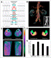
Multimodality evaluation with ankle-brachial indices (A), CT angiography (B), and hybrid 99mTc-tetrofosmin SPECT/CT (C) reveals impaired lower-extremity pressures and tissue perfusion in PVD patient with previously implanted aortoiliac stents (B). Segmentation of muscle groups into 3-dimensional regions of interest by CT attenuation images (D) confirmed differences in regional tissue perfusion between legs (E). Red 5 gastrocnemius; yellow 5 soleus; green 5 tibialis; purple 5 fibularis; ABI 5 ankle–brachial index; PPG 5 photoplethysmograph; PVR 5 pulse volume recording; TBI 5 toe–brachial index.
FIGURE 3.
FIGURE 3.
Lower-extremity PET H215O imaging of healthy subject and PVD patient during selective adenosine infusion into left leg (A). Baseline and adenosine stress blood flow was assessed, and flow reserve was expressed as ratio of adenosine flow to baseline flow (B). Flow reserve was significantly lower in PVD patients than in healthy subjects. (Reprinted from (32).).
FIGURE 4.
FIGURE 4.
PET/CT imaging with 64Cu-DOTA–C-type atrial natriuretic factor–comb (A) and 64Cu-DOTA–comb (B) in mouse model of hind limb ischemia, 7 d after femoral artery occlusion. 64Cu-DOTA–C-type atrial natriuretic factor–comb uptake was significantly higher than uptake of 64Cu-DOTA–comb in ischemic limb when percentage injected tracer dose per gram of tissue (C) or ischemic-to-nonischemic leg ratios (D) were examined. (Reprinted from (52).) CANF 5 C-type atrial natriuretic factor.
FIGURE 5.
FIGURE 5.
Analysis of mean (A) and maximum (B) 18F-FDG tumor-to-background ratios within multiple arterial regions on day 1 (solid bars) and 14 d later (hatched bars). No significant differences were observed within arterial regions across time. Carotid artery uptake was significantly higher than 18F-FDG uptake within lower-extremity arteries on days 1 and 14. 18F-FDG PET (C, middle) fused with CT imaging (C, right) revealed similar uptake in femoral arteries on days 1 and 14 (noted by arrows). CFA 5 common femoral artery; SFA 5 superficial femoral artery; TBR 5 tumor-to-background ratio. *P < 0.001. (Reprinted from (58).)

References

    1. Leeper NJ, Kullo IJ, Cooke JP Genetics of peripheral artery disease. Circulation. 2012;125:3220–3228. - PMC - PubMed
    1. Peach G, Griffin M, Jones KG, Thompson MM, Hinchliffe RJ. Diagnosis and management of peripheral arterial disease. BMJ. 2012;345:e5208. - PubMed
    1. Gregg EW, Sorlie P, Paulose-Ram R, et al. Prevalence of lower-extremity disease in the US adult population >=40 years of age with and without diabetes: 1999-2000 national health and nutrition examination survey. Diabetes Care. 2004;27:1591–1597. - PubMed
    1. Hooi JD, Stoffers HE, Kester AD, et al. Risk factors and cardiovascular diseases associated with asymptomatic peripheral arterial occlusive disease. The Limburg PAOD study. Peripheral Arterial Occlusive Disease. Scand J Prim Health Care. 1998;16:177–182. - PubMed
    1. Pollak AW, Norton PT, Kramer CM. Multimodality imaging of lower extremity peripheral arterial disease: current role and future directions. Circ Cardiovasc Imaging. 2012;5:797–807. - PMC - PubMed

MeSH terms

Substances

LinkOut - more resources