. 2013 Oct;25(12):661-78.
doi: 10.3109/08958378.2013.833660.

Toxicity of lunar dust assessed in inhalation-exposed rats

Affiliations

Toxicity of lunar dust assessed in inhalation-exposed rats

Chiu-wing Lam et al. Inhal Toxicol. 2013 Oct.

Abstract

Humans will again set foot on the moon. The moon is covered by a layer of fine dust, which can pose a respiratory hazard. We investigated the pulmonary toxicity of lunar dust in rats exposed to 0, 2.1, 6.8, 20.8 and 60.6 mg/m(3) of respirable-size lunar dust for 4 weeks (6 h/day, 5 days/week); the aerosols in the nose-only exposure chambers were generated from a jet-mill ground preparation of a lunar soil collected during the Apollo 14 mission. After 4 weeks of exposure to air or lunar dust, groups of five rats were euthanized 1 day, 1 week, 4 weeks or 13 weeks after the last exposure for assessment of pulmonary toxicity. Biomarkers of toxicity assessed in bronchoalveolar fluids showed concentration-dependent changes; biomarkers that showed treatment effects were total cell and neutrophil counts, total protein concentrations and cellular enzymes (lactate dehydrogenase, glutamyl transferase and aspartate transaminase). No statistically significant differences in these biomarkers were detected between rats exposed to air and those exposed to the two low concentrations of lunar dust. Dose-dependent histopathology, including inflammation, septal thickening, fibrosis and granulomas, in the lung was observed at the two higher exposure concentrations. No lesions were detected in rats exposed to ≤6.8 mg/m(3). This 4-week exposure study in rats showed that 6.8 mg/m(3) was the highest no-observable-adverse-effect level (NOAEL). These results will be useful for assessing the health risk to humans of exposure to lunar dust, establishing human exposure limits and guiding the design of dust mitigation systems in lunar landers or habitats.


Figures
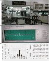
Figure 1
Figure 1
(A) Nose-only inhalation chamber systems and the associated dust generators, cyclones, dust monitors and data acquistion computers. (B) A typical 6-h chamber concentration recorded by the Casella Microdust Pro. (C) An example of a particle size profile recorded by the Quartz Crystal Microbalance Cascade Impactor.
Figure 2
Figure 2
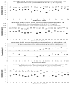
Lunar dust concentrations in each chamber were photometrically recorded by a Casella Microdust Pro real-time monitor continuously throughout the exposure period. Dust samples were collected on filter papers and the exposure concentrations were estimated on the basis of gravimetric determination. Plotted are average daily dust concentrations in each chamber for the two 4-week studies (20 exposures); Chambers 1A and 1B for 1st Study and Chambers 2A and 2B for 2nd Study.
Figure 3
Figure 3
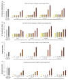
Cellular components in right-lung lavage fluids of lunar dust (LD) inhalation-exposed rats: total cell counts (A), macrophage counts (B), neutrophil counts (C) and lymphocyte counts (D). Control groups H-0 and 0, were exposed to air at the same time as other groups of rats were exposed to high concentrations (21 or 61 mg/m3) or low concentrations (2.1 or 6.8 mg/m3) of lunar dust, respectively. Groups of five rats each were instilled with 2.5 mg quartz 1 week before the day of lung lavage serving as positive controls for biomarker assays; QH (4 × 5 rats) and QL (4 × 5 rats) were lavaged on the same days as the groups exposed to high-concentrations or low concentrations of lunar dust, respectively. All the values from the groups exposed to 21 and 61 mg/m3 were significantly different from H-0 at the same time point.
Figure 4
Figure 4
Three groups of 20 rats each were exposed to 0, 2.1 or 6.8 mg/m3 of lunar dust for 4 weeks (6 h/day, 5 days/week). Five rats from each group were euthanized on 0- (day 1), 1-, 4- or 13-week post-exposure for assessment of toxicity biomarkers in lavage fluids from right lungs and for lesions in the left lungs. Plotted here are total cell counts (A), neutrophil counts (B), and lymphocyte counts (C) in lavage fluids. *Statistically significantly different from air-exposed group (0) at the same time point.
Figure 5
Figure 5
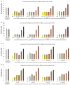
Acellular biomarkers of toxicity in right-lung lavage fluids of rats exposed by inhalation to lunar dust (LD). Control groups, H-0 and 0, were exposed to air at the same time as other groups of rats were exposed to high concentrations (21 or 61 mg/m3) or low concentrations (2.1 or 6.8 mg/m3) of lunar dust, respectively. Groups of five rats each were instilled with 2.5 mg quartz 1 week before the day of lung lavage serving as positive controls for biomarker assays; QH (4 × 5 rats) and QL (4 × 5 rats) were lavaged on the same days as high-concentration groups and low-concentration groups, respectively. LD: N = 5 rats. All the values from the groups exposed to 21 and 61 mg/m3 are significantly different from those of the H-0 groups at the same time point.
Figure 6
Figure 6
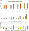
Among the indices assessed and shown in Table 2, the major histopathology indices with average scores in lungs of rats exposed to lunar dust are shown here. Scores for lungs of rats exposed to air or to 2.1 or 6.8 mg/m3 lunar dust were minimal (increase of macrophages) or 0 and are not plotted in the graphs. Histopathology scoring scales: 1, minimal; 2, mild; 3, moderate; 4, marked and 5, severe. Each bar is the average score for five rats.
Figure 7
Figure 7
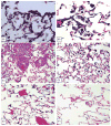
Lung tissues of rats exposed to lunar dust at 61 (HC) or 6.8 (LC) mg/m3 for 4 weeks. (A) HC: lunar dust particles visible in alveoli or within macrophages 1-day post-exposure. (B) HC: lung with thickened alveolar septa, macrophages containing particles, 1-week post-exposure. (C) HC: perivascular lymphocytes and macrophages in lung from a rat necropsied 4-week post-exposure. (D) HC: focal septal thickening, macrophages and inflammatory cells in alveoli 13-week post-exposure. (E) LC: lung tissue of a rat exposed to 6.8 mg/m3 lunar dust for 4 weeks and necropsied 1-week post-exposure showing normal structure like that in air-exposed rats. (F) LC: an alveolus at the center contains a cluster of macrophages containing dust particles. Image magnification: (A) and (B): 40×; (C–F): 20×.
Figure 8
Figure 8
Tracheobronchial lymph nodes from rats exposed to 61 mg/m3 for 4 weeks and examined 13 weeks after the last exposure. (A) Macrophages containing lunar dust particles in the lymph node of a rat necropsied 13-week post-exposure. (B) Higher magnification of (A) shows numerous lunar dust particles in granulomas. Image magnification: (A): 20×; (B): 40×.

References

    1. Bellmann B, Muhle H, Creutzenberg O, et al. Lung clearance and retention of toner, utilizing a tracer technique, during chronic inhalation exposure in rats. Fundam Appl Toxicol. 1991;17:300–13. Cited in Pauluhn 2012. - PubMed
    1. Bermudez E, Mangum JB, Asgharian B, et al. Long-term pulmonary responses of three laboratory rodent species to subchronic inhalation of pigmentary titanium dioxide particles. Toxicol Sci. 2002;70:86–97. - PubMed
    1. Bilder RB. A legal regime for the mining of helium-3 on the moon: U.S. policy options. Fordham Int Law J. 2009;33:243–98.
    1. Carpenter P, Sibille L, Meeker G, Wilson S. Characterization of standardized lunar regolith simulant materials, NASA Technical Report 20060019180. Washington, DC: National Aeronautics and Space Administration; 2006.
    1. Casella Measure. Belford; United Kingdom: 2013. [Last accessed: Jun 2013]. Available from: http://www.casellameasurement.com/downloadable-content/Handbooks/Microdu....

Publication types

LinkOut - more resources