Review
. 2014 Jan;224(1):61-73.
doi: 10.1111/joa.12106. Epub 2013 Sep 15.

Protein kinase C isoforms at the neuromuscular junction: localization and specific roles in neurotransmission and development

Affiliations
Review

Protein kinase C isoforms at the neuromuscular junction: localization and specific roles in neurotransmission and development

Maria A Lanuza et al. J Anat. 2014 Jan.

Abstract

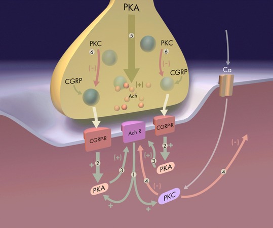
The protein kinase C family (PKC) regulates a variety of neural functions including neurotransmitter release. The selective activation of a wide range of PKC isoforms in different cells and domains is likely to contribute to the functional diversity of PKC phosphorylating activity. In this review, we describe the isoform localization, phosphorylation function, regulation and signalling of the PKC family at the neuromuscular junction. Data show the involvement of the PKC family in several important functions at the neuromuscular junction and in particular in the maturation of the synapse and the modulation of neurotransmission in the adult.

Keywords: electrical stimulation; immunofluorescence; isoforms; neuromuscular junction; neurotransmission; protein kinase C; synapse elimination.


Figures

Fig. 1
Fig. 1
Protein kinase C and NMJ. (A) Serine/threonine kinase C family. Schematic diagram of primary structures of PKC family members showing domain composition and activators. The PKC family of isozymes consists of three classes: the classical (α, βI, βII, γ), novel (δ, ε, η and θ) and atypical (ζ and ι/λ). (B) Adult NMJ image. Double immunofluorescence NMJs labelled with syntaxin/neurofilament-200 nerve terminal, NT, in green) and α-BTX (AChR in red). (C1–3) Semithin (0.5 μm) cross-sections of the NMJ stained with toluidine blue (C1) and with a triple immunofluorescence method (C3; syntaxin/neurofilament-200-NT, in green, S-100 Schwann cell, SC, in blue and α-BTX -AChR in red). In (C2), the cellular components in C1 are delineated. Reproduced with permission from Lanuza et al. (2007). (D) Immunohistochemical staining for cPKC isoform βI at the adult NMJ. cPKCβI are labelled in green, the AChRs in red and the Schwann cells (SC, S-100) or the nerve terminals (NT, neurofilament-200/syntaxin) in blue. (D1) NMJ from a whole muscle immunolabelled. (D2–D3) Semithin cross-sections from a whole-mount multiple-immunofluorescent stained muscle. cPKCβI was found to be localized to the presynaptic terminals. Reproduced with permission from Besalduch et al. (2010). (D4) The diagram summarizes the localization of the PKC isoforms in the three cellular components of the NMJ (nerve terminal, muscle cell and Schwann cell). Scale bars: 10 μm.
Fig. 2
Fig. 2
Effect of electrical stimulation and electrical stimulation -induced contraction on nPKCε, cPKCα and cPKC βI. (A) Time course of CaC effect on EPP amplitude when incubated solely (only CaC, 10 μm; dotted line) to the muscle, when a continuous stimulation at 1 Hz was applied previously (1 h) during the CaC incubation (1 : 1 Hz; 2 : CaC) and in the presence of high external calcium (1 : Ca2+; 2 : CaC). We can see also the EPP amplitude when continuous stimulation at 1 Hz was applied (only 1 Hz). Values are expressed as percentages (mean ± SE) with respect to initial amplitude. n = 10–15 single fibers by the kind of experiment. Points into the grey area: P < 0.05 with respect to initial values (0.0 min). On the right, raw data showing examples of the CaC effect on synaptic potentials with and without electrical stimulation. EPPs were recorded before and at 60 min of CaC application. Up: only CaC. Down: CaC with continuous stimulation at 1 Hz. Note that the following changes can be seen only in down: the EPP sizes were diminished and the EPP variances increased. Artefacts are modified for clarity. Scale bars: horizontal: 4 ms, vertical: 3 mV. (B) Drawing of the diaphragm muscle showing the innervated area of the muscle. Dotted lines indicate the place at which synaptic and extrasynaptic zones were separated. The fluorescent image shows the NMJs detected with TRITC-conjugated α-BTX. Note that NMJs are located at the synaptic area and no NMJs were detected in the extrasynaptic area. Western blotting analysis of cPKCα, βI, βII isoform immunoreactivity levels in diaphragm muscles from synaptic membranes under basal conditions (Control) and after electrical stimulation (1 Hz, 30 min) with (ES + C) and without (ES) muscle contraction. Experimental conditions resulted in a significant change (P < 0.05) from control values. Reproduced with permission from Besalduch et al. (2010).
Fig. 3
Fig. 3
Pre- and postsynaptic changes during developmental activity-dependent synapse elimination. Synaptic AChR cluster morphologies in the neuromuscular junctions of the rat LAL muscle from birth to adulthood. AChRs were stained with rhodamine-conjugated α-bungarotoxin. Postsynaptic AChR clusters are classified from M1 to M4 types according their morphologic maturation. The colour image shows a confocal image showing several NMJs immunostained in green with neurofilament-200 and in red with α-bungarotoxin. Scale bars: 10 μm. Reproduced with permission from Lanuza et al. (2002).
Fig. 4
Fig. 4
Presynaptic PKC signalling. (A) Functional relation between calcium inflows, voltage-dependent calcium channels (VDCC), presynaptic muscarinic acetylcholine receptors (mAChRs), and PKC activity in the modulation of the postnatal developmental activity dependent-synaptic elimination process. (B) Localization of neurotrophins (brain-derived neurotrophic factor (BDNF), neurotrophin (NT)-3 and NT-4 and its receptor proteins (p75NTR, trkB and trkC) in neonatal monoinnervated and multiple-innervated NMJ. Figure also shows that neurotrophins enhance neurotransmission and that this role is selective for the different types of nerve terminals depending on their developmental maturation. In the diagram the framed-neurotrophin name into a particular presynaptic component indicates that this neurotrophin is potentiating the ACh release in this specific nerve terminal type.
Fig. 5
Fig. 5
Postsynaptic PKC signalling. A spatially specific and opposing action of PKC and PKA may result in activity-dependent alterations to synaptic connectivity at both the nerve inputs and the postsynaptic AChR clusters in the initial process of synapse elimination. This balance between PKC and PKA actions could mediate the retention of active neural inputs and the loss of the inactive (or less active) inputs located over the common postsynaptic cell. The diagram represents these mechanisms: (1) Cholinergic activation of muscle produces an increase in PKC and PKA activity. (2) Muscle activation, due to peptidergic mechanisms (CGRP), produces an increase in PKA activity, which has a positive effect, increasing efficacy and producing a local synapse stabilization effect (3). (4) PKC activity has a general negative effect; it is widely distributed within the postsynaptic cell and acts on all synapses to reduce postsynaptic responsiveness. The positive, synapse-stabilizing effect, mediated by PKA (2–3), is more localized, counteracts the PKC effect and tends to preserve only stimulated inputs because of this local action. These effects of both PKA and PKC may be due, at least in part, to direct action of the kinases in phosphorylating the AChR at the posynaptic site. In addition, neural activity produces both positive (5) and negative (6) effects on transmitter and neuropeptide output on presynaptic site (Li et al., 2001).
Fig. 6
Fig. 6
Confocal and electron microscopy images of NMJs from newborn and adult. Confocal microphotographs from wild type (WT) and knockout in nPKCθ (KO) NMJs at neonatal postnatal 4-day (P4, polyinnervated NMJs), postnatal 8-day (P8, monoinnervated NMJs) and adult were stained for AChRs (red) and axons and nerve terminals (neurofilament-200 protein in green). The selected examples of polyinnervated and monoinnervated NMJs from developing WT and KO muscles (P4, P8) show differences in the pre- and postsynaptic components that are in accordance with the quantitative measurements of the cluster morphology. Scale bars: 10 μm. The ultrastructure images of the KO NMJs during development and in the adult have the same structure than NMJs with comparable levels of maturation in WT muscles at P3, P6, P13 and adult. The NMJ from a P3 KO muscle shows the coexistence of several nerve terminal boutons (marked by asterisks in the drawing at the right of the picture) on a poorly defined, low-density, postsynaptic membrane without gutters. The NMJ from a P6 KO muscle shows an intermediary stage of axon separation. The NMJ from a P13 KO muscle shows advanced gutter formation and nerve terminal segregation and elimination. Nerve terminals (marked with # in the accompanying drawing) are engulfed by Schwann cell processes that also contain membrane debris. The right column shows drawings in which the three cellular components of the NMJ (axon terminal, Schwann cells and postsynaptic membrane density) have been delineated. Scale bar: 200 nm. Reproduced with permission from Lanuza et al. (2010) and Besalduch et al. (2011).

References

    1. Abbs ET, Joseph DN. The effects of atropine and oxotremorine on acetylcholine release in rat phrenic nerve diaphragm preparations. Br J Pharmacol. 1981;73:481–483. - PMC - PubMed
    1. Antipenko A, Frías JA, Parra J, et al. Effect of chronic electrostimulation of rabbit skeletal muscle on calmodulin level and protein kinase activity. Int J Biochem Cell Biol. 1999;31:303–310. - PubMed
    1. Anwyl R. Metabotropic glutamate receptor-dependent long-term potentiation. Neuropharmacology. 2009;56:735–740. - PubMed
    1. Arakawa M, Mizoguchi A, Masutani M, et al. Ultrastructural localization of protein kinase C beta-subspecies in the axon terminal of rat neuromuscular junction. Neurosci Res. 1993;16:125–130. - PubMed
    1. Arenson MS. Muscarinic inhibition of quantal transmitter release from the magnesium-paralysed frog sartorius muscle. Neuroscience. 1989;30:827–836. - PubMed

Publication types

MeSH terms

LinkOut - more resources