. 2014 Jan;15(1):12-21.
doi: 10.1111/tra.12128. Epub 2013 Oct 31.

Dissecting functions of the conserved oligomeric Golgi tethering complex using a cell-free assay

Affiliations
Free PMC article

Dissecting functions of the conserved oligomeric Golgi tethering complex using a cell-free assay

Nathanael P Cottam et al. Traffic. 2014 Jan.
Free PMC article

Abstract

Vesicle transport sorts proteins between compartments and is thereby responsible for generating the non-uniform protein distribution along the eukaryotic secretory and endocytic pathways. The mechanistic details of specific vesicle targeting are not yet well characterized at the molecular level. We have developed a cell-free assay that reconstitutes vesicle targeting utilizing the recycling of resident enzymes within the Golgi apparatus. The assay has physiological properties, and could be used to show that the two lobes of the conserved oligomeric Golgi tethering complex play antagonistic roles in trans-Golgi vesicle targeting. Moreover, we can show that the assay is sensitive to several different congenital defects that disrupt Golgi function and therefore cause glycosylation disorders. Consequently, this assay will allow mechanistic insight into the targeting step of vesicle transport at the Golgi, and could also be useful for characterizing some novel cases of congenital glycosylation disorders.

Keywords: Golgi apparatus; cell-free reconstitution; congenital disorders of glycosylation; conserved oligomeric Golgi complex; glycosylation enzyme sorting; vesicle tethering.


Figures

Figure 1
Figure 1
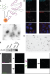
Setup of a cell-free assay using fluorescently tagged GalT as a marker. A) Schematic view of the assay in which Golgi and vesicle fractions are isolated from HEK293 cells stably expressing CFP-tagged (magenta) or YFP-tagged (green) GalT. Following their coincubation under assay conditions, colocalization is determined using fluorescence microscopy. B) Confocal fluorescent micrographs of stable GalT-CFP (green)-expressing HEK293 cells immunostained for TGN46 (left, red) or GM130 (right, red). Blue in the merge image shows DAPI staining, scale bar is 2 µm. C and D) Negatively stained electron micrographs of Golgi (C) and vesicle (D) fractions. Scale bars are 500 nm (C) and 200 nm (D). E) Immunoblot against Sec22b of equivalent Golgi and vesicle amounts. The two lanes on the right contain 2.5-fold more membrane material than the left ones. F) Typical imageJ-based data analysis workflow after image acquisition. Following noise reduction and background subtraction, binary images were overlaid and the particles containing at least one colocalizing pixel exported to be counted. For details, see Materials and Methods. Scale bar, 10 µm.
Figure 2
Figure 2
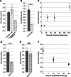
Testing the assay under physiological conditions. A) Assays in the presence of membranes, cytosol and energy were performed (grey bar) in the presence of the protease inhibitor Pefabloc (dotted bar) and in the presence of Pefabloc added following a 30-min incubation on ice with Proteinase K (striped bar). Error bars show SEM for n = 2. B) Assays performed in the presence of membranes and cytosol with (grey bar) or without (dotted bar) energy source. Error bars show SEM for n = 3. C) Assay activity in the presence of increasing amount of cytosol. Error bars show SD for n = 2. Note that standard assays used 57 µg cytosol to allow the addition of other factors without increasing osmolarity beyond 310 mOsm. D and E) Activity in the presence of the chelators EDTA (D, n = 6) or BAPTA (E, n = 5). Error bars represent SEM. F) Assay activity in the presence of increasing amounts of RabGDI. Full assay mix was preincubated with the indicated amount of RabGDI for 20 min on ice before addition of the energy regeneration system and normal assay incubation. Error bars represent SEM for n = 3 (0 and 25 µg RabGDI) or n = 2 (10 µg RabGDI). Note that the value of 1.0 is the activity observed in the ice-control sample. Statistical significance was determined as described in the Materials and Methods section, *p < 0.05; ns, not significant.
Figure 3
Figure 3
Functional distinction between lobe A and lobe B of the COG complex. A) Transport activity with cytosol from fibroblasts of COG-deficient patients as well as a novel CDG-X patient with an unknown mutation. Because of differences in cytosol and membrane concentrations between different experiments the bars show the average of the ratio between the given cytosol's activity and WT cytosol activity. The value of 1.0 therefore represents WT activity in this graph. Escherichia coli cytosol was used in the control without cytosol. SEM calculated for no cytosol, COG6 and COG7 n = 4, CDG-X n = 3 and complemented COG6 and human fibroblast n = 2. See Figure S1, Supporting Information for the individual assays used for calculating the ratios for the COG7-deficient cytosol as an example. B) Transport activity in the presence of cytosol isolated from COG mutant CHO cell lines. ldlB cells (horizontal stripes) are COG1 deficient, whereas ldlC cells (slanted stripes) are COG2 deficient. All details as in ‘A’, activities are shown in ratio to WT CHO cytosol. Error bars represent SEM for n = 4. Although E. coli cytosol was used as a protein mix in ‘A’ to suppress non-specific membrane aggregation, we found that omission of this did not cause a difference, and therefore conducted some of the experiments in ‘B’ without this additive.

Similar articles

Cited by

References

    1. Barlowe C, Orci L, Yeung T, Hosobuchi M, Hamamoto S, Salama N, Rexach MF, Ravazzola M, Amherdt M, Schekman R. Copii – a membrane coat formed by sec proteins that drive vesicle budding from the endoplasmic-reticulum. Cell. 1994;77:895–907. - PubMed
    1. Reinhard C, Schweikert M, Wieland FT, Nickel W. Functional reconstitution of COPI coat assembly and disassembly using chemically defined components. Proc Natl Acad Sci U S A. 2003;100:8253–8257. - PMC - PubMed
    1. Weber T, Zemelman BV, McNew JA, Westermann B, Gmachl M, Parlati F, Sollner TH, Rothman JE. SNAREpins: minimal machinery for membrane fusion. Cell. 1998;92:759–772. - PubMed
    1. Brandhorst D, Zwilling D, Rizzoli SO, Lippert U, Lang T, Jahn R. Homotypic fusion of early endosomes: SNAREs do not determine fusion specificity. Proc Natl Acad Sci U S A. 2006;103:2701–2706. - PMC - PubMed
    1. Balch WE, Dunphy WG, Braell WA, Rothman JE. Reconstitution of the transport of protein between successive compartments of the Golgi measured by the coupled incorporation of N-acetylglucosamine. Cell. 1984;39:405–416. - PubMed

Publication types

MeSH terms

Substances

LinkOut - more resources