Review
. 2014 Feb:308:50-9.
doi: 10.1016/j.heares.2013.09.017. Epub 2013 Oct 6.

Thalamocortical mechanisms for integrating musical tone and rhythm

Affiliations
Review

Thalamocortical mechanisms for integrating musical tone and rhythm

Gabriella Musacchia et al. Hear Res. 2014 Feb.

Abstract

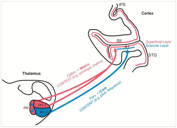
Studies over several decades have identified many of the neuronal substrates of music perception by pursuing pitch and rhythm perception separately. Here, we address the question of how these mechanisms interact, starting with the observation that the peripheral pathways of the so-called "Core" and "Matrix" thalamocortical system provide the anatomical bases for tone and rhythm channels. We then examine the hypothesis that these specialized inputs integrate acoustic content within rhythm context in auditory cortex using classical types of "driving" and "modulatory" mechanisms. This hypothesis provides a framework for deriving testable predictions about the early stages of music processing. Furthermore, because thalamocortical circuits are shared by speech and music processing, such a model provides concrete implications for how music experience contributes to the development of robust speech encoding mechanisms.


Figures

Figure 1
Figure 1. A model for integration content and context cues in music based on “Core” and “Matrix” divisions of the auditory thalamocortical projection pathways
Parvalbumin-positive (Parv. +) “Core” thalamocortical neurons (blue) lying mostly in the ventral division of the Medial Geniculate body (MGv) receive frequency-tuned inputs from the tonotopically organized internal nuclear portions of the inferior colliculus. These neurons terminate in the granular cortical layers of Core regions of primary auditory cortex (A1), giving rise to the relatively precise tonotopic representation observed there (Kimura et al., 2003; Hashikawa et al., 1995; Pandya et al., 1994). In contrast, calbindin positive (Calbin. +) “Matrix” thalamocortical neurons (red) lying mainly in the Magnocellular (mc) and dorsal (d) Medial Geniculate divisions receive broadly-tuned (diffuse) inputs input from the non-tonotopic external nuclear portions of the inferior colliculus. These inputs project broadly to secondary/tertiary (e.g., auditory belt and parabelt), as well as primary regions, terminating in the superficial cortical layers (Kimura et al., 2003; Hashikawa et al., 1995; Pandya et al., 1994). As detailed in the text, we propose that that activity conveyed by thalamocortical Matrix projections, and entrained to the accent/beat pattern of a musical rhythm, dynamically reset and “modulate” the phase of ongoing cortical rhythms, so that the cortical rhythms themselves synchronize with the rhythmic “contextual” pattern of the music. Because oscillatory phase translates to excitability state in a neuronal ensemble, a potential consequence of the modulatory phase resetting that underlies synchrony induction and maintenance is that responses “driven” by specific “content” that is associated with contextual musical accents (e.g., notes or words) will be amplified, relative to content that occurs off the beat. Abbreviations: IPS: Intrapariaetal Sulcus; 3b: Area 3b, Primary somatosensory cortex; SII: Secondary somatosenaory cortex; STG: Superior Temporal Gyrus
Figure 2
Figure 2. Oscillations control neuronal excitability
Panel A. On the left, four aspects of neural activity are shown: the low pass filtered local field potential (LFP) (A1), the amplitude activity (B1), phase activity (C1) and multiunit activity (MUA) (D1) (Slezia et al., 2011). A2-D2 shows the same indices for an oscillation that increases in frequency. Panel B. MUA bursting of cat parietal corticothalamic neurons (top trace) occurs in the Delta frequency range and co-occurs with slower (0.3–0.4 Hz) LFP rhythms (middle trace) and scalp recorded electroencephalographic (EEG) activity (bottom trace) (Steriade et al., 1993). Panel C. In macaque (Lakatos et al., 2005), a current source density (CSD) profile for the supragranular layers of auditory cortex (i) sampled using a multielectrode (left) shows Theta-band (5–9 Hz) oscillatory activity (superimposed black line). Drop lines from ii shows coincidence of simultaneously recorded MUA activity. Reproduced, with permission, from [33] (A), [38] (B), [77] (C).
Figure 3
Figure 3. Contrasting laminar profiles and physiology of “driving” and “modulatory” inputs
A) A schematic of the multi-contact electrode and positioned astride the cortical laminae is shown on the right. Box-plots show quantified onset latencies of the best-frequency pure tone (blue) and somatosensory stimulus (red) related current source density (CSD) response in supragranular (S), granular (G) and infragranular (I) layers across experiments. Lines in the boxes depict the lower quartile, median, and upper quartile values. Notches in boxes depict 95% confidence interval for medians of each distribution. Brackets indicate the significant differences(Games-Howell post-hoc test, p<0.01). B) Quantified multi-unit activity (MUA) amplitudes (over 38 cases) from the representative supragranular, granular and infragranular channels (S, G, and I) over the 15–60 ms time interval for the same conditions as in A, along with a bimodal stimulation condition. Brackets indicate the significant differences (Games-Howell, p<0.01). C) left. Quantified (n=38) post-/pre-stimulus single trial oscillatory amplitude ratios (0 – 250 ms / −500 – −250 ms) for different frequency bands (different colors) of the auditory, somatosensory and bimodal supragranular responses. Stars denote the amplitude ratios significantly different from 1 (one-sample t-tests, p<0.01). right. Quantified (n=38) intertrial coherence (ITC) expressed as a vector quantity (mean resultant length), measured at 15 ms post-stimulus (i.e., at the initial peak response). For somatosensory events, increase in phase concentration only occurs in the low-delta (1–2.2 Hz), theta (4.8–9.3 Hz) and gamma (25–49 Hz) bands, indicated by colored arrows on the right. Adapted, with permission, from (Lakatos et al., 2007).

References

    1. Abrams DA, Nicol T, Zecker S, Kraus N. A possible role for a paralemniscal auditory pathway in the coding of slow temporal information. Hear Res. 2011;272:125–134. - PMC - PubMed
    1. Alluri V, Toiviainen P, Jaaskelainen IP, Glerean E, Sams M, Brattico E. Large-scale brain networks emerge from dynamic processing of musical timbre, key and rhythm. Neuroimage. 2012;59:3677–3689. - PubMed
    1. Anderson LA, Wallace MN, Palmer AR. Identification of subdivisions in the medial geniculate body of the guinea pig. Hear Res. 2007;228:156–167. - PubMed
    1. Bailey JA, Penhune VB. Rhythm synchronization performance and auditory working memory in early- and late-trained musicians. Exp Brain Res. 2010;204:91–101. - PubMed
    1. Benasich AA, Gou Z, Choudhury N, Harris KD. Early cognitive and language skills are linked to resting frontal gamma power across the first 3 years. Behav Brain Res. 2008;195:215–222. - PMC - PubMed