. 2013 Oct;25(10):4110-22.
doi: 10.1105/tpc.113.115113. Epub 2013 Oct 8.

Morphine biosynthesis in opium poppy involves two cell types: sieve elements and laticifers

Affiliations

Morphine biosynthesis in opium poppy involves two cell types: sieve elements and laticifers

Akpevwe Onoyovwe et al. Plant Cell. 2013 Oct.

Abstract

Immunofluorescence labeling and shotgun proteomics were used to establish the cell type-specific localization of morphine biosynthesis in opium poppy (Papaver somniferum). Polyclonal antibodies for each of six enzymes involved in converting (R)-reticuline to morphine detected corresponding antigens in sieve elements of the phloem, as described previously for all upstream enzymes transforming (S)-norcoclaurine to (S)-reticuline. Validated shotgun proteomics performed on whole-stem and latex total protein extracts generated 2031 and 830 distinct protein families, respectively. Proteins corresponding to nine morphine biosynthetic enzymes were represented in the whole stem, whereas only four of the final five pathway enzymes were detected in the latex. Salutaridine synthase was detected in the whole stem, but not in the latex subproteome. The final three enzymes converting thebaine to morphine were among the most abundant active latex proteins despite a limited occurrence in laticifers suggested by immunofluorescence labeling. Multiple charge isoforms of two key O-demethylases in the latex were revealed by two-dimensional immunoblot analysis. Salutaridine biosynthesis appears to occur only in sieve elements, whereas conversion of thebaine to morphine is predominant in adjacent laticifers, which contain morphine-rich latex. Complementary use of immunofluorescence labeling and shotgun proteomics has substantially resolved the cellular localization of morphine biosynthesis in opium poppy.


Figures

Figure 1.
Figure 1.
Biosynthesis of Morphine in Opium Poppy from the Tyr Derivatives Dopamine and 4-Hydroxyphenylacetaldehyde. Corresponding cDNAs have been isolated for all enzymes shown in blue. The dotted arrow refers to a conversion catalyzed by unknown enzymes. Chemical conversions catalyzed by each enzyme are shown in red. Compounds in bold are major accumulating alkaloids in opium poppy latex.
Figure 2.
Figure 2.
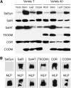
Immunoblot Analysis of Morphine Pathway Enzymes in Opium Poppy. (A) Immunoblots showing the relative abundance of the final six morphine biosynthetic enzymes in the opium poppy chemotypes T and 40. Equal amounts (50 µg) of total protein extracts from different organs were separated by SDS-PAGE. Protein blots were probed with polyclonal antibodies specific for each enzyme. (B) Immunoblots showing the occurrence of the final six morphine biosynthetic enzymes in the latex of opium poppy chemotype 40. The blots were probed simultaneously with MLP antibodies as a gel loading and autoradiogram exposure control. Data are representative of three independent experiments.
Figure 3.
Figure 3.
Immunolocalization of Morphine Biosynthetic Enzymes in Different Opium Poppy Organs. Polyclonal antibodies were raised against: SalSyn ([E] to [H]), SalR ([I] to [L]), SalAT ([M] to [P]), T6ODM ([O] to [T]), COR ([U] to [X]), and CODM ([Y] to [ZZ]). Serial cross sections of resin-embedded tissues from opium poppy chemotype 40 were 0.5 µm in thickness. One serial section for each organ was stained with toluidine blue O to show the anatomical organization of the phloem, with several laticifers indicated by red asterisks ([A] to [D]). Immunofluorescence labeling of laticifers was performed using MLP polyclonal antibodies ([AA] to [DD]). Bars = 25 µm.
Figure 4.
Figure 4.
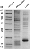
SDS-PAGE of Whole-Stem and Latex Protein Extracts Used for Shotgun Proteomics Analysis. Lanes containing whole-stem and latex proteins were sectioned into 48 and 41 segments, respectively, which were individually subjected to in-gel digestion with trypsin. Numbers on the left show the molecular masses of protein markers in kilodaltons.
Figure 5.
Figure 5.
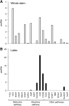
Relative Abundance of Alkaloid Biosynthetic Enzymes in Opium Poppy Based on emPAI Scores. The emPAI scores were derived from shotgun proteomics performed on whole-stem (A) and latex (B) total protein extracts.
Figure 6.
Figure 6.
Relative Abundance of Gene Transcripts Encoding the Final Six Enzymes of Morphine Biosynthesis in Opium Poppy. Quantitative RT-PCR was performed using total RNA isolated from the whole stem and latex of opium poppy chemotypes T (A) and 40 (B). The experiment was performed in triplicate and produced similar results each time.
Figure 7.
Figure 7.
Immunoblot Analysis of Opium Poppy Latex Proteins Separated by 2D SDS-PAGE Showing Numerous Charge Isoforms of T6ODM and CODM. Latex proteins (50 µg) were subjected to isoelectric focusing followed by SDS-PAGE and transferred to nitrocellulose membranes. Duplicate protein blots were probed with polyclonal antibodies raised against T6ODM or CODM.
Figure 8.
Figure 8.
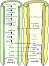
Model Summarizing the Localization and Relative Abundance of Morphine Biosynthetic Enzymes in Sieve Elements and Laticifers of Opium Poppy. The cellular localization of biosynthetic enzymes was based on immunolocalization and shotgun proteomics data. The font size used for each enzyme shown in blue was adjusted to reflect the estimated relative abundance in sieve elements and laticifers. The thickness of vertical arrows suggests the proposed relative flux through various conversions in each cell type. The dashed vertical arrow represents an unknown enzyme. Horizontal arrows indicate alkaloids that are putatively transported between sieve elements and laticifers with thebaine suggested as the major translocated intermediate.

References

    1. Alcantara J., Bird D.A., Francheschi V.R., Facchini P.J. (2005). Sanguinarine biosynthesis is associated with the endoplasmic reticulum in cultured opium poppy cells after elicitor treatment. Plant Physiol. 138: 173–183 - PMC - PubMed
    1. Bird D.A., Franceschi V.R., Facchini P.J. (2003). A tale of three cell types: Alkaloid biosynthesis is localized to sieve elements in opium poppy. Plant Cell 15: 2626–2635 - PMC - PubMed
    1. Brochmann-Hanssen E. (1984). A second pathway for the terminal steps in the biosynthesis of morphine. Planta Med. 50: 343–345 - PubMed
    1. Cho W.K., Chen X.Y., Rim Y., Chu H., Jo Y., Kim S., Park Z.-Y., Kim J.Y. (2010). Extended latex proteome analysis deciphers additional roles of the lettuce laticifer. Plant Biotechnol. Rep. 4: 311–319
    1. D’Amato A., Bachi A., Fasoli E., Boschetti E., Peltre G., Sénéchal H., Sutra J.P., Citterio A., Righetti P.G. (2010). In-depth exploration of Hevea brasiliensis latex proteome and “hidden allergens” via combinatorial peptide ligand libraries. J. Proteomics 73: 1368–1380 - PubMed

Publication types