. 2014 Feb 15;23(4):980-91.
doi: 10.1093/hmg/ddt494. Epub 2013 Oct 8.

Recessive TTN truncating mutations define novel forms of core myopathy with heart disease

Affiliations

Recessive TTN truncating mutations define novel forms of core myopathy with heart disease

Claire Chauveau et al. Hum Mol Genet. .

Abstract

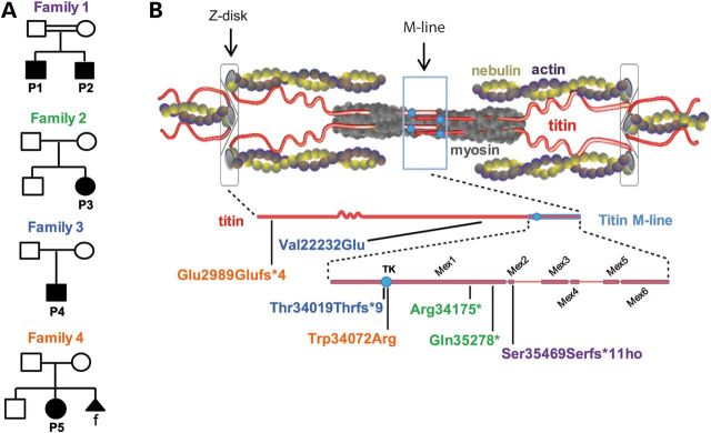
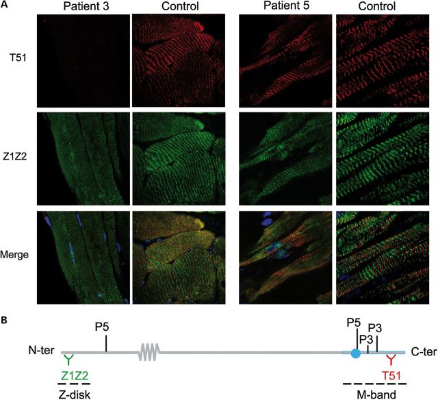
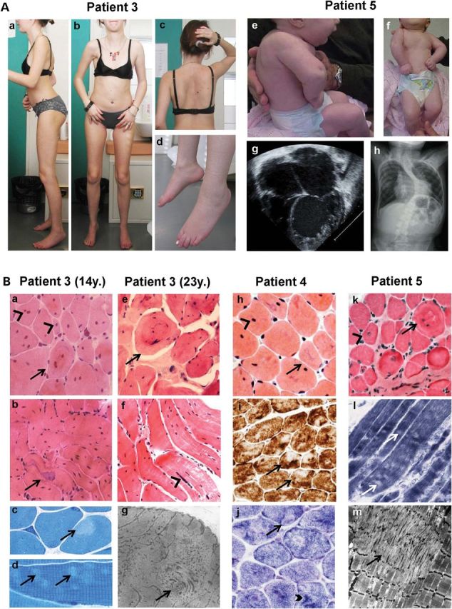
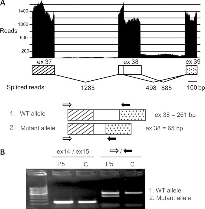
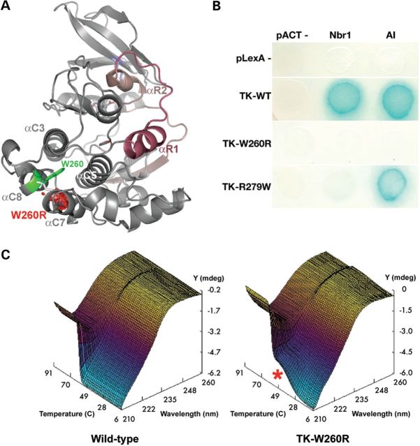
Core myopathies (CM), the main non-dystrophic myopathies in childhood, remain genetically unexplained in many cases. Heart disease is not considered part of the typical CM spectrum. No congenital heart defect has been reported, and childhood-onset cardiomyopathy has been documented in only two CM families with homozygous mutations of the TTN gene. TTN encodes titin, a giant protein of striated muscles. Recently, heterozygous TTN truncating mutations have also been reported as a major cause of dominant dilated cardiomyopathy. However, relatively few TTN mutations and phenotypes are known, and titin pathophysiological role in cardiac and skeletal muscle conditions is incompletely understood. We analyzed a series of 23 families with congenital CM and primary heart disease using TTN M-line-targeted sequencing followed in selected patients by whole-exome sequencing and functional studies. We identified seven novel homozygous or compound heterozygous TTN mutations (five in the M-line, five truncating) in 17% patients. Heterozygous parents were healthy. Phenotype analysis identified four novel titinopathies, including cardiac septal defects, left ventricular non-compaction, Emery-Dreifuss muscular dystrophy or arthrogryposis. Additionally, in vitro studies documented the first-reported absence of a functional titin kinase domain in humans, leading to a severe antenatal phenotype. We establish that CM are associated with a large range of heart conditions of which TTN mutations are a major cause, thereby expanding the TTN mutational and phenotypic spectrum. Additionally, our results suggest titin kinase implication in cardiac morphogenesis and demonstrate that heterozygous TTN truncating mutations may not manifest unless associated with a second mutation, reassessing the paradigm of their dominant expression.


Figures

Figure 1.
Figure 1.
TTN mutations and distribution. (A) Pedigrees from Families 1 to 4. Solid symbols denote affected subjects. Double lines signify consanguinity. Triangular shape indicates pregnancy termination; f = female. (B) Schema of a sarcomere with its main proteins (top) including titin (middle) and zoom on the titin M-line region (bottom). TK = titin kinase. Patient's mutations are indicated using colors corresponding to their pedigrees.
Figure 2.
Figure 2.
Expression and sarcomere integration of mutant titin in Patients 3 and 5. (A) Quadriceps from P3 and control (left panel, 63×) and myocardium from P5 and control (right panel, 63 × 4); longitudinal cryosections. Note total loss of labeling with T51 in P3. Dapi (blue) stains nuclei. (B) Recognition sites of Z1Z2 and T51 anti-titin antibodies, respectively, upstream and downstream of P5 and P3 mutations.
Figure 3.
Figure 3.
Clinical and histopathological findings. (A) P3 (23 years, left) and P5 (3 months, right). P3 showed global amyotrophy, relatively bulky calf muscles, severe elbow and heel contractures, flat retractile thorax (a, b), scoliosis with dorsal lordosis (c, after arthrodesis) and foot drop (d). Neck flexor weakness and cervical rigidity require head support when standing (c). P5 presented with shoulder, elbow, wrist, hand and lower limb contractures, short neck, low-set ears, retrognathia and kyphoscoliosis (e, f) which subsequently progressed (h). Echocardiography (four-chamber view) showed severe LVNC at 1 month (g). (B) Muscle biopsies from P3 (at 14 (a–d) and 23 years (e–g)), P4 (h–j) and P5 (k–m). A massive (a, b, e, f) or moderate (h, k) number of nuclear centralizations (arrowheads), sometimes forming nuclear chains (f, arrowhead) were associated with ring-like fibers (a, e, k; arrows) and irregular-shaped basophilic areas (b, h; arrows). Minicores were visible as small light foci of mitochondria depletion (i, j, l) and sarcomere disorganization (c, d, g, m). In some cases, they coexisted with dark areas visible on oxidative stainings (j, thick arrowhead). Endomysial fibrosis was absent (h) or mild (k). Sequential quadriceps biopsies from P3 at 2, 14 and 23 years were comparable. Transverse (a, e, h–k) and semi-longitudinal (b, f) cryosections stained with HE (a, b, e, f, h, k), Cox (i), SDH (j) or NADH-TR (l) (40× b, f, i; 60× a, e, h, j–l); transverse (c) and longitudinal (d) semithin sections; electron microscopy (g, m).
Figure 4.
Figure 4.
Consequences of P5 splice site mutation. ESE finder predicted major loss of strength of the mutant donor site (down to 7, 75%) and two alternative sites in exon 38. Use of one of these leads to coexistence of full-length and shorter exon 38 transcripts as revealed by deep sequencing (A) and cDNA amplification of TTN exon 38 (B). P and C: patient and control, respectively. A few reads suggested minor intron 38 retention (A).
Figure 5.
Figure 5.
Biochemical characterization of the TK-W260R mutant. (A) Molecular model of TK (PDB entry 1TKI) with the regulatory tail shown in red. The mutated tryptophane residue, Trp34072 or W260 in the TK sequence (TK-W260), occupies a hydrophobic pocket formed by alpha-helices C3, C5, C7 and C8. Mutation to arginine replaces this conserved hydrophobic residue with a large hydrophilic residue and results also in a predicted steric clash with alphaC7 (red volume), likely destabilizing part of the catalytic domain. (B) Mutations in TK abrogate Nbr1 binding. Yeast-two-hybrid reporter gene assay (growth on histidine-free media and activation of beta-galactosidase activity, blue color reaction). Wild-type TK (TK-WT) interacts with its own autoinhibitory tail (AI) and with Nbr1 in trans. Both interactions are abrogated in the TK-W260R mutant, whereas the TK-R279W mutant maintains AI interaction. (C) CD spectra recorded from 200 to 260 nm over 6–94°C reveal a single sharp secondary structure transition in wild-type TK with loss of negative ellipticity (Y) at 209 nm (Z) of ∼59°C (X). In contrast, the TK-W260R mutant shows partial melting (red asterisk) ∼42°C followed by a second transition at 57°C (C).

References

    1. Ferreiro A., Estournet B., Chateau D., Romero N.B., Laroche C., Odent S., Toutain A., Cabello A., Fontan D., dos Santos H.G., et al. Multi-minicore disease: searching for boundaries. Phenotypical analysis of 38 cases. Ann. Neurol. 2000;48:745–757. - PubMed
    1. Ferreiro A., Monnier N., Romero N.B., Leroy J.-P., Bönnemann C., Haenggeli C.-A., Straub V., Voss W.D., Nivoche Y., Jungbluth H., et al. A recessive form of central core disease, transiently presenting as multi-minicore disease, is associated with a homozygous mutation in the ryanodine receptor type 1 gene. Ann. Neurol. 2002;51:750–759. - PubMed
    1. Ferreiro A., Quijano-Roy S., Pichereau C., Moghadaszadeh B., Goemans N., Bönnemann C., Jungbluth H., Straub V., Villanova M., Leroy J.-P., et al. Mutations of the selenoprotein N gene, which is implicated in rigid spine muscular dystrophy, cause the classical phenotype of multiminicore disease: reassessing the nosology of early-onset myopathies. Am. J. Hum. Genet. 2002;71:739–749. doi:10.1086/342719. - DOI - PMC - PubMed
    1. Boyden S.E., Mahoney L.J., Kawahara G., Myers J.A., Mitsuhashi S., Estrella E.A., Duncan A.R., Dey F., DeChene E.T., Blasko-Goehringer J.M., et al. Mutations in the satellite cell gene MEGF10 cause a recessive congenital myopathy with minicores. Neurogenetics. 2012;13:115–124. doi:10.1007/s10048-012-0315-z. - DOI - PMC - PubMed
    1. Carmignac V., Salih M.A.M., Quijano-Roy S., Marchand S., Al Rayess M.M., Mukhtar M.M., Urtizberea J.A., Labeit S., Guicheney P., Leturcq F., et al. C-terminal titin deletions cause a novel early-onset myopathy with fatal cardiomyopathy. Ann. Neurol. 2007;61:340–351. doi:10.1002/ana.21089. - DOI - PubMed

Publication types

MeSH terms