The effects of testosterone deprivation and supplementation on proteasomal and autophagy activity in the skeletal muscle of the male mouse: differential effects on high-androgen responder and low-androgen responder muscle groups
- PMID: 24105483
- PMCID: PMC3836062
- DOI: 10.1210/en.2013-1004
The effects of testosterone deprivation and supplementation on proteasomal and autophagy activity in the skeletal muscle of the male mouse: differential effects on high-androgen responder and low-androgen responder muscle groups
Abstract
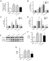
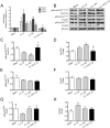
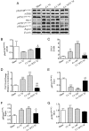
Men with prostate cancer who receive androgen deprivation therapy show profound skeletal muscle loss. We hypothesized that the androgen deficiency activates not only the ubiquitin-proteasome systems but also the autophagy and affects key aspects of the molecular cross talk between protein synthesis and degradation. Here, 2-month-old male mice were castrated and treated with either testosterone (T) propionate or vehicle for 7 days (short term) or 43 days (long term), and with and without hydroxyflutamide. Castrated mice showed rapid and profound atrophy of the levator ani muscle (high androgen responder) at short term and lesser atrophy of the triceps muscle (low androgen responder) at long term. Levator ani and triceps muscles of castrated mice showed increased level of autophagy markers and lysosome enzymatic activity; only the levator ani showed increased proteasomal enzymatic activity. The levator ani muscle of the castrated mice showed increased level and activation of forkhead box protein O3A, the inhibition of mechanistic target of rapamicyn, and the activation of tuberous sclerosis complex protein 2 and 5'-AMP-activated protein kinase. Similar results were obtained in the triceps muscle of castrated mice. T rescued the loss of muscle mass after orchiectomy and inhibited lysosome and proteasome pathways dose dependently and in a seemingly IGF-I-dependent manner. Hydroxyflutamide attenuated the effect of T in the levator ani muscle of castrated mice. In conclusion, androgen deprivation in adult mice induces muscle atrophy associated with proteasomal and lysosomal activity. T optimizes muscle protein balance by modulating the equilibrium between mechanistic target of rapamicyn and 5'-AMP-activated protein kinase pathways.
Figures


Similar articles
-
Regulation of Akt-mTOR, ubiquitin-proteasome and autophagy-lysosome pathways in response to formoterol administration in rat skeletal muscle.Int J Biochem Cell Biol. 2013 Nov;45(11):2444-55. doi: 10.1016/j.biocel.2013.07.019. Epub 2013 Aug 2. Int J Biochem Cell Biol. 2013. PMID: 23916784
-
Testosterone regulation of Akt/mTORC1/FoxO3a signaling in skeletal muscle.Mol Cell Endocrinol. 2013 Jan 30;365(2):174-86. doi: 10.1016/j.mce.2012.10.019. Epub 2012 Oct 29. Mol Cell Endocrinol. 2013. PMID: 23116773 Free PMC article.
-
Testosterone represses ubiquitin ligases atrogin-1 and Murf-1 expression in an androgen-sensitive rat skeletal muscle in vivo.J Appl Physiol (1985). 2010 Feb;108(2):266-73. doi: 10.1152/japplphysiol.00490.2009. Epub 2009 Nov 19. J Appl Physiol (1985). 2010. PMID: 19926828
-
Protein breakdown in muscle wasting: role of autophagy-lysosome and ubiquitin-proteasome.Int J Biochem Cell Biol. 2013 Oct;45(10):2121-9. doi: 10.1016/j.biocel.2013.04.023. Epub 2013 May 7. Int J Biochem Cell Biol. 2013. PMID: 23665154 Free PMC article. Review.
-
Recent progress toward understanding the molecular mechanisms that regulate skeletal muscle mass.Cell Signal. 2011 Dec;23(12):1896-906. doi: 10.1016/j.cellsig.2011.07.013. Epub 2011 Jul 23. Cell Signal. 2011. PMID: 21821120 Free PMC article. Review.
Cited by
-
Overcoming resistance to anabolic SARM therapy in experimental cancer cachexia with an HDAC inhibitor.EMBO Mol Med. 2020 Feb 7;12(2):e9910. doi: 10.15252/emmm.201809910. Epub 2020 Jan 13. EMBO Mol Med. 2020. PMID: 31930715 Free PMC article.
-
Sex differences in the response to oxidative and proteolytic stress.Redox Biol. 2020 Apr;31:101488. doi: 10.1016/j.redox.2020.101488. Epub 2020 Mar 9. Redox Biol. 2020. PMID: 32201219 Free PMC article. Review.
-
Androgen-mediated regulation of skeletal muscle protein balance.Mol Cell Endocrinol. 2017 May 15;447:35-44. doi: 10.1016/j.mce.2017.02.031. Epub 2017 Feb 22. Mol Cell Endocrinol. 2017. PMID: 28237723 Free PMC article. Review.
-
Testosterone exacerbates neutrophilia and cardiac injury in myocardial infarction via actions in bone marrow.Nat Commun. 2025 Feb 5;16(1):1142. doi: 10.1038/s41467-025-56217-x. Nat Commun. 2025. PMID: 39910039 Free PMC article.
-
BCAA metabolism: the Achilles' heel of ovarian cancer, polycystic ovary syndrome, and premature ovarian failure.Front Endocrinol (Lausanne). 2025 Jul 4;16:1579477. doi: 10.3389/fendo.2025.1579477. eCollection 2025. Front Endocrinol (Lausanne). 2025. PMID: 40687583 Free PMC article. Review.
References
-
- Basaria S, Muller DC, Carducci MA, Egan J, Dobs AS. Hyperglycemia and insulin resistance in men with prostate carcinoma who receive androgen-deprivation therapy. Cancer. 2006;106:581–588 - PubMed
-
- Biolo G, Maggi SP, Williams BD, Tipton KD, Wolfe RR. Increased rates of muscle protein turnover and amino acid transport after resistance exercise in humans. Am J Physiol. 1995;268:E514–E520 - PubMed
-
- Thomason DB, Booth FW. Atrophy of the soleus muscle by hindlimb unweighting. J Appl Physiol. 1990;68:1–12 - PubMed
Publication types
MeSH terms
Substances
Grants and funding
LinkOut - more resources
Full Text Sources
Other Literature Sources

