. 2013 Nov 22;288(47):34041-34051.
doi: 10.1074/jbc.M113.518019. Epub 2013 Oct 8.

Phosphorylation of angiomotin by Lats1/2 kinases inhibits F-actin binding, cell migration, and angiogenesis

Affiliations

Phosphorylation of angiomotin by Lats1/2 kinases inhibits F-actin binding, cell migration, and angiogenesis

Xiaoming Dai et al. J Biol Chem. .

Abstract

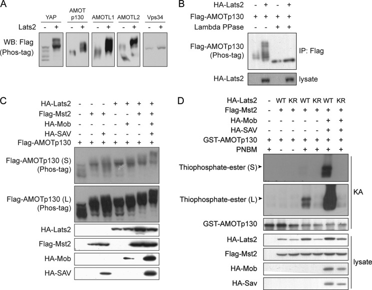
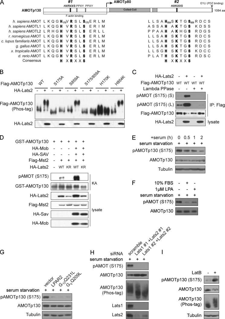
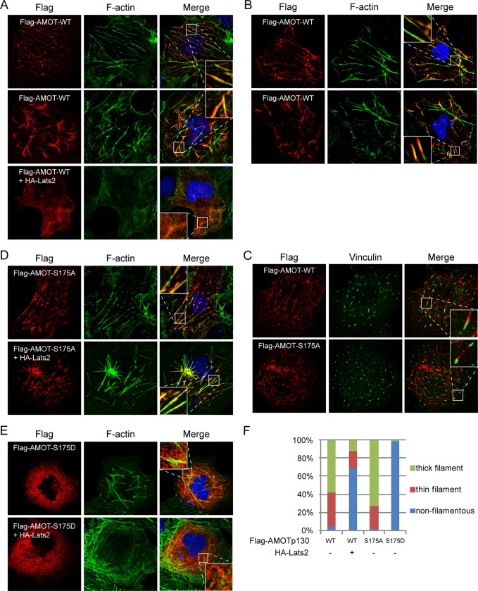
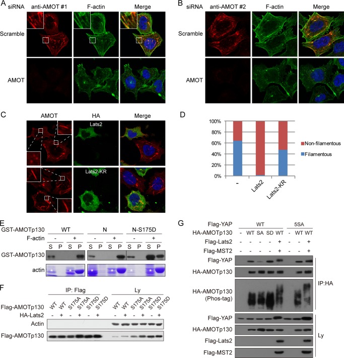
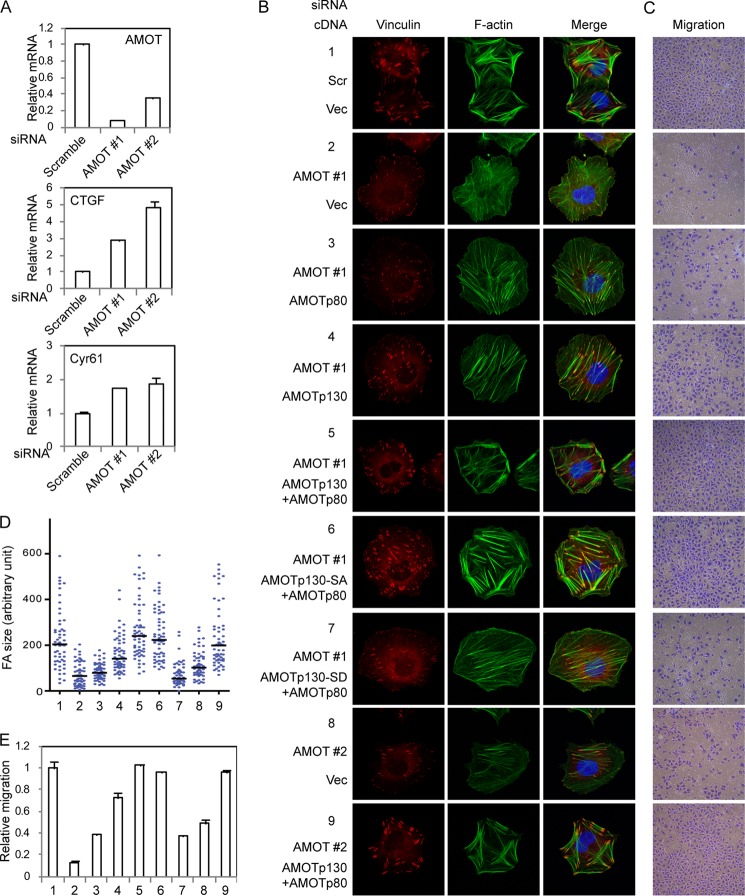
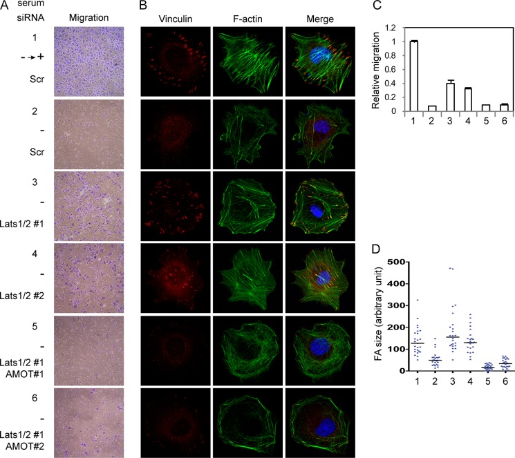
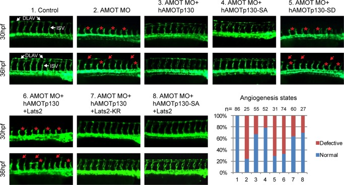
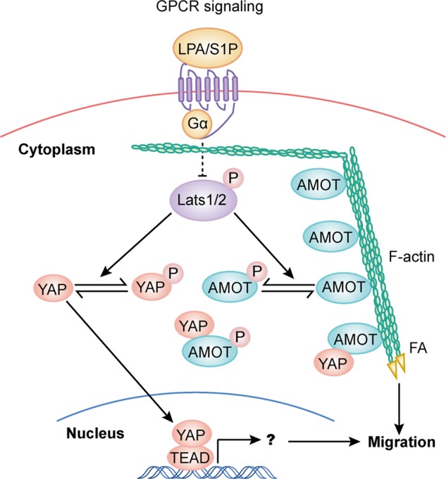
The Hippo tumor suppressor pathway plays important roles in organ size control through Lats1/2 mediated phosphorylation of the YAP/TAZ transcription co-activators. However, YAP/TAZ independent functions of the Hippo pathway are largely unknown. Here we report a novel role of the Hippo pathway in angiogenesis. Angiomotin p130 isoform (AMOTp130) is phosphorylated on a conserved HXRXXS motif by Lats1/2 downstream of GPCR signaling. Phosphorylation disrupts AMOT interaction with F-actin and correlates with reduced F-actin stress fibers and focal adhesions. Furthermore, phosphorylation of AMOT by Lats1/2 inhibits endothelial cell migration in vitro and angiogenesis in zebrafish embryos in vivo. Thus AMOT is a direct substrate of Lats1/2 mediating functions of the Hippo pathway in endothelial cell migration and angiogenesis.

Keywords: AMOT; Actin; Angiogenesis; Cell Migration; Lats; Protein Kinase; Protein Kinases; Protein Phosphorylation; the Hippo Pathway.


Figures

FIGURE 1.
FIGURE 1.
The Hippo pathway phosphorylates angiomotin family proteins. A, Lats2 induces mobility shift of AMOT family proteins. HEK293 cells were transfected with plasmids encoding Flag-YAP or AMOT family proteins with or without Lats2 co-transfection. Cell lysates were resolved on Phos-tag-containing gels. Vps34 is a negative control. B, phosphatase eliminates AMOT mobility shift induced by Lats2. Flag-AMOTp130 was immunoprecipitated from transfected cells and treated with lambda protein phosphatase as indicated. Samples were analyzed as that in A. C, hippo pathway proteins synergistically induces AMOTp130 mobility shift. Lysates of transfected cells were resolved on Phos-tag-containing gels for mobility shift of Flag-AMOTp130. D, Lats2 phosphorylates AMOTp130 in vitro. In vitro phosphorylation of GST-AMOTp130 by immunoprecipitated Lats2 was performed. (S), short exposure; (L), long exposure.
FIGURE 2.
FIGURE 2.
AMOTp130 is phosphorylated on Ser-175 by Lats1/2 in response to GPCR signaling. A, domain organization of AMOTp130 and alignment of AMOT family proteins from different species. B, phosphorylation of Ser-175 leads to Lats2-induced AMOTp130 up-shift. Lysates of transfected HEK293 cells were analyzed by Western blots. C, Ser-175 of AMOTp130 is phosphorylated by Lats2. AMOTp130 phosphorylation was detected by pAMOT (S175)-specific antibody. D, Lats2 phosphorylates AMOTp130 on Ser-175 in vitro. Experiments were similar to that in Fig. 1D except that regular ATP was used. E, serum inhibits phosphorylation of endogenous AMOTp130 on Ser-175. Serum-starved 293T cells were stimulated with 10% FBS for the indicated time before harvest. F, LPA inhibits phosphorylation of endogenous AMOTp130 on Ser-175. Serum-starved 293T cells were stimulated with serum or LPA for 1 h before harvest. G, GPCR signaling inhibits Ser-175 phosphorylation of endogenous AMOTp130. 293T cells were transfected with indicated plasmids and serum-starved overnight before harvest. H, knockdown of Lats1 and Lats2 represses endogenous AMOTp130 phosphorylation on Ser-175. HEK293T cells were transfected with scramble or Lats1- and Lats2-specific siRNAs. Cells were serum-starved overnight before harvest. I, disruption of F-actin induces phosphorylation of AMOTp130. HEK293T cells cultured in serum-rich medium were treated with 1 μg/ml Latrunculin B (LatB) for 1 h as indicated before being harvested for Western blot analysis.
FIGURE 3.
FIGURE 3.
Phosphorylation of AMOTp130 on Ser-175 inhibits interaction with F-actin. A, Lats2 inhibits colocalization of AMOTp130 with F-actin. Transfected COS7 cells were stained with anti-Flag antibody for localization of AMOTp130, AlexaFluor488-Phalloidin for F-actin, and DAPI for cell nuclei. B, colocalization of AMOTp130 with F-actin in digitonin-treated cells. Flag-AMOTp130 transfected Cos7 cells were treated with digitonin before fixation and stained as that in A. C, AMOT-F-actin filaments are anchored to focal adhesions. COS7 cells expressing wild-type or mutant AMOTs were co-stained with anti-Flag for AMOT and anti-vinculin to visualize focal adhesions. D, phospho-deficient mutant of AMOTp130 constitutively colocalizes with F-actin. Experiments were similar to these in A except the use of S175A mutant. E, phospho-mimetic mutant of AMOTp130 loses colocalization with F-actin. Experiments were similar to these in A except the use of S175D mutant. F, quantification of AMOT-F-actin colocalization. 120 cells were quantified for each transfection. Thick filament is similar to Fig. 3A, middle panel; thin filament is similar to that in Fig. 3A, top panel; non filamentous is similar to that in Fig. 3E, top panel.
FIGURE 4.
FIGURE 4.
Phosphorylation inhibits endogenous AMOTp130-F-actin interaction and direct AMOTp130-F-actin interaction in vitro. A, endogenous AMOT colocalizes with F-actin. Scramble or AMOT-specific siRNA transfected HEK293T cells were stained with anti-AMOT antibody #1 for localization of endogenous AMOTp130, AlexaFluor488-Phalloidin for F-actin, and DAPI for cell nuclei. B, endogenous AMOT colocalizes with F-actin. Experiments were similar to these in A except that endogenous AMOT was stained with anti-AMOT antibody #2. C, Lats2 inhibits AMOT colocalization with F-actin in a kinase-dependent manner. HA-Lats2 wild-type or KR transfected HEK293T cells were stained with anti-AMOT antibody #1 for localization of endogenous AMOTp130 and anti-HA for Lats2 expression. D, quantification of filamentous localization of endogenous AMOT. Cells with or without filamentous AMOT in A and C were quantified. 120 cells were quantified for each sample. E, phospho-mimetic mutation inhibits AMOTp130 association with F-actin in vitro. Recombinant GST-AMOT wild-type or N-terminal fragment (N) were subjected to in vitro actin spin-down assay. (S), supernatant; (P), pellet. Actin was examined by Coomassie Blue staining and GST-AMOT was determined by Western blots. Asterisks denote nonspecific proteins. F, AMOTp130 does not co-immunoprecipitate with G-actin. HEK293 cells were transfected as indicated. Transfection amount was adjusted to make soluble AMOT levels comparable. Lysates were immunoprecipitated with anti-Flag antibody. The presence of endogenous actin and Flag-AMOTp130 in immunoprecipitates and lysates were examined by Western blots. G, phosphorylation of wild-type AMOTp130 by Lats2 does not affect interaction between AMOTp130 and YAP. HEK293 cells were transfected as indicated. AMOTp130 was immunoprecipitated with anti-HA antibody. Co-immunoprecipitation of YAP wild-type or 5SA mutant was examined by anti-Flag Western blots. AMOTp130 phosphorylation was shown by electrophoretic mobility shift on Phos-tag containing gel.
FIGURE 5.
FIGURE 5.
Phosphorylation of AMOTp130 on Ser-175 inhibits endothelial cell migration. A, knockdown efficiency of AMOT siRNAs. AMOT, CTGF, and Cyr61 levels in siRNA transfected HUVEC cells were determined by realtime RT-PCR. B, phospho-mimetic mutant of AMOT is unable to compensate endogenous AMOT in actin stress fiber and focal adhesion formation. Control or AMOT-expressing stable cells transfected with siRNAs were serum-starved overnight and then seeded on fibronectin-coated coverslips for 2 h in serum-containing medium. Focal adhesions and F-actin were stained with anti-vinculin and AlexaFluor488-Phalloidin, respectively. C, phospho-mimetic AMOT mutant could not rescue cell migration defects caused by loss of AMOT. Cells the same as these in B were examined in transwell migration assay. D, size of 60 FAs from the staining of each stable cell as these in B was quantified by ImageJ and drawn into scatter plot. The median number was indicated. E, number of migrated cells in C was quantified. Experiments performed were in duplicates.
FIGURE 6.
FIGURE 6.
Knockdown of Lats1/2 induces cell migration, actin stress fiber and focal adhesion in an AMOT-dependent manner. A, Lats1/2 plays a role in serum deprivation-induced migration retardation by repressing AMOT. siRNA-transfected HUVEC cells were serum starved and then seeded in transwell inserts in serum-free medium. The lower chambers were filled with serum-containing or serum-free medium as indicated. Cells on bottom sides of the transwells were stained after 12 h. B, Lats1/2 plays a role in serum deprivation-induced stress fiber and focal adhesion deformation by repressing AMOT. Cells were the same as these in A. Cells were serum starved overnight and then seeded on fibronectin-coated coverslips for 2 h either in serum-containing medium or serum-free medium as indicated. Focal adhesions and F-actin were stained with anti-vinculin and AlexaFluor488-Phalloidin respectively. C, number of migrated cells in A was quantified. Experiments were in duplicates. D, size of 25 FAs from the staining of each stable cell as these in B was quantified by ImageJ and drawn into scatter plot. The median number was indicated.
FIGURE 7.
FIGURE 7.
Phosphorylation of AMOTp130 by Lats1/2 regulates angiogenesis in zebrafish. The fluorescent microscopy analyses revealed the development of ISV and DLAV at 30 hpf and 36 hpf in the trunk region of control Tg(flk:GFP) embryos and embryos injected with AMOT-MO or a combination of AMOT-MO and mRNAs encoding human AMOTp130, Lats2 or their mutants. Red asterisk: defective ISV; red arrow: defective DLAV. ISV: intersegmental vessels. DLAV: dorsal longitudinal anastomotic vessels. Embryos were orientated with anterior to the left. Histogram depicted the quantification of normal and angiogenesis defective embryos at 30 hpf. n, embryo number.
FIGURE 8.
FIGURE 8.
A model of Lats1/2 in regulation of cell migration through phosphorylation of AMOT and YAP. Phosphorylation of AMOT by Lats1/2 inhibits F-actin association and then actin stress fiber and focal adhesion formation. These effects of AMOT on cell structure and cell adhesion may work independently or in cooperation with YAP-dependent gene transcription alteration downstream of Lats1/2 to regulate cell migration.

References

    1. Zhao B., Li L., Lei Q., Guan K. L. (2010) The Hippo-YAP pathway in organ size control and tumorigenesis: an updated version. Genes Dev. 24, 862–874 - PMC - PubMed
    1. Dong J., Feldmann G., Huang J., Wu S., Zhang N., Comerford S. A., Gayyed M. F., Anders R. A., Maitra A., Pan D. (2007) Elucidation of a universal size-control mechanism in Drosophila and mammals. Cell 130, 1120–1133 - PMC - PubMed
    1. Zhou D., Conrad C., Xia F., Park J. S., Payer B., Yin Y., Lauwers G. Y., Thasler W., Lee J. T., Avruch J., Bardeesy N. (2009) Mst1 and Mst2 maintain hepatocyte quiescence and suppress hepatocellular carcinoma development through inactivation of the Yap1 oncogene. Cancer Cell 16, 425–438 - PMC - PubMed
    1. Camargo F. D., Gokhale S., Johnnidis J. B., Fu D., Bell G. W., Jaenisch R., Brummelkamp T. R. (2007) YAP1 increases organ size and expands undifferentiated progenitor cells. Curr. Biol. 17, 2054–2060 - PubMed
    1. Wu S., Huang J., Dong J., Pan D. (2003) hippo encodes a Ste-20 family protein kinase that restricts cell proliferation and promotes apoptosis in conjunction with salvador and warts. Cell 114, 445–456 - PubMed

Publication types

MeSH terms