

Avoiding the pitfalls of single particle cryo-electron microscopy: Einstein from noise
- PMID: 24106306
- PMCID: PMC3831464
- DOI: 10.1073/pnas.1314449110
Avoiding the pitfalls of single particle cryo-electron microscopy: Einstein from noise
Abstract
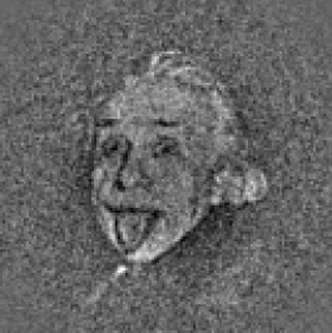
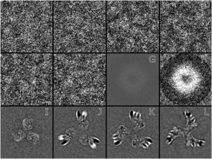
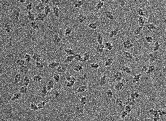
Single particle cryo-electron microscopy is currently poised to produce high-resolution structures of many biological assemblies, but several pitfalls can trap the unwary. This critique highlights one problem that is particularly relevant when smaller structures are being studied. It is known as "Einstein from noise," in which the experimenter honestly believes they have recorded images of their particles, whereas in reality, most if not all of their data consist of pure noise. Selection of particles using cross-correlation methods can then lead to 3D maps that resemble the model used in the initial selection and provide the illusion of progress. Suggestions are given about how to circumvent the problem.
Keywords: 3DEM; EMDB; SPEM; cryo-EM; validation.
Conflict of interest statement
The author declares no conflict of interest.
Figures
Comment in
-
Reply to Subramaniam, van Heel, and Henderson: Validity of the cryo-electron microscopy structures of the HIV-1 envelope glycoprotein complex.Proc Natl Acad Sci U S A. 2013 Nov 5;110(45):E4178-82. doi: 10.1073/pnas.1316666110. Proc Natl Acad Sci U S A. 2013. PMID: 24350339 Free PMC article. No abstract available.
Comment on
-
Molecular architecture of the uncleaved HIV-1 envelope glycoprotein trimer.Proc Natl Acad Sci U S A. 2013 Jul 23;110(30):12438-43. doi: 10.1073/pnas.1307382110. Epub 2013 Jun 11. Proc Natl Acad Sci U S A. 2013. PMID: 23757493 Free PMC article.
References
-
- Adrian M, Dubochet J, Lepault J, McDowall AW. Cryo-electron microscopy of viruses. Nature. 1984;308(5954):32–36. - PubMed
-
- Henderson R. The potential and limitations of neutrons, electrons and X-rays for atomic resolution microscopy of unstained biological molecules. Q Rev Biophys. 1995;28(2):171–193. - PubMed
-
- Henderson R. Realizing the potential of electron cryo-microscopy. Q Rev Biophys. 2004;37(1):3–13. - PubMed
Publication types
MeSH terms
Substances
Grants and funding
LinkOut - more resources
Full Text Sources
Other Literature Sources
