

High-resolution restoration of 3D structures from widefield images with extreme low signal-to-noise-ratio
- PMID: 24106307
- PMCID: PMC3808589
- DOI: 10.1073/pnas.1315675110
High-resolution restoration of 3D structures from widefield images with extreme low signal-to-noise-ratio
Abstract
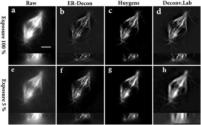
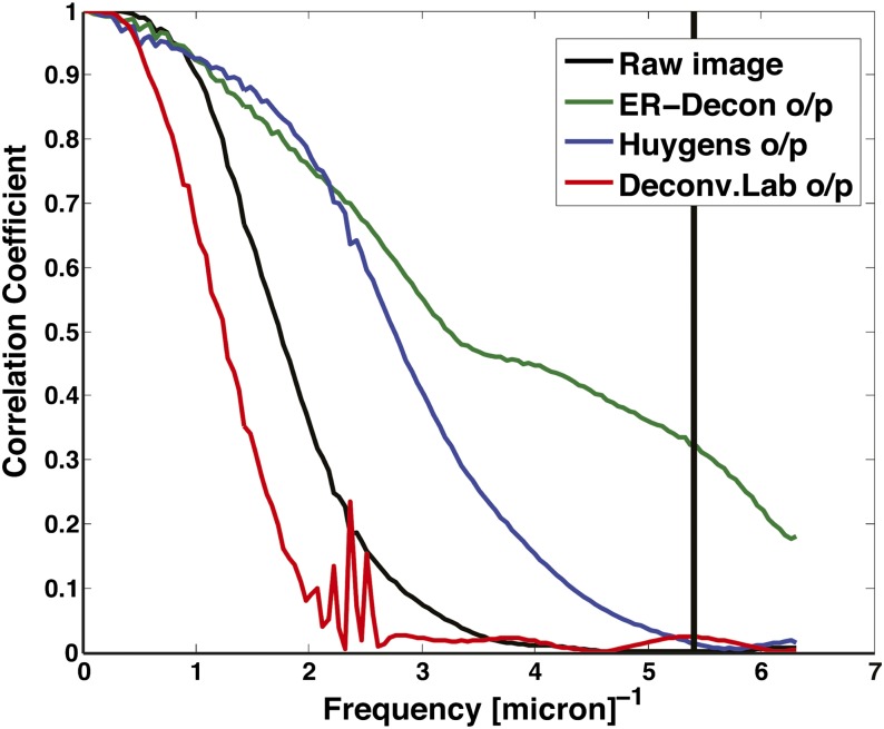
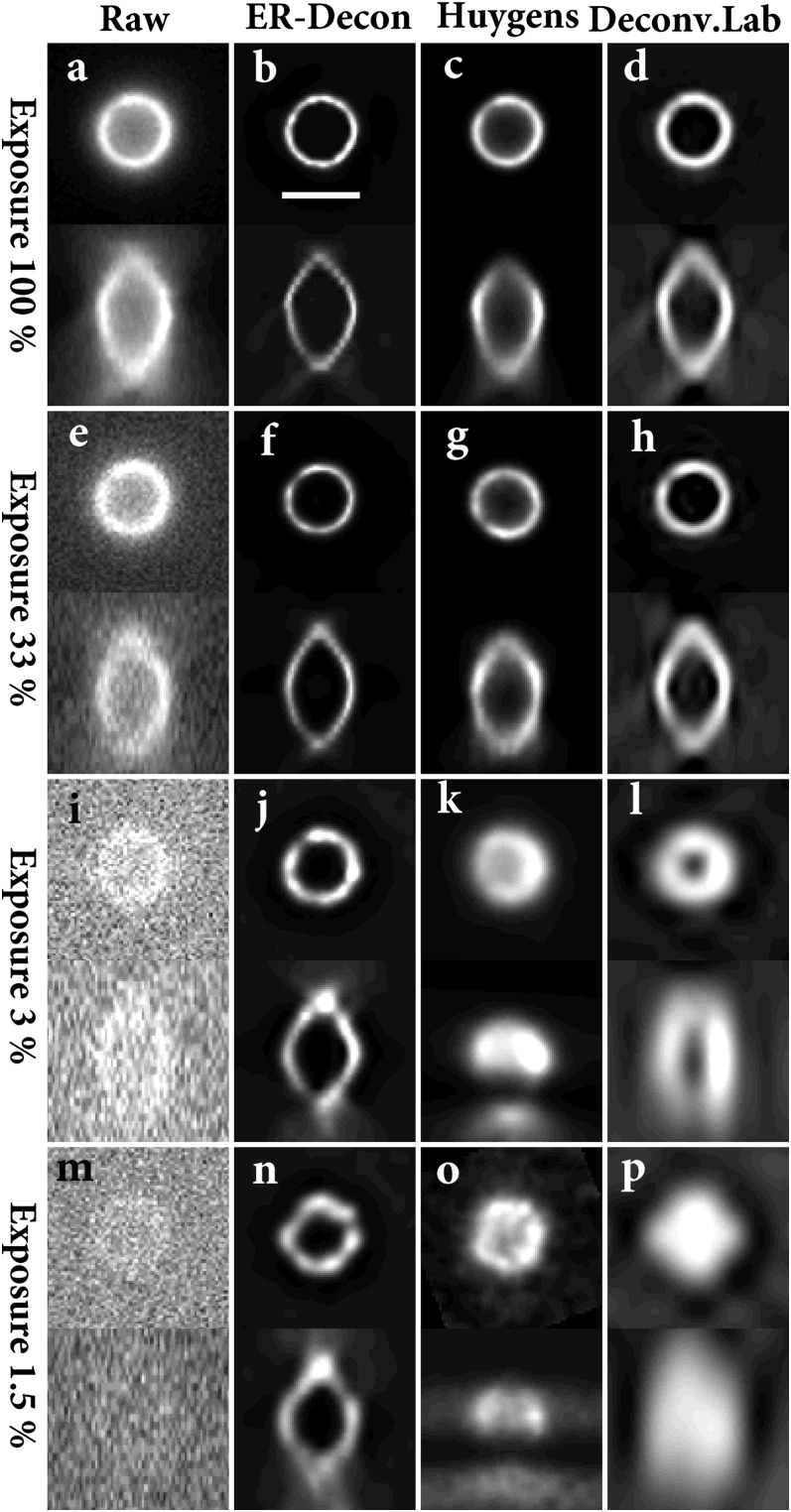
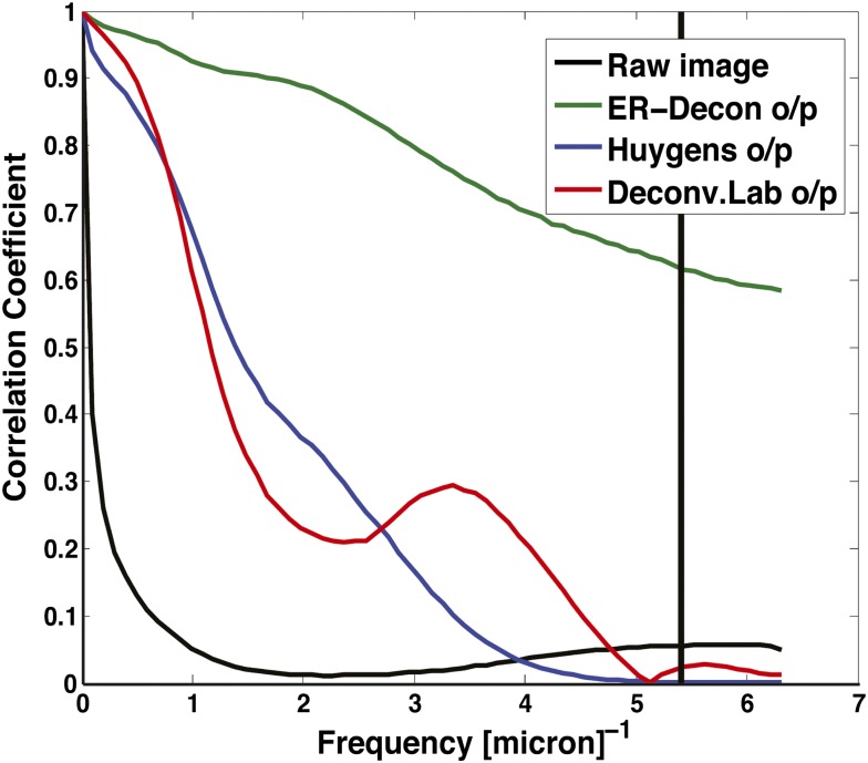
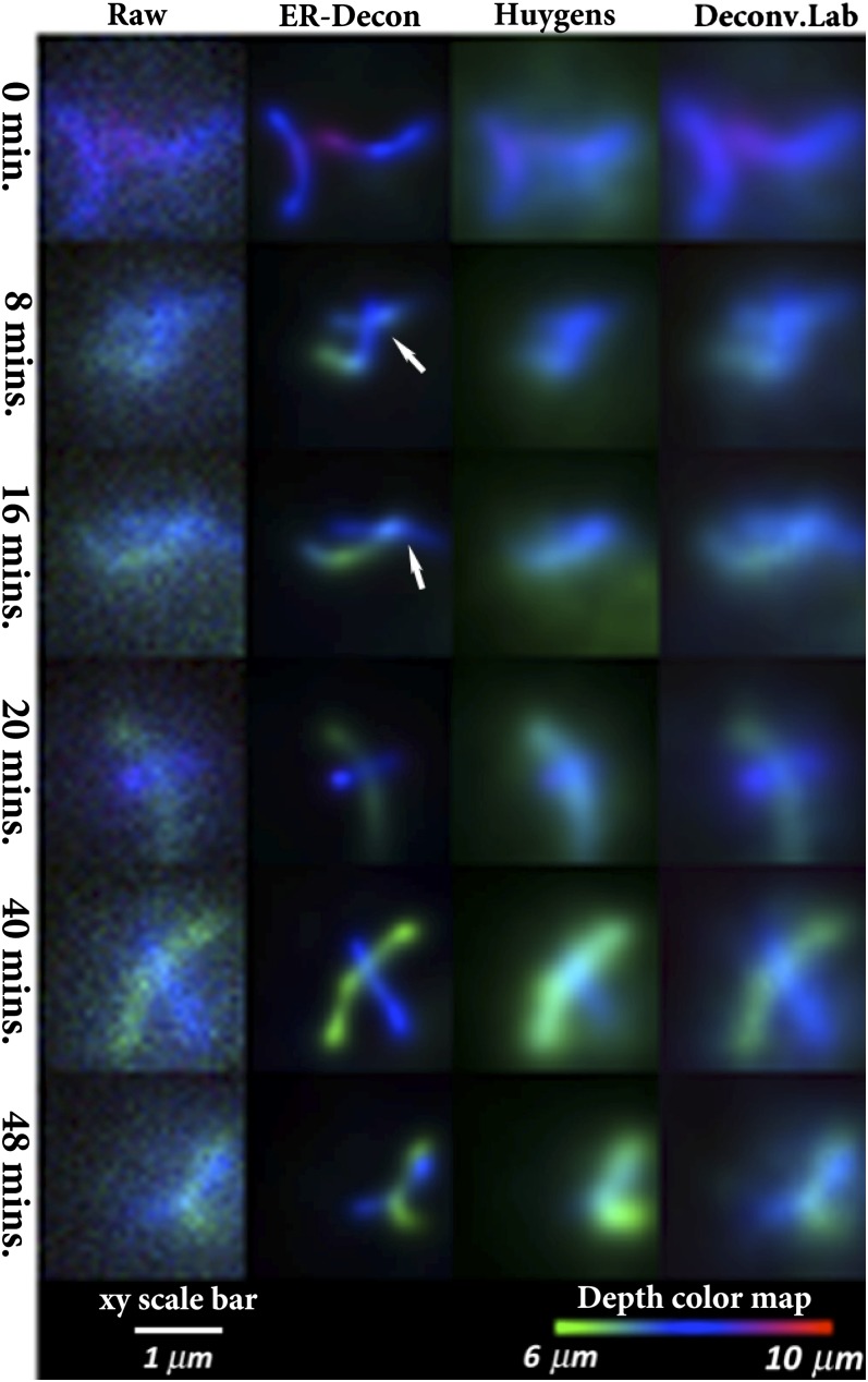
Four-dimensional fluorescence microscopy--which records 3D image information as a function of time--provides an unbiased way of tracking dynamic behavior of subcellular components in living samples and capturing key events in complex macromolecular processes. Unfortunately, the combination of phototoxicity and photobleaching can severely limit the density or duration of sampling, thereby limiting the biological information that can be obtained. Although widefield microscopy provides a very light-efficient way of imaging, obtaining high-quality reconstructions requires deconvolution to remove optical aberrations. Unfortunately, most deconvolution methods perform very poorly at low signal-to-noise ratios, thereby requiring moderate photon doses to obtain acceptable resolution. We present a unique deconvolution method that combines an entropy-based regularization function with kernels that can exploit general spatial characteristics of the fluorescence image to push the required dose to extreme low levels, resulting in an enabling technology for high-resolution in vivo biological imaging.
Keywords: 4D microscopy; low dose microscopy; noise-suppressing regularization.
Conflict of interest statement
The authors declare no conflict of interest.
Figures

References
-
- Lane R. Methods for maximum-likelihood deconvolution. J Opt Soc Am A Opt Image Sci Vis. 1996;13(10):1992–1998. - PubMed
-
- Conchello JA, Hansen EW. Enhanced 3-D reconstruction from confocal scanning microscope images. 1: Deterministic and maximum likelihood reconstructions. Appl Opt. 1990;29(26):3795–3804. - PubMed
-
- Conchello JA, Kim JJ, Hansen EW. Enhanced three-dimensional reconstruction from confocal scanning microscope images. II. Depth discrimination versus signal-to-noise ratio in partially confocal images. Appl Opt. 1994;33(17):3740–3750. - PubMed
-
- Conchello JA. Superresolution and convergence properties of the expectation-maximization algorithm for maximum-likelihood deconvolution of incoherent images. J Opt Soc Am A Opt Image Sci Vis. 1998;15(10):2609–2619. - PubMed
