. 2013 Sep 10:2013:261297.
doi: 10.1155/2013/261297. eCollection 2013.

Ultrastructural changes of airway in murine models of allergy and diet-induced metabolic syndrome

Affiliations

Ultrastructural changes of airway in murine models of allergy and diet-induced metabolic syndrome

Geeta Devi Leishangthem et al. ISRN Allergy. .

Abstract

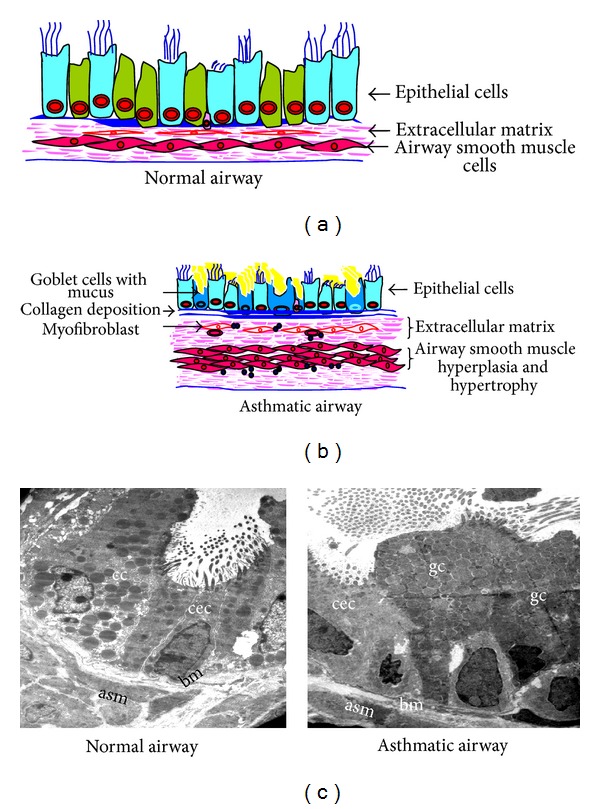
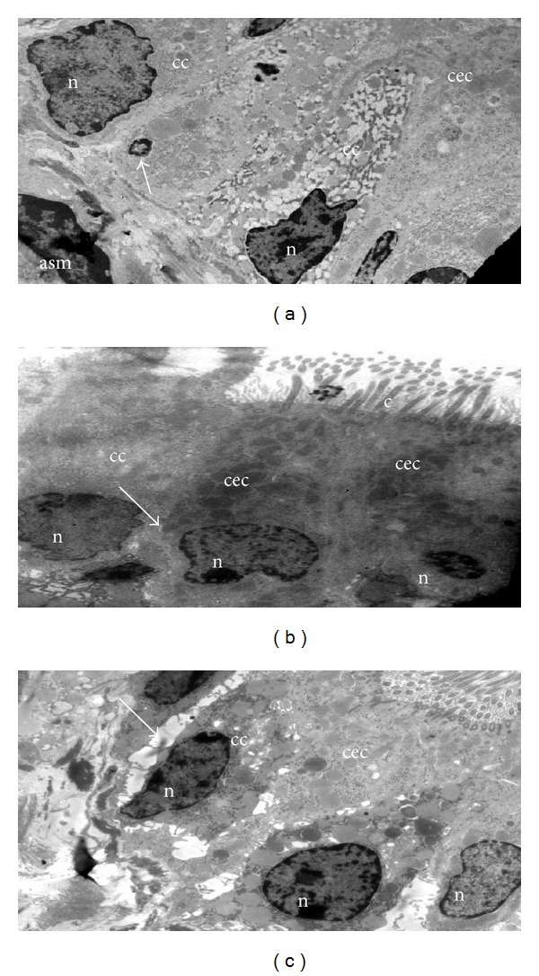
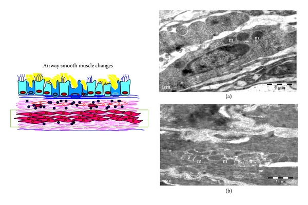
Studying ultrastructural changes could reveal novel pathophysiology of obese-asthmatic condition as existing concepts in asthma pathogenesis are based on the histological changes of the diseased airway. While asthma is defined in functional terms, the potential of electron microscopy (EM) in providing cellular and subcellular detail is underutilized. With this view, we have performed transmission EM in the lungs from allergic mice that show key features of asthma and high-fat- or high-fructose-fed mice that mimicked metabolic syndrome to illustrate the ultrastructural changes. The primary focus was epithelial injury and metaplasia, which are cardinal features of asthma and initiate airway remodeling. EM findings of the allergically inflamed mouse lungs correlate with known features of human asthma such as increased mitochondria in airway smooth muscle, platelet activation and subepithelial myofibroblasts. Interestingly, we found a clear and unambiguous evidence to suggest that ciliated cells can become goblet cells using immunoelectron microscopy. Additionally, we show for the first time the stressed mitochondria in the bronchial epithelia of high-fat- or high-fructose-fed mice even without allergen exposure. These results may stimulate interest in using EM in understanding novel pathological mechanisms for different subtypes of asthma including obese asthma.


Figures

Figure 1
Figure 1
Components of structural changes of asthmatic airway. (a) and (b) Schematic diagrams show major components of normal and asthmatic airways. (c) Transmission electron microscopy (TEM) of lung to show the components of airway remodeling. Normal airway shows almost equal portions of ciliated epithelial cells (cec) and Clara cells (cc) and occasional basal cells and no mucous cell, thin basement membrane (bm), attenuated fibroblast sheath, and thin layer of airway smooth muscle (asm). Asthmatic airway shows the predominance of goblet cells (gc), thick basement membrane (bm), thickened (myo) fibroblast sheath, and hypertrophy and hyperplasia of airway smooth muscle (asm). Images are at 880X magnification.
Figure 2
Figure 2
Eosinophil activity and platelet sequestration in asthmatic airway. TEM of eosinophil shows the peculiar granules with electron dense core surrounded by electron lucent matrix. Asthmatic mice of subacute (a) model show more granules (white arrows) with loss of either core or matrix indicating more degranulation and thus more activation, and in contrast asthmatic mice of chronic (b) model show more intact granules indicating less degranulation and thus less activation. (c) Numerous platelets (pl) are seen in and around the blood vessel in asthmatic mice, and higher magnification (d) shows the process of platelet sequestration (circle), n, nucleus. Images are at 3200X magnification.
Figure 3
Figure 3
Different types of major epithelial cells present in the airway and Goblet cell metaplasia. (a) Ciliated: columnar shaped with elongated nuclei (n) near the base, and electron lucent cytoplasm. (b) Clara: dome-shaped with circular electron dense secretory granules (sg). (c) Goblet: mucus containing membrane bound electron lucent vacuoles in cytoplasm and (d) Basal cell: triangle shaped cell located just above the basement membrane with small basal nuclei (n). (e) TEM of the bronchial epithelia of asthmatic mice showed an abundance of goblet cells (gc) with numerous secretory granules (sg) containing mucus, c, cilia. (f) Clara (cc) and (g) Ciliated epithelial cells (cec) also show secretory granules. (h) Immunogold labeling with Muc5ac show the positive localization of Muc5ac as shown by the presence of gold particles (15 nm) in secretory granules (sg) of ciliated epithelia of asthmatic mice (bbc—basal bodies condensation). Images are at 880X (a)–(f) 3200X (g)-(h) magnifications.
Figure 4
Figure 4
Epithelial injury. (a) Asthmatic mice show the shrinkage of bronchial epithelia (arrow) with pyknotic nucleus and marginated heterochromatin. Normal airways (b) show thin and regular intercellular space as compared to that of asthmatic airways. (c) Widening of intercellular space; cc, clara cells; asm, airway smooth muscle; cec, ciliated epithelial cells; n, nucleus; c, cilia. Arrows in (b) and (c) indicate the intercellular space between adjacent bronchial epithelia. Images are at 880X magnification.
Figure 5
Figure 5
Mitochondrial ultrastructural changes in bronchial epithelia of high-fat- or high-fructose-fed mice. TEM images of airway epithelia of normal murine lung (a) show normal mitochondria (green arrows) with well-developed cristae and dense matrix compared to airway epithelia of high-fat- or high-fructose-fed mice which show the reduced number or loss of cristae (green arrows). Images are at 10500X magnification.
Figure 6
Figure 6
Subepithelial fibrosis. TEM of normal airway (a) shows the regular and thin basement membrane (bm), and asthmatic airways (b) and (c) show the thickened basement membrane (bm) with bundles of collagen (col) in subepithelial region and airway smooth muscle (asm) hyperplasia. ec, epithelial cell and mf, myofibroblast. Images are at 1950X magnification.
Figure 7
Figure 7
Airway Smooth muscle changes. TEM of normal airway smooth muscle (a) shows few mitochondria (m) as compared to asthmatic airway smooth muscles (asm) (b) numerous mitochondria (m). Images are at 1950X magnification.

References

    1. Kay AB. Pathology of mild, severe, and fatal asthma. American Journal of Respiratory and Critical Care Medicine. 1996;154(2):S66–S69. - PubMed
    1. Davies DE. The role of the epithelium in airway remodeling in asthma. Proceedings of the American Thoracic Society. 2009;6(8):678–682. - PMC - PubMed
    1. Lin TY, Poon AH, Hamid Q. Asthma phenotypes and endotypes. Current Opinion in Pulmonary Medicine. 2013;19(1):18–23. - PubMed
    1. Wenzel S. Severe asthma: from characteristics to phenotypes to endotypes. Clinical and Experimental Allergy. 2012;42(5):650–658. - PubMed
    1. Lötvall J, Akdis CA, Bacharier LB, et al. Asthma endotypes: a new approach to classification of disease entities within the asthma syndrome. Journal of Allergy and Clinical Immunology. 2011;127(2):355–360. - PubMed

LinkOut - more resources