Review
. 2013:2013:658270.
doi: 10.1155/2013/658270. Epub 2013 Sep 11.

"Zebrafishing" for novel genes relevant to the glomerular filtration barrier

Affiliations
Review

"Zebrafishing" for novel genes relevant to the glomerular filtration barrier

Nils Hanke et al. Biomed Res Int. 2013.

Abstract

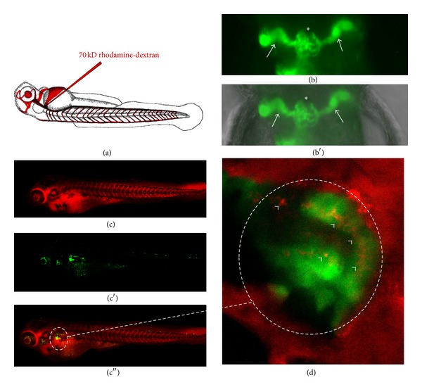
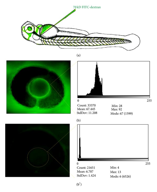
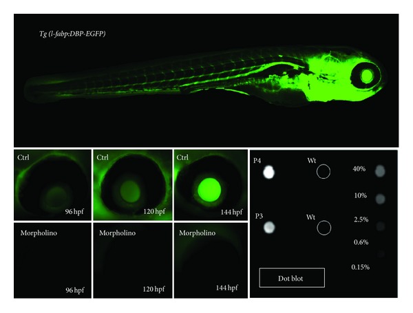
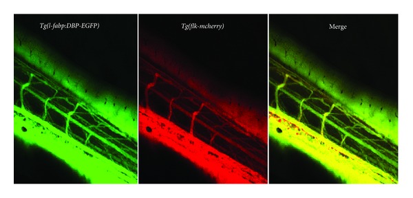
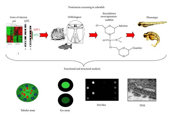
Data for genes relevant to glomerular filtration barrier function or proteinuria is continually increasing in an era of microarrays, genome-wide association studies, and quantitative trait locus analysis. Researchers are limited by published literature searches to select the most relevant genes to investigate. High-throughput cell cultures and other in vitro systems ultimately need to demonstrate proof in an in vivo model. Generating mammalian models for the genes of interest is costly and time intensive, and yields only a small number of test subjects. These models also have many pitfalls such as possible embryonic mortality and failure to generate phenotypes or generate nonkidney specific phenotypes. Here we describe an in vivo zebrafish model as a simple vertebrate screening system to identify genes relevant to glomerular filtration barrier function. Using our technology, we are able to screen entirely novel genes in 4-6 weeks in hundreds of live test subjects at a fraction of the cost of a mammalian model. Our system produces consistent and reliable evidence for gene relevance in glomerular kidney disease; the results then provide merit for further analysis in mammalian models.


Figures

Figure 1
Figure 1
Edema as a sign of kidney failure in zebrafish. Edema in zebrafish is detected and rated as different grades of swelling in zebrafish embryos. (a) Modified fish (e.g., by morpholino knockdown) are examined at 120 hours postfertilization (hpf) and are compared to control (e.g., scrambled morpholino injected) fish. At that developmental stage a clear pericardial effusion (black arrow) and an edema of the yolk sac (asterisk) are visible. (b) Edema can be graded in four stages. Stage I: no signs of edema; stage II: mild edema; stage III: intermediate stage of edema; and stage IV: severe edema. Other features of the embryo such as curved or arched back deformities of the head structure, an absent or present swim bladder, and a sign of variable developmental delay, are highly variable, depend on the morpholino used, and need to be evaluated separately.
Figure 2
Figure 2
Tubular detection of proteinuria. The normal glomerular filter in a zebrafish embryo has a similar size selectivity compared to the glomerular filter in a mammalian kidney. Excessive amounts of high-molecular-weight proteins in the tubules would indicate a loss of this size selectivity and damage to the filter unit. To examine this we inject a 70 kD rhodamine-red-fluorescent labelled dextran (a) at 48 hours after fertilization into the cardinal vein of the control and morpholino-injected wt1b-transgenic fish. These fish express a green-fluorescent protein (b) in the glomerulus (asterisk in (b)) and in the proximal tubular part of the pronephros (white arrows in (b)) ((b) fluorescence view of a normal pronephros; (b′) fluorescent view merged with brightfield picture indicating the localization of the center of the pronephros in the pectoral fin region). The combination of both ((c)–(c′′), merged view enlarged in (d)) can be used to visualize reuptake of filtered high-molecular-weight dextran in the proximal tubular region (white arrowheads in (d)). Examinations can be performed in living, anaesthetized fish larvae allowing for serial examinations of the same animal over time.
Figure 3
Figure 3
Functional assay for glomerular filter integrity measuring systemic fluorescence over the retinal vessel plexus (FITC-eye-assay). 70 kD FITC-labelled dextran is injected in the cardinal vein of anaesthetized morpholino and control-injected fish at 48 hours after fertilization (a). FITC-dextran level in the circulation is measured 24 hours after cardinal vein injection using ImageJ software as baseline value (b) and is additionally measured 48 hours after injection. If leakage of the 70 kD protein occurs at the filtration barrier, it is detectable by the significant loss of fluorescent dextran measured over time (b′), whereas in fish without leakage the protein is not lost and the measured fluorescence remains constant.
Figure 4
Figure 4
Eye assay and dot blot for measuring glomerular filter integrity using transgenic l-fabp:DBP-eGFP zebrafish. The l-fabp:DBP-eGFP transgenic zebrafish produces a green-fluorescent plasma protein. The transgene expression is driven by the fabp-liver promoter and leads to expression of a vitamin D binding protein fused with eGFP. The promoter becomes active at 2 days postfertilization that leads to the production and accumulation of fluorescent plasma protein that can be monitored over the retinal vessel plexus. If a morpholino injection leads to a compromise of the glomerular filtration barrier, fluorescence accumulation measured over the retinal plexus does not occur, and the eGFP that is lost via the kidney can be detected in the fish water, for example, using a dot blot approach.
Figure 5
Figure 5
The Tg(l-fabp:DBP-eGFP/flk-mcherry) fish line is used to examine the integrity of the general vascular system of the fish. Normal development of the blood vessels can be examined as well as DBP-eGFP-fluorescence. If no vascular leakage is detected, the two fluorescent markers demonstrate a perfect overlap in the merged image.
Figure 6
Figure 6
TEM analysis to detect defects of the glomerular filtration barrier. It is essential to perform a detailed structural analysis after proteinuria or leakage of the glomerular filtration barrier is detected, since any part of the filtration barrier (podocytes, GBM, or endothelial cells) could be affected. Experimental and control morpholino-injected embryos are embedded in epon blocks and trimmed to the glomerular region (white arrow in (a)). When the region is reached, ultrathin sections are prepared. Under normal conditions (b) the glomerular filtration barrier displays all features of a mammalian kidney with elaborate podocyte foot processes (red in (b′)) connected by slit diaphragms (green in (b′)) and a normal glomerular basement membrane and a fenestrated endothelium (blue in (b′), asterisk depicts fenestrae). Pathologic features after, for example, knockdown (c) include loss of elaborate foot process interdigitations and slit diaphragms (podocyte effacement) and/or loss of endothelial fenestrations (c′).
Figure 7
Figure 7
Schematic overview of proteinuria screening in zebrafish embryos. After identifying the zebrafish orthologue of the gene of interest, a morpholino knockdown is designed and injected in embryos. Phenotype analysis then offers the first indication of how the genetic manipulation affects the kidney. With functional assays and ultrastructural analysis compromise of the filtration barrier can be detected, and the affected structures can be identified.

References

    1. Summerton J, Weller D. Morpholino antisense oligomers: design, preparation, and properties. Antisense and Nucleic Acid Drug Development. 1997;7(3):187–195. - PubMed
    1. Hudziak RM, Barofsky E, Barofsky DF, Weller DL, Huang S-B, Weller DD. Resistance of morpholino phosphorodiamidate oligomers to enzymatic degradation. Antisense and Nucleic Acid Drug Development. 1996;6(4):267–272. - PubMed
    1. Yan YL, Miller CT, Nissen RM, et al. A zebrafish sox9 gene required for cartilage morphogenesis. Development. 2002;129(21):5065–5079. - PubMed
    1. Buchardt O, Egholm M, Berg RH, Nielsen PE. Peptide nucleic acids and their potential applications in biotechnology. Trends in Biotechnology. 1993;11(9):384–386. - PubMed
    1. de Costa NT, Heemstra JM. Evaluating the effect of ionic strength on duplex stability for PNA having negatively or positively charged side chains. PLoS One. 2013;8(3)e58670 - PMC - PubMed

Publication types

LinkOut - more resources