

The role and prognostic value of apoptosis in colorectal carcinoma
- PMID: 24106912
- PMCID: PMC3852032
- DOI: 10.1186/1472-6890-13-24
The role and prognostic value of apoptosis in colorectal carcinoma
Abstract
Background: Alterations to apoptosis are a common occurrence in human tumours. The aim of our study was to determine the influence of apoptotic variations on the carcinogenesis and prognosis of colorectal carcinomas (CRCs).
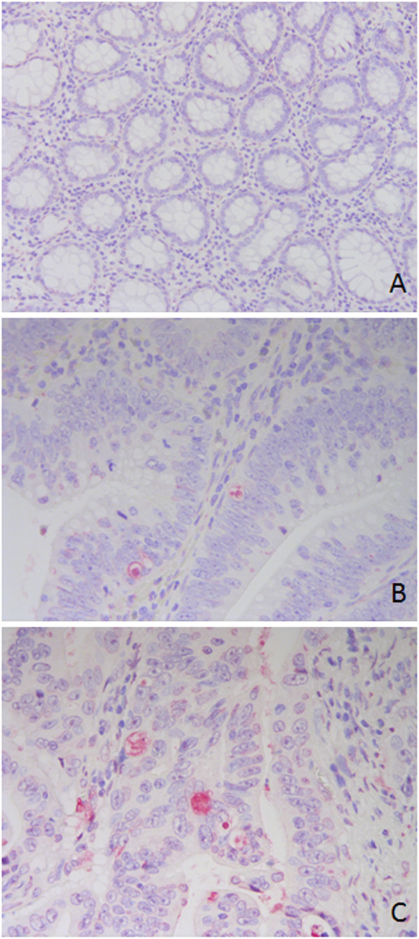
Methods: A TUNEL assay was performed on archival material from 103 colorectal carcinomas, 26 adenomas and 20 samples of normal epithelia.
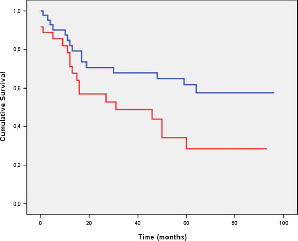
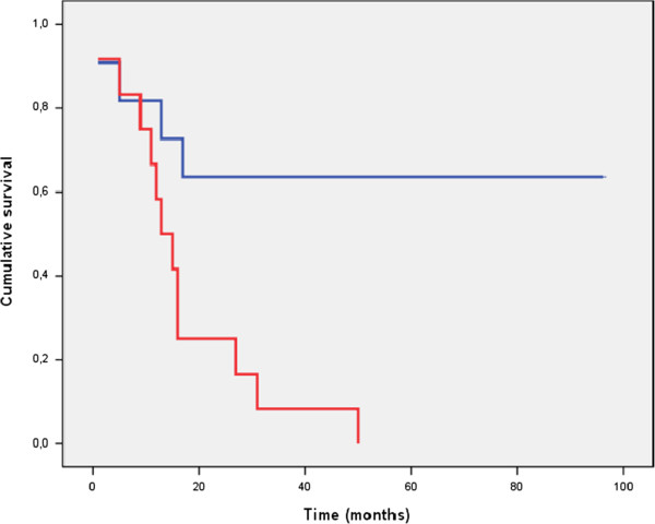
Results: The number of apoptotic cells was higher in CRCs (1.09 ± 0.13) than in adenomas (0.38 ± 0.23, p = 0.059) and normal epithelium (0.06 ± 0.04, p = 0.001). In addition, the apoptotic index (AI) was greater in metastatic disease (stage IV) than in other stages (p = 0.017). No relationship was found between apoptotic rates and age, gender or tumour grade. However, patients with tumours that showed higher AI values had a significantly lower disease-free survival (DFS) and overall survival (OS) than those with tumours that had lower AIs (p = 0.020 and p = 0.027). In a multivariate Cox proportional hazards model, AI remained a significant independent predictor of survival.
Conclusions: We conclude that disregulated apoptosis is an important event during CRC development and progression. Higher AIs are associated with more aggressive tumours and a poorer prognosis for patients with CRC.
Figures
References
-
- Bedi A, Pasricha PJ, Akhtar AJ, Barber JP, Bedi GC, Giardiello FM, Zehnbauer BA, Hamilton SR, Jones RJ. Inhibition of apoptosis during development of colorectal cancer. Cancer Res. 1995;55:1811–1816. - PubMed
-
- Aotake T, Lu CD, Chiba Y, Muraoka R, Tanigawa N. Changes of angiogenesis and tumor cell apoptosis during colorectal carcinogenesis. Clin Cancer Res. 1999;5:135–142. - PubMed
-
- Baretton GB, Diebold J, Christoforis G, Vogt M, Müller C, Dopfer K, Schneiderbanger K, Schmidt M, Löhrs U. Apoptosis and immunohistochemical bcl-2 expression in colorectal adenomas and carcinomas. Aspects of carcinogenesis and prognostic significance. Cancer. 1996;77:255–264. doi: 10.1002/(SICI)1097-0142(19960115)77:2<255::AID-CNCR6>3.0.CO;2-L. - DOI - PubMed
LinkOut - more resources
Full Text Sources
Other Literature Sources
Medical
