. 2013 Oct 9;33(41):16236-48.
doi: 10.1523/JNEUROSCI.3319-12.2013.

A critical role for macrophages near axotomized neuronal cell bodies in stimulating nerve regeneration

Affiliations

A critical role for macrophages near axotomized neuronal cell bodies in stimulating nerve regeneration

Jon P Niemi et al. J Neurosci. .

Abstract

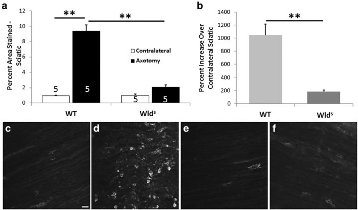
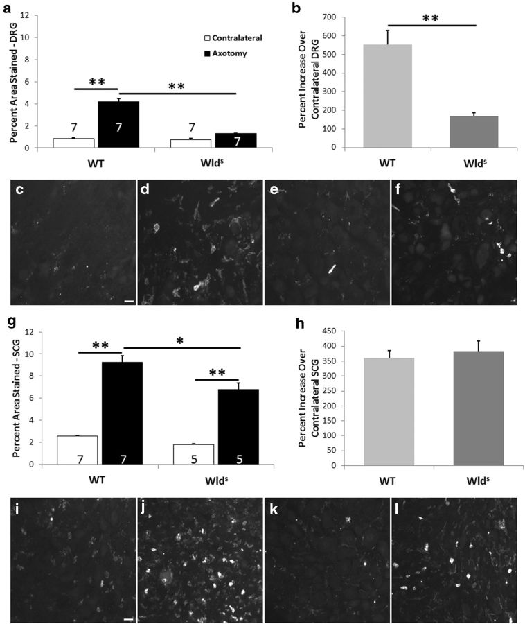
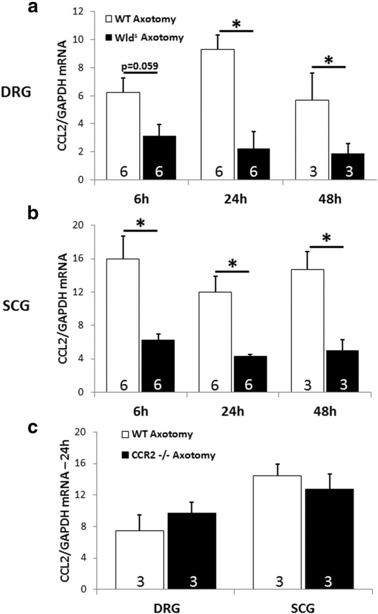
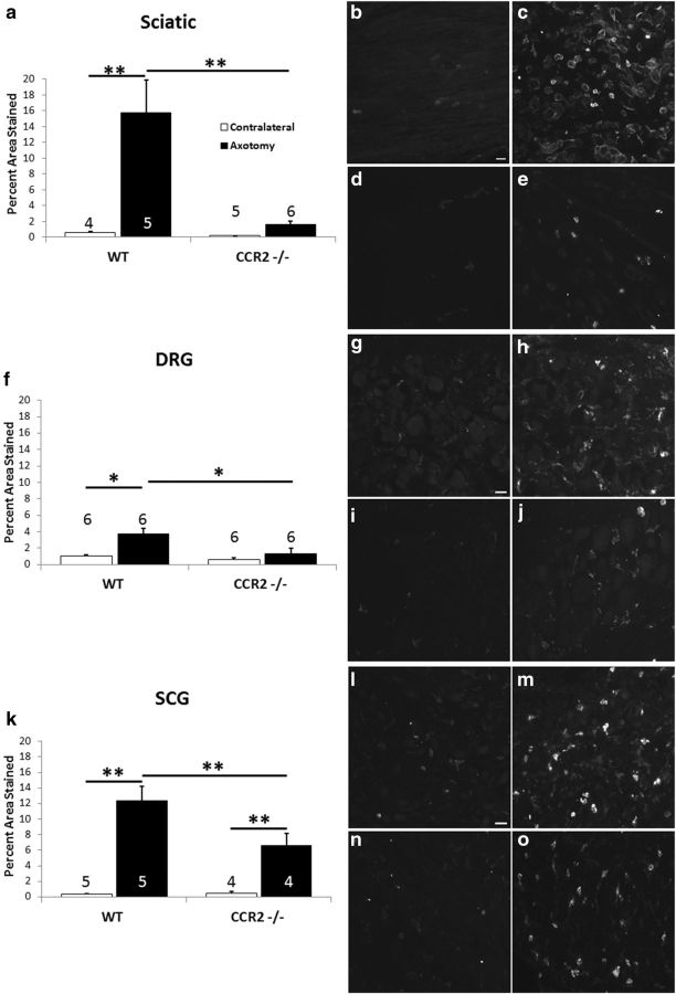
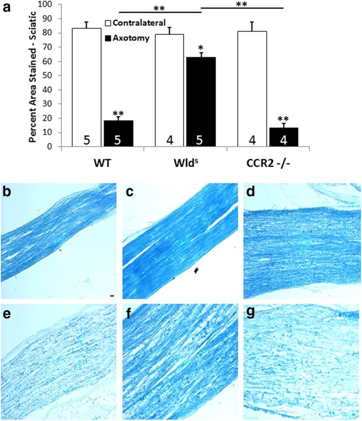
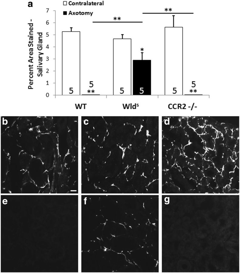
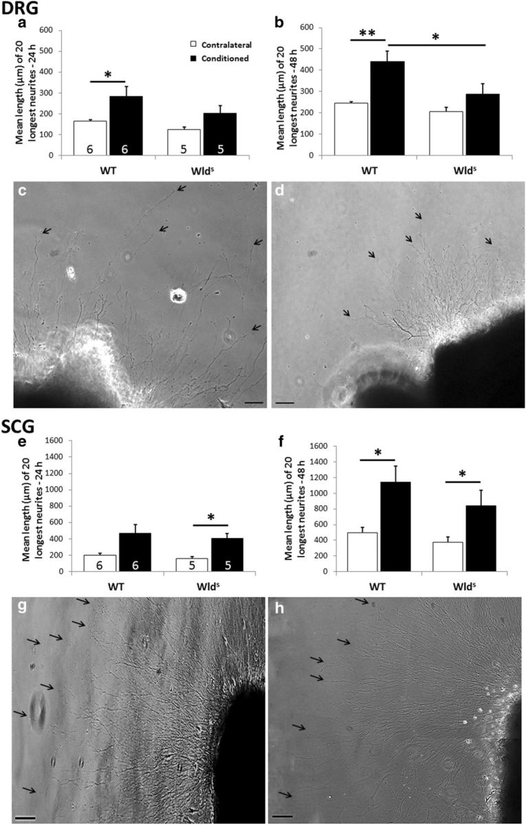
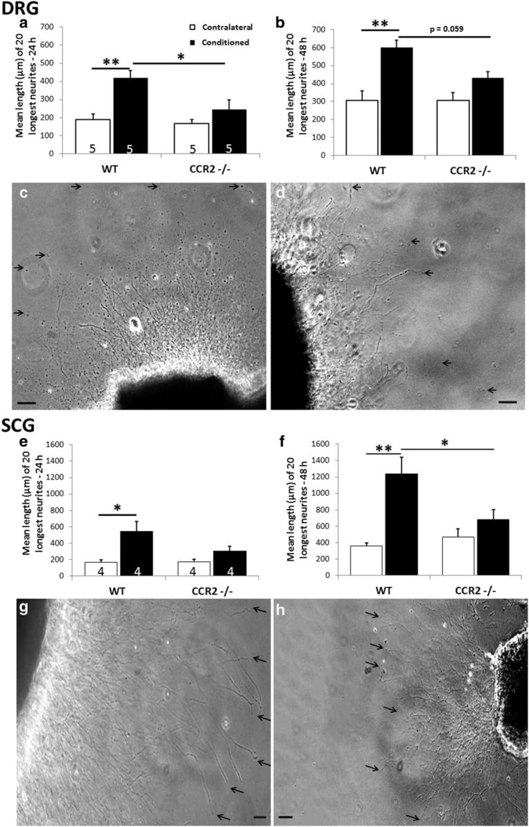
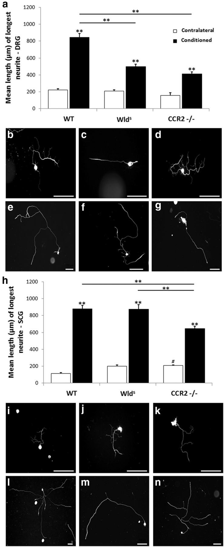
Macrophages have been implicated in peripheral nerve regeneration for some time, supposedly through their involvement in Wallerian degeneration, the process by which the distal nerve degenerates after axotomy and is cleared by phagocytosis. Thus, in several studies in which macrophage accumulation in the distal nerve was reduced and Wallerian degeneration inhibited, regeneration was delayed. However, this interpretation ignores the more recent findings that macrophages also accumulate around axotomized cell bodies. The function of macrophage action at this second site has not been clear. In two mutant strains of mice, the slow Wallerian degeneration (Wld(s)) mouse and the chemokine receptor CCR2 knock-out mouse, we report that macrophage accumulation after axotomy was abolished in both the dorsal root ganglion (DRG) and the distal sciatic nerve. To measure neurite outgrowth, DRG neurons were given a conditioning lesion, and outgrowth was measured in vitro 7 d later in the absence of the distal nerve segment. The increased growth normally seen after a conditioning lesion did not occur or was reduced in Wld(s) or CCR2(-/-) mice. In the superior cervical ganglion (SCG), particularly in Wld(s) mice, macrophage accumulation was reduced but not abolished after axotomy. In SCG neurons from Wld(s) mice, the conditioning lesion response was unchanged; however, in CCR2(-/-) mice in which the effect on macrophage accumulation was greater, SCG neurite outgrowth was significantly reduced. These results indicate that macrophages affect neurite outgrowth by acting at the level of peripheral ganglia in addition to any effects they might produce by facilitation of Wallerian degeneration.


Figures

Figure 1.
Figure 1.
Macrophage accumulation is significantly increased in the distal segment of the transected sciatic nerve in WT mice but not in Wlds mice. The sciatic nerve in WT and Wlds mice was transected unilaterally (Axotomy), and, 7 d later, the nerve segment distal to the site of transection was immunostained with a CD11b antibody to visualize macrophages. A comparable segment of the contralateral nerve was also examined. The extent of staining was quantitated as a percentage of the section area that was stained (a). The number of nerves examined in each group is given within each bar of the histogram. The staining in the axotomized nerve is also expressed as a mean ratio of staining in the ipsilateral and contralateral nerves for each animal × 100 (b). Micrographs are shown for contralateral (c, e) and axotomized (d, f) nerves from WT (c, d) and Wlds (e, f) animals. The data presented here and in all subsequent figures are means ± SEM. **p < 0.001. Scale bar, 20 μm.
Figure 2.
Figure 2.
The macrophage accumulation in L5 DRGs and SCGs after unilateral axotomy is significantly diminished in Wlds mice compared with WT mice. The tissues examined were from the same groups of animals. Seven days after unilateral transection of the relevant nerves, the ipsilateral and contralateral ganglia were removed and immunostained for CD11b-positive cells. The data are presented as both the percentage of the section area that was stained (a, g) and the mean ratio of staining in the ipsilateral and contralateral ganglia for each animal × 100 (b, h). Micrographs are shown for contralateral DRGs (c, e) and SCGs (i, k) and axotomized DRGs (d, f) and SCGs (j, l) from WT (c, d, i, j) and Wlds (e, f, k, l) mice. *p < 0.05, **p < 0.001. Scale bars, 20 μm.
Figure 3.
Figure 3.
The axotomy-induced induction of CCL2 is diminished in DRGs (a) and SCGs (b) in Wlds mice compared with WT mice. Six, 24, and 48 h after transection of the relevant nerves, axotomized and contralateral L5 DRGs and SCGs were extracted, and CCL2 mRNA was measured by RT-PCR. No detectable CCL2 mRNA was found in the contralateral ganglia. CCL2 mRNA was also measured in axotomized DRGs and SCGs from WT and CCR2−/− mice 24 h after axotomy. No differences were seen (c). The data are normalized to GAPDH mRNA for each sample. *p < 0.05.
Figure 4.
Figure 4.
The axotomy-induced accumulation of macrophages in the sciatic nerve, DRG, and SCG is diminished in CCR2−/− mice compared with WT mice. The data show the extent of CD11b staining in sciatic nerves from WT and CCR2−/− mice 7 d after unilateral axotomy (a). No effect of axotomy was seen in the CCR2−/− animals. Representative micrographs are shown from contralateral (b, d) and axotomized (c, e) sciatic nerves from WT (b, c) and CCR2−/− (d, e) animals. Axotomy-induced macrophage accumulation was abolished also in L5 DRGs from the same animals (f). Micrographs are shown from contralateral (g, i) and axotomized (h, j) DRGs from WT (g, h) and CCR2−/− (i, j) animals. Axotomy-induced macrophage accumulation is lessened, but not abolished, in the SCGs from CCR2−/− mice (k). Micrographs are shown from contralateral (l, n) and axotomized (m, o) SCGs from WT (l, m) and CCR2−/− (n, o) animals. *p < 0.05, **p < 0.001. Scale bars, 20 μm.
Figure 5.
Figure 5.
Seven days after the sciatic nerve was unilaterally transected, changes in reactivity for myelin proteins were determined in nerves from WT, Wlds, and CCR2−/− mice by staining with LFB. The distal nerve segments from WT and CCR2−/− mice showed significantly less myelin staining compared with contralateral nerves, whereas axotomized nerves from Wlds mice retained >80% of myelin reactivity compared with contralateral nerves (a). The micrographs represent sections from the ipsilateral (e–g) and contralateral (b–d) nerves from WT, Wlds, and CCR2−/− mice, respectively. *p < 0.05, **p < 0.001. Scale bar, 20 μm.
Figure 6.
Figure 6.
Sympathetic nerve fibers in an autonomic target in the Wlds mouse, but not the CCR2−/− mouse, exhibit slow Wallerian degeneration (a). To determine whether sympathetic neurons in vivo in the Wlds and CCR2−/− mouse exhibit slow Wallerian degeneration, the internal and external carotid nerves were transected, and, 7 d later, the ipsilateral and contralateral submandibular glands were stained for TH in WT, Wlds, and CCR2−/− mice. The micrographs represent sections from the ipsilateral (e–g) and contralateral (b–d) nerves from WT, Wlds, and CCR2−/− mice, respectively. *p < 0.05, **p < 0.001. Scale bar, 20 μm.
Figure 7.
Figure 7.
The conditioning lesion effect is abolished in DRG, but not SCG, explants from Wlds mice. Seven days after unilateral sciatic nerve transection, DRGs were placed in explant culture, and neurite outgrowth was measured after 24 h (a) and 48 h (b). The phase micrographs are of individual conditioned DRG explants from WT (c) and Wlds (d) mice at 48 h. The arrows point to the endings of individual neurites. SCGs were examined 7 d after transection of the internal and external carotid nerves. Phase micrographs are shown for conditioned SCG explants from WT (g) and Wlds (h) mice after 48 h. In the SCG, no significant difference in neurite outgrowth was seen between the two genotypes (e, f). *p < 0.05, **p < 0.001. Scale bars, 100 μm.
Figure 8.
Figure 8.
The conditioning lesion effect is abolished in DRG and SCG explants from CCR2−/− mice. Seven days after unilateral sciatic nerve transection, DRGs were placed in explant culture, and neurite outgrowth was measured after 24 h (a) and 48 h (b). The representative phase micrographs are of individual conditioned DRG explants from WT (c) and CCR2−/− (d) mice at 48 h. The arrows point to the endings of individual neurites. Examination of SCG explants from CCR2−/− mice 7 d after transection of the internal and external carotid nerves revealed no conditioning lesion effect at 24 h (e) or 48 h (f). Representative micrographs of individual conditioned SCGs are shown for WT (g) and CCR2−/− (h) mice at 48 h. *p < 0.05, **p < 0.001. Scale bars, 100 μm.
Figure 9.
Figure 9.
Neurite outgrowth from dissociated cells after 24 h in culture from DRG (a–g) and SCG (h–n) neurons. The sciatic nerve and the internal and external carotid nerves were transected unilaterally in WT, Wlds, and CCR2−/− mice. Seven days later, the DRGs and SCGs were removed, dissociated, and cultured for 24 h. Neurite outgrowth was measured and expressed as mean length of the longest neurite from each neuron. Greater than 50 neurons were measured for each group, with the exception of the CCR2−/− sham DRG group in which 21 neurons were measured. A conditioning lesion response was observed in all three genotypes, but in DRGs, the response was significantly less than WT in both mutants (a). In SCGs, the magnitude of conditioning lesion response was similar in Wlds and WT mice but was significantly less in CCR2−/− mice (h). Representative micrographs of single DRG neurons from contralateral (b–d) and conditioned (e–g) ganglia from WT (b, e), Wlds (c, f), and CCR2−/− (d, g) mice. Representative micrographs of single SCG neurons from contralateral (i–k) and conditioned (l–n) ganglia from WT (i, l), Wlds (j, m), and CCR2−/− (k, n) mice. **p < 0.001. # indicates that the mean length of CCR2−/− contralateral SCG neurons was significantly longer than WT (p = 0.04). Scale bars, 100 μm.

References

    1. Abbadie C, Lindia JA, Cumiskey AM, Peterson LB, Mudgett JS, Bayne EK, DeMartino JA, MacIntyre DE, Forrest MJ. Impaired neuropathic pain responses in mice lacking the chemokine receptor CCR2. Proc Natl Acad Sci U S A. 2003;100:7947–7952. doi: 10.1073/pnas.1331358100. - DOI - PMC - PubMed
    1. Agius E, Cochard P. Comparison of neurite outgrowth induced by intact and injured sciatic nerves: a confocal and functional analysis. J Neurosci. 1998;18:328–338. - PMC - PubMed
    1. Barrette B, Hébert MA, Filali M, Lafortune K, Vallières N, Gowing G, Julien JP, Lacroix S. Requirement of myeloid cells for axon regeneration. J Neurosci. 2008;28:9363–9376. doi: 10.1523/JNEUROSCI.1447-08.2008. - DOI - PMC - PubMed
    1. Barrette B, Calvo E, Vallières N, Lacroix S. Transcriptional profiling of the injured sciatic nerve of mice carrying the Wld(S) mutant gene: identification of genes involved in neuroprotection, neuroinflammation, and nerve regeneration. Brain Behav Immun. 2010;24:1254–1267. doi: 10.1016/j.bbi.2010.07.249. - DOI - PubMed
    1. Bedi KS, Winter J, Berry M, Cohen J. Adult rat dorsal root ganglion neurons extend neurites on predegenerated but not on normal peripheral nerves in vitro. Eur J Neurosci. 1992;4:193–200. doi: 10.1111/j.1460-9568.1992.tb00867.x. - DOI - PubMed

Publication types

LinkOut - more resources