. 2013 Oct 9;33(41):16383-93.
doi: 10.1523/JNEUROSCI.1731-13.2013.

Parceling human accumbens into putative core and shell dissociates encoding of values for reward and pain

Affiliations

Parceling human accumbens into putative core and shell dissociates encoding of values for reward and pain

Marwan N Baliki et al. J Neurosci. .

Abstract

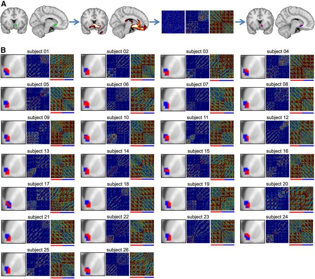
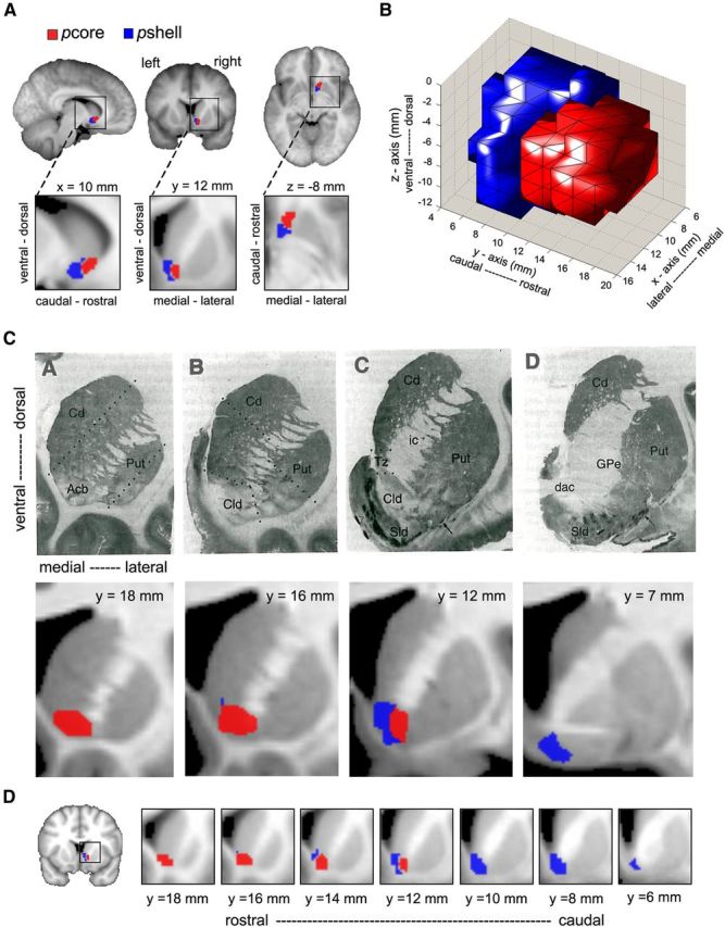
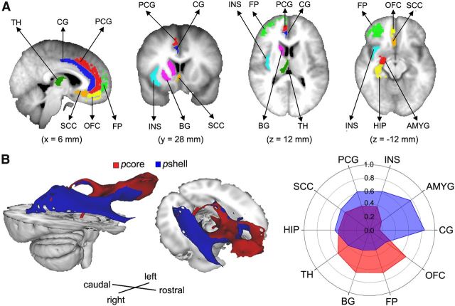
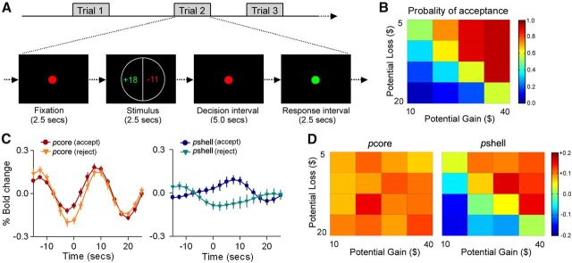
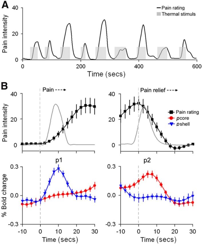
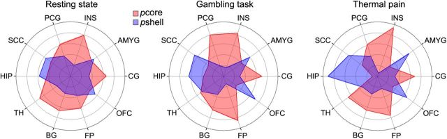
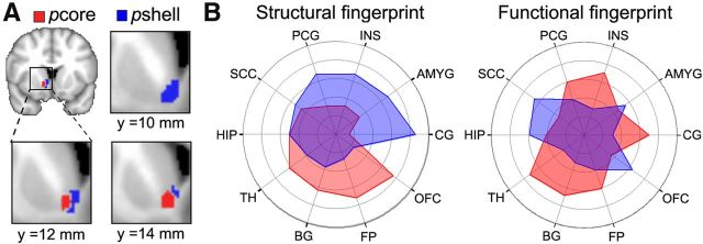
In addition to their well-established role in signaling rewarding outcomes and reward-predictive cues and in mediating positive reinforcement, there is growing evidence that nucleus accumbens (NAc) neurons also signal aversive events and cues that predict them. Here we use diffusion tractography to subdivide the right NAc into lateral-rostral (putative core, pcore) and medial-caudal (putative shell, pshell) subdivisions in humans. The two subregions exhibited differential structural connectivity, based on probabilistic tractography, to prefrontal cortical and subcortical limbic regions. We also demonstrate unique roles for each of the two subdivisions for monetary reward and thermal pain perception tasks: pshell signaling impending pain and value predictions for monetary gambles and pcore activating with anticipation of cessation of thermal pain (signaling reward value of analgesia). We examined functional connectivity for resting state, monetary reward, and thermal pain tasks, and for all three conditions observed that pcore and pshell of right NAc exhibit distinct patterns of synchrony (functional connectivity) to prefrontal cortical and subcortical limbic targets within the right hemisphere. To validate the NAc segregation, we mirrored the coordinates of right NAc pcore and pshell onto the left hemisphere and examined structural and resting state connectivity in the left hemisphere. This latter analysis closely replicated target-specific connections we obtained for the right hemisphere. Overall, we demonstrate that the human NAc can be parceled based on structural and functional connectivity, and that activity in these subdivisions differentially encodes values for expected pain relief and for expected monetary reward.


Figures

Figure 1.
Figure 1.
DTI tractography parcellation identifies two distinct subdivisions in the right NAc in individual subjects. A, Summary of the steps leading to the NAc clustering in an example subject. Left brain images, NAc region (green) and resulting tracks (red). Middle panels, Cross-correlation matrices indicating the degree of similarity in connectivity pattern between the NAc voxels. Leftmost matrix, Cross-correlation from all NAc voxels. Middle, Matrix reorganized based on the k-means cluster algorithms. Rightmost panel, Matrix normalized by distance. This final matrix was used to delineate the two different clusters indicated by red and blue bars beneath the matrix. Right brain images, Two resulting clusters (blue and red) projected back on the brain. B, Results of the connectivity-based clustering for all 26 subjects. For each subject: left panel, part of coronal slice at y = 12 mm and the corresponding NAc clusters; middle and right panels, reorganized and normalized cross-correlation matrices, respectively.
Figure 2.
Figure 2.
The human NAc can be divided into two subdivisions. A, Group-averaged DTI-based clustering of the right NAc into a medial–caudal (blue, putative shell, pshell) and a lateral–rostral (red, putative core, pcore) subdivision. Group-averaged clusters were determined as regions showing 80% overlap across individual subjects (n = 27). B, Three-dimensional rendering of pcore and pshell, observed from a right, lateral view. C, Top row represents a series of coronal sections through the striatum from a single human brain, illustrating the DAMGO binding at four rostrocaudal levels. Images taken from Voorn et al. (1996), in which they identify the accumbens (Acb), its Core-like division (Cld), and shell-like division (Sld). The two most rostral slices (A, B) contain only Cld, delineated by broken lines in B, whereas D identifies only Sld. Bottom row, Group average DTI-based parcellation of the NAc into the pcore and pshell. The pcore (red) and pshell (blue) subdivisions approximate Cld and Sld, respectively. D, Detailed rostrocaudal coronal slices of group-averaged clustering of the human NAc (higher values correspond to rostral slices and lower values to caudal slices).
Figure 3.
Figure 3.
The NAc subdivisions exhibit distinct structural connectivity patterns. A, Brain slices showing the cortical and subcortical targets of interest, which included the TH, BG, CG, PCG, HIP, AMYG, INS, SCC, OFC, and FP. The targets were all identified from the Harvard-Oxford cortical and subcortical structural atlas and were restricted to gray matter within the right hemisphere. B, Brain images show the group average white matter tracts for the pcore (red) and pshell (blue) in the right hemisphere. Polar plot represents the structural connectivity fingerprints in the right hemisphere from right NAc subdivisions. The values indicate relative connection probabilities of white matter tracks, based on DTI tractography for pshell and pcore to the targets.
Figure 4.
Figure 4.
The NAc subdivisions exhibit specific activity during a monetary gambling task. A, The monetary gambling task design, derived from Tom et al. (2007). During each trial, a display showing the size of the potential gain (in green) and loss (in red) was presented for 2.5 s. This was followed by a 5 s decision interval and a 2.5 s response interval. After an accept or reject response, a variable interval was presented (10–15 s) to allow for adequate deconvolution of fMRI responses. B, Color-coded heatmap matrix of group-averaged probability of gamble acceptance at each level of accept/reject: red represents high willingness to accept the gamble; blue represents low willingness to accept the gamble). C, Group average time course of BOLD responses (% change from baseline), for the pcore and pshell of right NAc, during accepted and rejected trials. The pcore showed similarly increased responses for both accepted and rejected choices, whereas the pshell showed differential increased responses for acceptances and decreased responses for rejections. D, Heatmaps were created by averaging %BOLD change versus baseline BOLD, within the pcore and pshell of right NAc, for each of the 16 cells in the potential gain/loss matrix; color coding reflects the strength of neural response for each condition: dark red represents the strongest activation; dark blue represents the strongest deactivation. The pcore did not exhibit any differential activation, whereas the pshell reflected decision values.
Figure 5.
Figure 5.
NAc subdivisions show differential activation for onset and offset of acute thermal pain. A, The thermal pain task. Average pain ratings (black) and the time course of the painful heat (gray) applied to the skin. Data are from Baliki et al. (2010). B, Top panels, Mean ± SEM time course of the pain rating (convolved with hemodynamic function) during start (left) and end (right) of thermal stimulus. The time courses were averaged across all stimulation epochs where subjects reported pain. The gray lines indicate absolute value of the derivative, |d/dt|, for the stimulus. Bottom panels, Average BOLD response time course for right NAc, pcore (red) and pshell (blue) for the same time periods. Data are mean ± SEM. Anticipation of impending pain transiently activates the pshell (p1), whereas anticipation of pain relief transiently activates the pcore (p2).
Figure 6.
Figure 6.
The NAc subdivisions exhibit distinct functional connectivity patterns. Polar plots represent the functional connectivity fingerprints in the right hemisphere for different scans. The values indicate relative functional connection for pshell and pcore of the right NAc to the 10 targets. The NAc subdivisions exhibit similar functional connectivity patterns for resting state, gambling, and thermal pain scans. Target abbreviations are listed in Figure 3.
Figure 7.
Figure 7.
Left and right hemisphere NAc subdivisions exhibit similar, within hemisphere, connectivity properties. A, Panels represent the left NAc subdivisions, derived by flipping the coordinates of right NAc subdivisions. B, Polar plots represent the structural and resting state functional connectivity fingerprints in the left hemisphere to the subdivisions of left NAc. The values indicate relative structural (left plot) and functional connections (right plot) for pshell and pcore to the 10 targets. Overall, the left NAc subdivisions showed similar structural and functional connectivity to their right counterparts.

References

    1. Alexander GE, DeLong MR, Strick PL. Parallel organization of functionally segregated circuits linking basal ganglia and cortex. Annu Rev Neurosci. 1986;9:357–381. doi: 10.1146/annurev.ne.09.030186.002041. - DOI - PubMed
    1. Ambroggi F, Ishikawa A, Fields HL, Nicola SM. Basolateral amygdala neurons facilitate reward-seeking behavior by exciting nucleus accumbens neurons. Neuron. 2008;59:648–661. doi: 10.1016/j.neuron.2008.07.004. - DOI - PMC - PubMed
    1. Apkarian AV. Pain perception in relation to emotional learning. Curr Opin Neurobiol. 2008;18:464–468. doi: 10.1016/j.conb.2008.09.012. - DOI - PMC - PubMed
    1. Badrinarayan A, Wescott SA, Vander Weele CM, Saunders BT, Couturier BE, Maren S, Aragona BJ. Aversive stimuli differentially modulate real-time dopamine transmission dynamics within the nucleus accumbens core and shell. J Neurosci. 2012;32:15779–15790. doi: 10.1523/JNEUROSCI.3557-12.2012. - DOI - PMC - PubMed
    1. Baldo BA, Kelley AE. Discrete neurochemical coding of distinguishable motivational processes: insights from nucleus accumbens control of feeding. Psychopharmacology. 2007;191:439–459. doi: 10.1007/s00213-007-0741-z. - DOI - PubMed

Publication types