. 2013 Oct 31;502(7473):637-43.
doi: 10.1038/nature12612. Epub 2013 Oct 9.

Arteriolar niches maintain haematopoietic stem cell quiescence

Affiliations

Arteriolar niches maintain haematopoietic stem cell quiescence

Yuya Kunisaki et al. Nature. .

Abstract

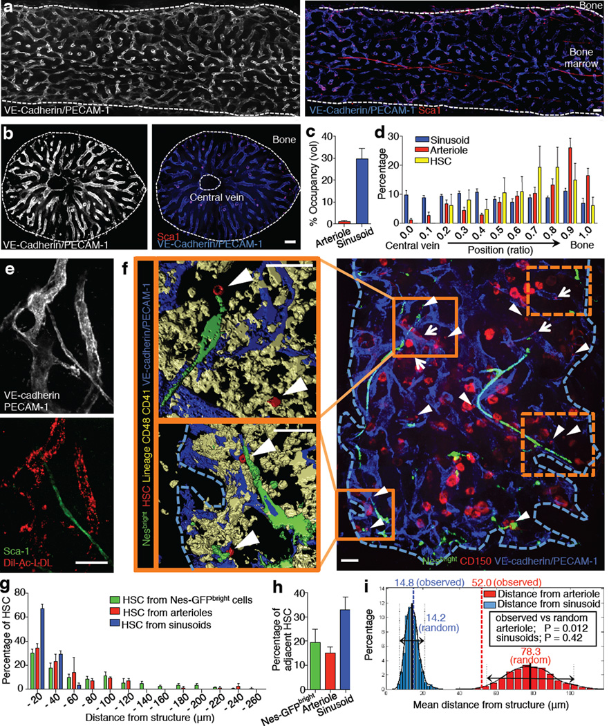
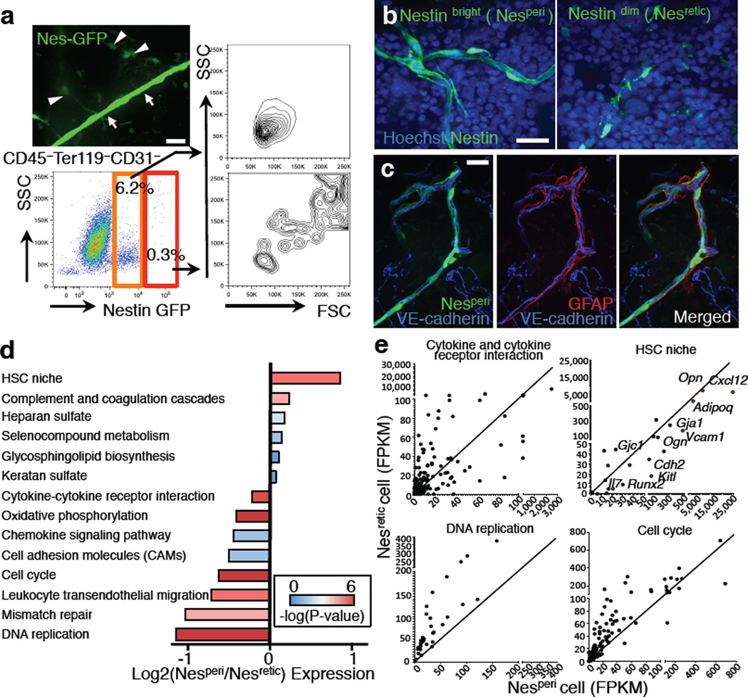
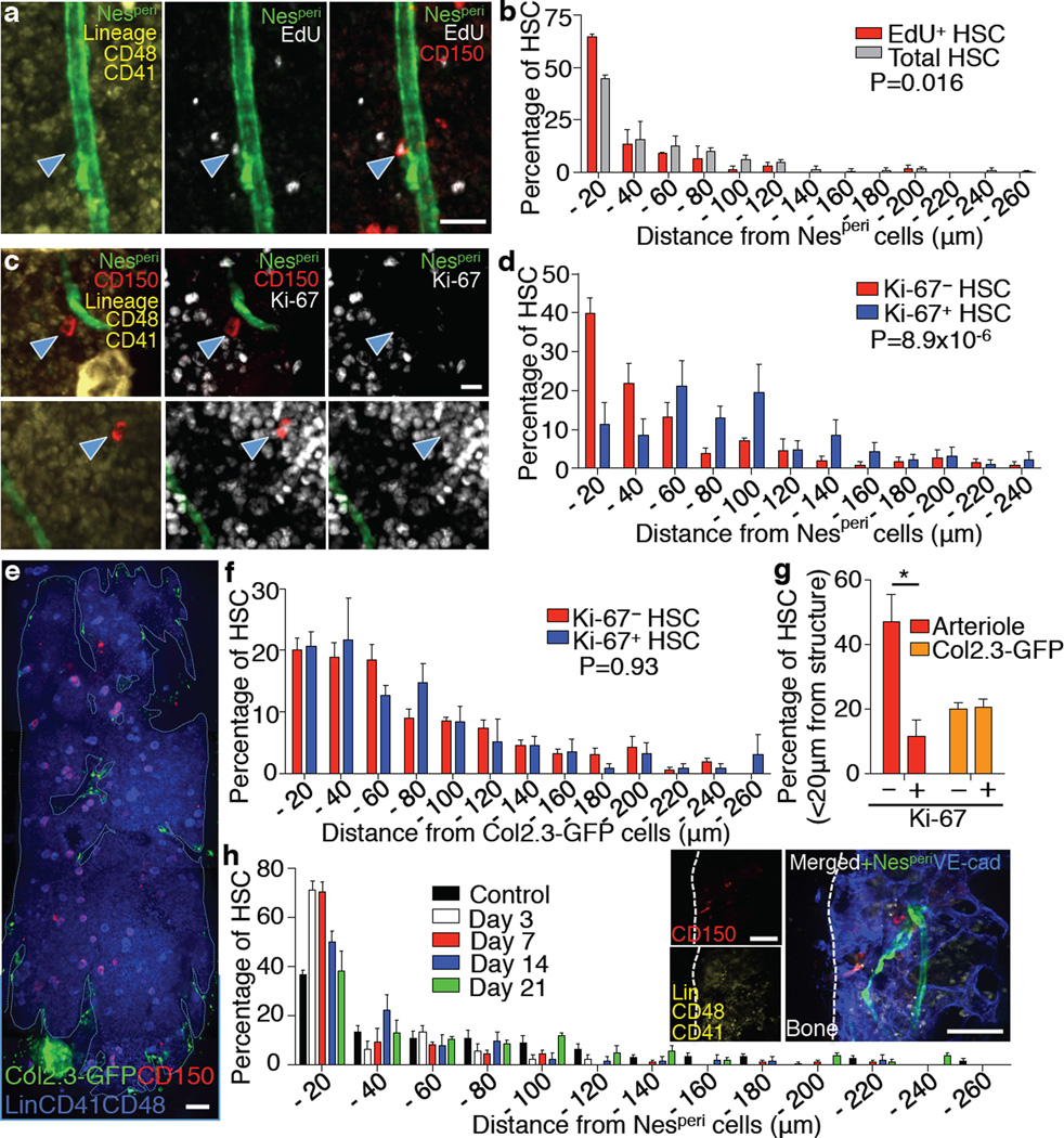
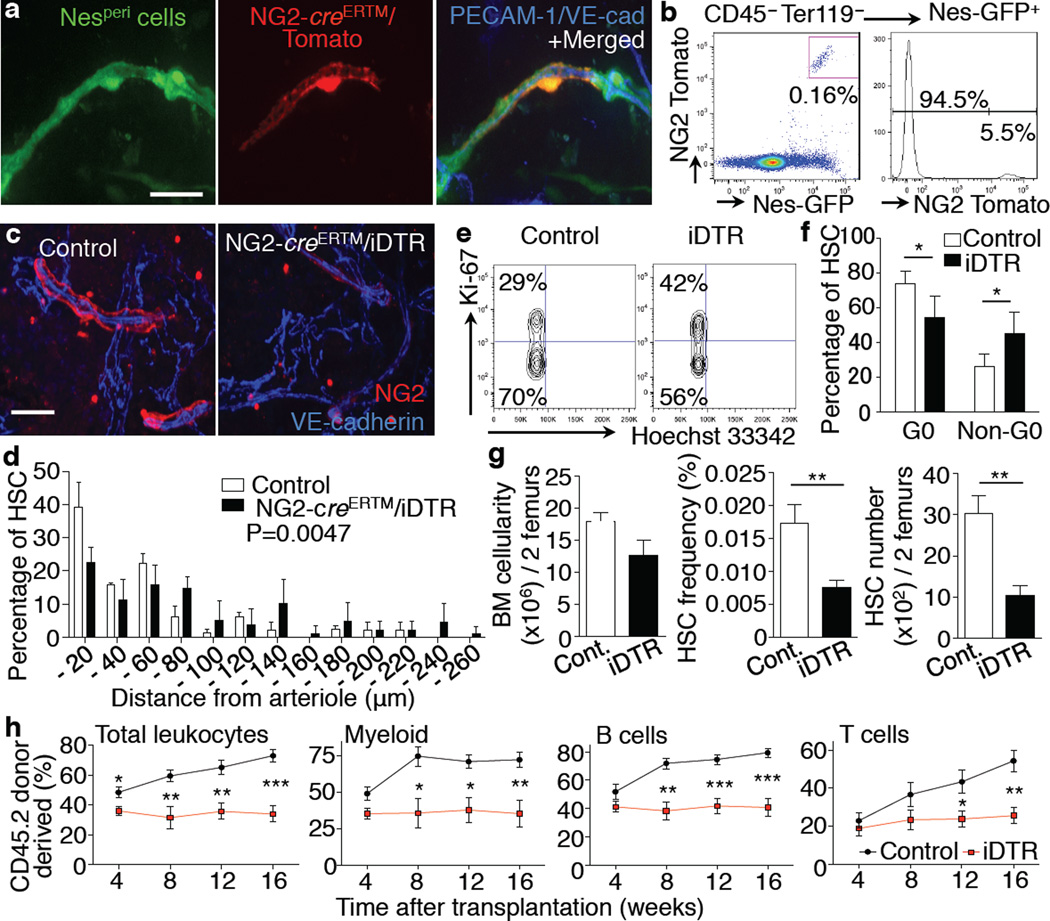
Cell cycle quiescence is a critical feature contributing to haematopoietic stem cell (HSC) maintenance. Although various candidate stromal cells have been identified as potential HSC niches, the spatial localization of quiescent HSCs in the bone marrow remains unclear. Here, using a novel approach that combines whole-mount confocal immunofluorescence imaging techniques and computational modelling to analyse significant three-dimensional associations in the mouse bone marrow among vascular structures, stromal cells and HSCs, we show that quiescent HSCs associate specifically with small arterioles that are preferentially found in endosteal bone marrow. These arterioles are ensheathed exclusively by rare NG2 (also known as CSPG4)(+) pericytes, distinct from sinusoid-associated leptin receptor (LEPR)(+) cells. Pharmacological or genetic activation of the HSC cell cycle alters the distribution of HSCs from NG2(+) periarteriolar niches to LEPR(+) perisinusoidal niches. Conditional depletion of NG2(+) cells induces HSC cycling and reduces functional long-term repopulating HSCs in the bone marrow. These results thus indicate that arteriolar niches are indispensable for maintaining HSC quiescence.


Figures

Figure 1
Figure 1. Spatial relationships between HSCs and the bone marrow vasculature
a,b, Longitudinally (a) and transverse-shaved (b) whole-mount images of the mouse femoral BM stained with anti-PECAM-1, anti-VE-cadherin and anti-Sca-1 antibodies. Scale bar: 100 µm. c, BM volumes occupied by arterioles or sinusoids. n = 6 areas from 3 mice. d, Distribution of sinusoids, arterioles and HSCs in the femoral BM. n = 6 mice. e, Whole-mount images of sternum stained with anti-VE-cadherin, anti-PECAM-1, anti-Sca-1 antibodies and Dil-Ac-LDL. Scale bar: 50 µm. f, Illustrative example of whole-mount sternal BM with 3D reconstructed images. Dashed squares are shown in Extended Data Fig. 3b. Arrowheads denote HSCs, arrows show CD150+ Lin/CD48/CD41+ cells. Scale bar: 50 µm. g,h, Distances between HSCs and Nes-GFPbright cells (n = 98 HSCs from 5 mice), arterioles or sinusoids (n = 119 HSCs from 5 mice) (g) and percentages of adjacent HSCs (distance = 0) (h) in the sternal BM. i, Probability distributions of mean distances from simulations of randomly positioned HSCs on maps of sternal BM in relation to sinusoids (blue) or arterioles (red). Mean distances observed in situ (dashed) are shown in relation to the grand mean ± 2 s.d. (solid and dotted lines).
Figure 2
Figure 2. Nestin+ cell subsets define distinct vascular structure
a, A whole-mount image of sternal BM with Nes-GFPbright (arrows) and Nes-GFPdim cells (arrowheads) and FACS analysis of BM stromal cells from Nes-GFP mice. Data were reproducible in at least 5 mice. b,c, Whole-mount images of sternum from Nes-GFP mice stained with anti-GFP and Hoechst 33342 (b) or anti-GFAP antibody (c). d,e, RNA-seq analysis of Nesretic and Nesperi cells. d, Differentially expressed pathways. Mean of log fold change of Nesperi/Nesretic expression was plotted for each pathway with colour-coded P-values. e, Dot plots showing gene expression levels in FPKM for representative pathways and functional groups.
Figure 3
Figure 3. Quiescent arteriolar niche cells are protected from myeloablation
a, Whole-mount immunostaining with anti-Ki-67 antibody of sterna from Nes-GFP mice. Scale bar: 50 µm. b, Q-PCR analyses of Nesperi, Nesretic and CD45 Ter119 CD31 Nes-GFP stromal cells. n = 5 independent experiments. c,d, Cell cycle analyses by FACS using anti-Ki-67 and Hoechst 33342 staining. Representative plots (c) and quantification (d). n = 3 mice per group. e–g, Whole mount images of sternal BM (e) and the kinetics of absolute numbers (f) and frequencies (g) of Nesperi and Nesretic cells in the long-bone BM analysed by FACS after 5FU treatment. n = 10, 9, 9, 5, 6 mice per time point. Scale bar: 100 µm.
Figure 4
Figure 4. Spatial relationship between arterioles and quiescent HSCs
a,b, Localization of dormant HSCs accessed by long-term label retention with EdU. Representative images (a) and distribution of total and EdU+ CD150+ CD48 CD41 Lineage HSCs in femoral BM sections (b). n = 144, 52 HSCs from 3 mice. Two-sample Kolmogorov-Smirnov test; P = 0.016. Scale bar: 25 µm. c,d, Localization of Ki-67 and Ki-67+ HSCs in the sternal BM. Ki-67 / Ki-67+: 39.7±3.9% / 11.4±5.6% in 0–20µm proximity, 21.5±5.3% / 47.9±6.5% > 80µm distance. Representative images (c) and distances from Nesperi cells (d). n = 116, 64 HSCs from 7 mice. Two-sample Kolmogorov-Smirnov test; P = 8.9×10⁻⁶. Scale bar: 10 µm. e–g, Localization of HSCs in relation to osteoblasts. e, Whole-mount images of sternum from Col2.3-GFP mice with HSC staining. Scale bar: 100 µm. f, Distances of Ki-67 and Ki-67+ HSCs from Col2.3-GFP cells. n = 80, 41 HSCs from 3 mice. Two-sample Kolmogorov-Smirnov test; P = 0.93. g, Percentage of Ki-67 and Ki-67+ HSCs located within 20µm distance from arterioles or Col2.3-GFP cells. n = 7, 3 mice per group. h, Localization of HSCs relative to Nesperi cells after 5FU treatment. Inset shows a representative image of sternal BM on day 7. n = 98, 39, 70, 55, 112 HSCs from 5, 9, 7, 4, 4 mice per group. Two-sample Kolmogorov-Smirnov test; Day 3 P =2.8×10⁻⁴, Day 7 P =1.0×10⁻⁴, Day 14 P =0.0013, Day 21 P =0.23 compared to control. Scale bar: 50 µm.
Figure 5
Figure 5. NG2+ periarteriolar cells are an essential constituent of the HSC niche promoting HSC quiescence
a,b, Whole-mount images of sternal BM (a) and FACS analysis of femoral BM (b) from NG2-creERTM / loxp-Tomato / Nes-GFP Tg mice. Representative data of 4 mice. c–h, Analysis of HSCs after specific deletion of NG2+ cells using NG2-creERTM/iDTR mice. c, Whole-mount immunostaining of sternum with anti-NG2 antibody. Representative images of 5 mice per group. d, HSC localization relative to arterioles in the sternal BM. n = 69, 71 HSCs from 3, 4 mice per group. Two-sample Kolmogorov-Smirnov test; P = 0.0047. e,f, Cell cycle analysis of HSCs using Ki-67 and Hoechst 33342 staining. Representative plots (CD150+ CD48 Sca-1+ c-kit+ Lineage gated) (e) and quantifications (f). n = 5 mice per group. g, BM cellularity, frequency and number of HSCs in the BM. n = 6 mice per group. h, Quantification of long-term reconstituting HSCs by competitive reconstitution assays. n = 6, 9 mice per group. Scale bar: 25 µm.

Comment in

References

    1. Orkin SH, Zon LI. Hematopoiesis: an evolving paradigm for stem cell biology. Cell. 2008;132:631–644. - PMC - PubMed
    1. Li L, Clevers H. Coexistence of quiescent and active adult stem cells in mammals. Science. 2010;327:542–545. - PMC - PubMed
    1. Hsu YC, Pasolli HA, Fuchs E. Dynamics between stem cells, niche, and progeny in the hair follicle. Cell. 2011;144:92–105. - PMC - PubMed
    1. Orford KW, Scadden DT. Deconstructing stem cell self-renewal: genetic insights into cell-cycle regulation. Nat Rev Genet. 2008;9:115–128. - PubMed
    1. Cheng T, et al. Hematopoietic stem cell quiescence maintained by p21cip1/waf1. Science. 2000;287:1804–1808. - PubMed

References (ON LINE ONLY)

    1. Kawamoto T. Use of a new adhesive film for the preparation of multi-purpose fresh-frozen sections from hard tissues, whole-animals, insects and plants. Arch Histol Cytol. 2003;66:123–143. - PubMed
    1. Chiang EY, Hidalgo A, Chang J, Frenette PS. Imaging receptor microdomains on leukocyte subsets in live mice. Nat Methods. 2007;4:219–222. - PubMed
    1. Miller CL, Dykstra B, Eaves CJ. Characterization of mouse hematopoietic stem and progenitor cells. Curr Protoc Immunol. 2008;Chapter 22(Unit 22B):22. - PubMed

Publication types

Associated data