. 2013 Dec;24(23):3663-74.
doi: 10.1091/mbc.E13-06-0302. Epub 2013 Oct 9.

Syntaxin 16 is a master recruitment factor for cytokinesis

Affiliations

Syntaxin 16 is a master recruitment factor for cytokinesis

Hélia Neto et al. Mol Biol Cell. 2013 Dec.

Abstract

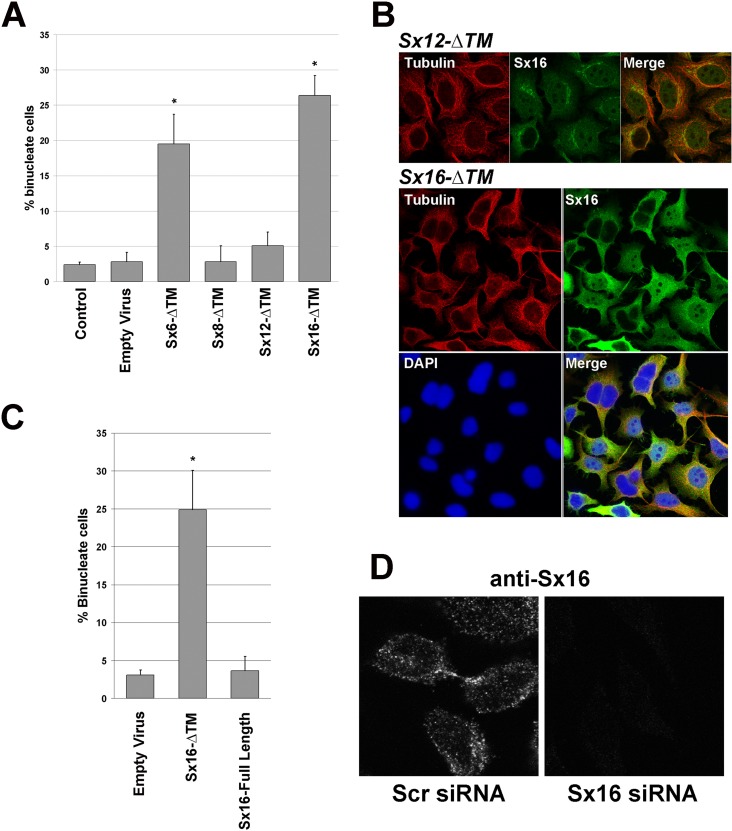
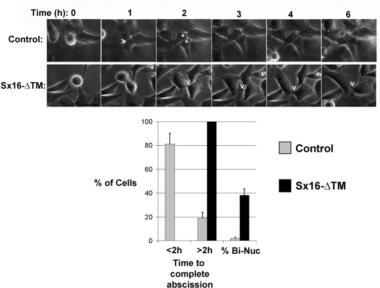
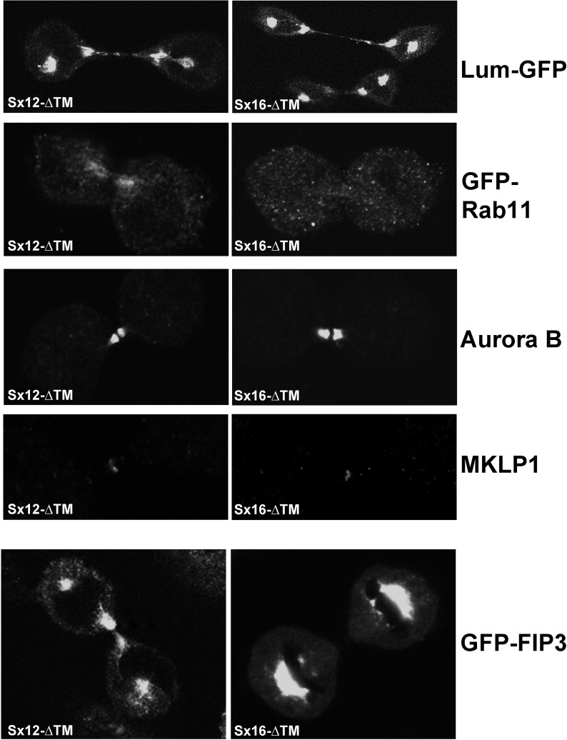
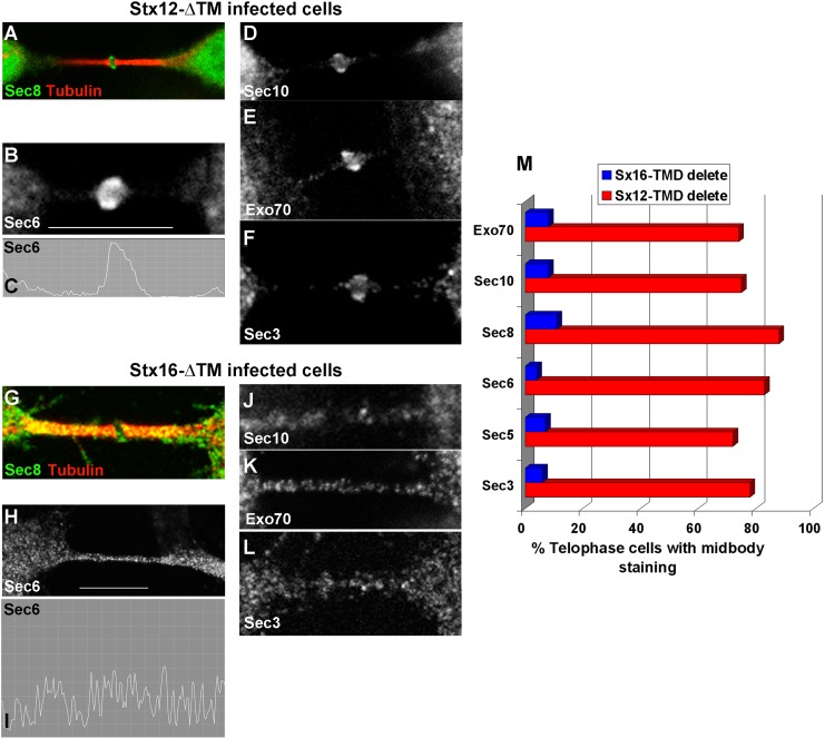
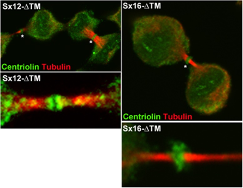
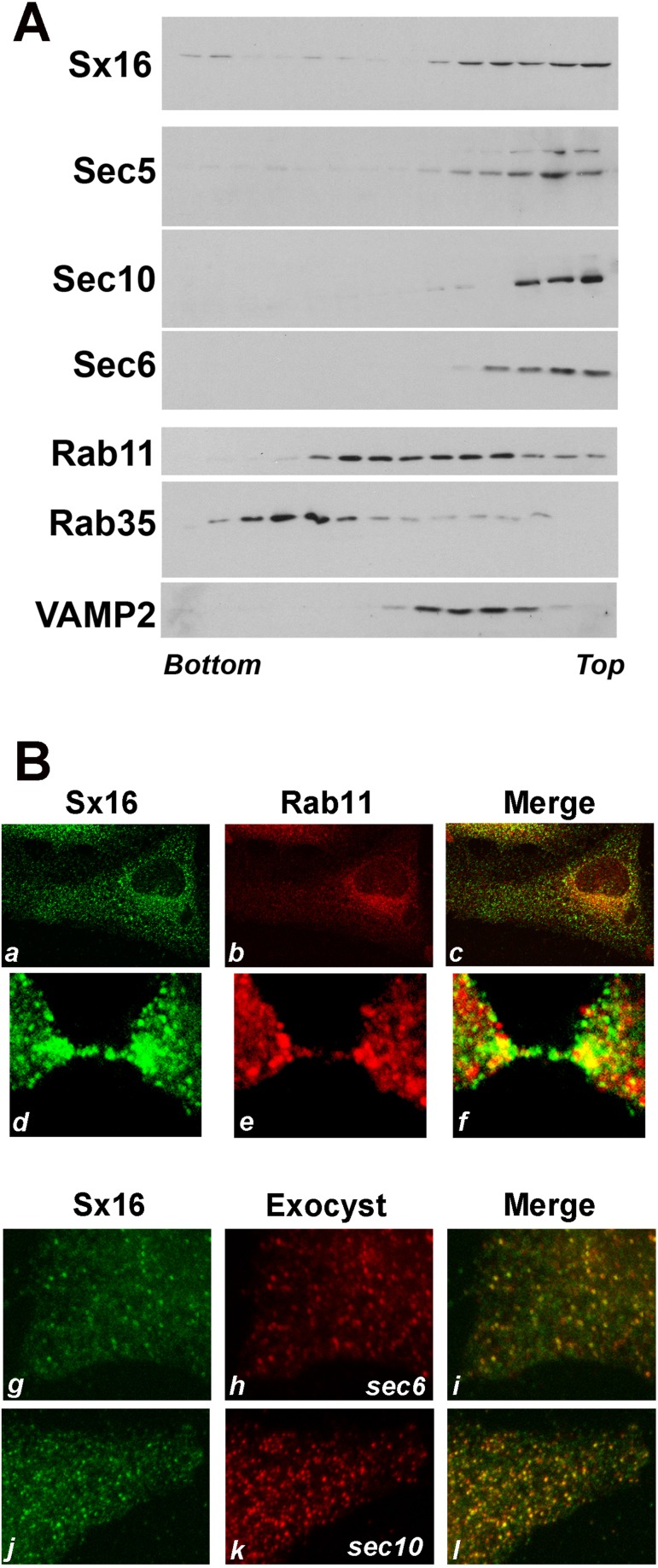
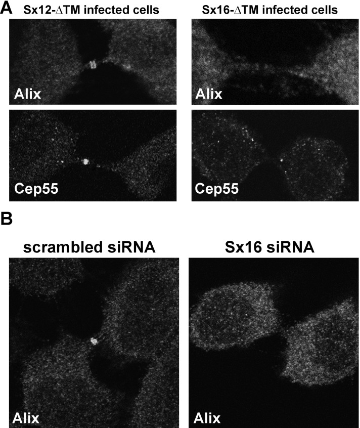
Recently it was shown that both recycling endosome and endosomal sorting complex required for transport (ESCRT) components are required for cytokinesis, in which they are believed to act in a sequential manner to bring about secondary ingression and abscission, respectively. However, it is not clear how either of these complexes is targeted to the midbody and whether their delivery is coordinated. The trafficking of membrane vesicles between different intracellular organelles involves the formation of soluble N-ethylmaleimide-sensitive factor attachment protein receptor (SNARE) complexes. Although membrane traffic is known to play an important role in cytokinesis, the contribution and identity of intracellular SNAREs to cytokinesis remain unclear. Here we demonstrate that syntaxin 16 is a key regulator of cytokinesis, as it is required for recruitment of both recycling endosome-associated Exocyst and ESCRT machinery during late telophase, and therefore that these two distinct facets of cytokinesis are inextricably linked.


Figures

FIGURE 1:
FIGURE 1:
Syntaxin 16 is required for cytokinesis. HeLa cells were incubated with adenovirus designed to express the indicated Sx-ΔTM construct at identical multiplicity of infection (in the experiment shown, 30:1), empty virus or not infected (control), as described in Materials and Methods. At 48 h later, cells were fixed and stained with anti-tubulin antibodies and DAPI and the frequency of binucleate cells counted. (A) Quantification of five independent experiments of this type. Sx6-ΔTM and Sx16-ΔTM significantly increased the frequency of binucleate cells (*p < 0.05), using at least three different batches of virus. (B) Typical fields of cells infected with Sx12-ΔTM or Sx16-ΔTM as indicated and stained for tubulin (red; tubulin), Sx16 (green; Sx16) or DNA (blue; DAPI). Note that for Sx16-ΔTM–infected cells, the high level of overexpression of Sx16-ΔTM required the use of reduced gain/laser power during collection of the image. (C) Comparison of the frequency of binucleate cells in cells infected with empty virus, Sx16-ΔTM, or Sx16-full length. (D) HeLa cells were transfected with an siRNA SmartPool designed to knock down Sx16 or a scrambled siRNA SmartPool control and then incubated for 48 h after transfection before fixation and staining with anti-Sx16. Data from a typical experiment. Left, typical cell in telophase, with endogenous Sx16 present near the midbody. See Figure 3 for Sx16 protein levels after knockdown. C and D are representative of three or more experiments of this type.
FIGURE 2:
FIGURE 2:
Sx16 is involved in abscission. Images from a typical time course of HeLa cell division, revealing delayed abscission in Sx16-ΔTM–expressing cells (bottom) compared with control cells (top). In control cells, a furrow is indicated by >, and cytokinesis is complete ∼1 h thereafter (two daughter cells indicated by asterisks in the next image). In Sx16-ΔTM–expressing cells, a long-lived midbody is indicated by the v; in the experiment shown, the cells remained connected for >4 h from the first clear appearance of the midbody (shown in the 2-h image). Data from >25 cells from three or more experiments in each group were binned according to the time taken to complete abscission after the start of furrowing into two groups, those taking <2 h, or those taking >2 h. The frequency of binucleate cells in this subgroup is also presented. Cells expressing Sx16-ΔTM consistently took longer to complete abscission, or failed abscission at a significantly greater rate, than their control counterparts.
FIGURE 3:
FIGURE 3:
Sx16 and mVps45 knockdown perturbs cytokinesis. HeLa cells were transfected with siRNA SmartPools against either scrambled siRNA (control), GAPDH, Sx6, Sx16, or mVps45 and were incubated for 48 h after transfection before fixation and staining for tubulin/DAPI to allow quantification of the frequency of binucleate cells. (A) Quantification of three experiments of this type. Asterisk indicates significant increase compared with control (p < 0.05). (B) Typical immunoblot analysis to confirm the extent and specificity of knockdown. Lysates were prepared in parallel to the analysis shown in A and immunoblotted with the antibodies shown. The approximate positions of molecular weight markers are shown.
FIGURE 4:
FIGURE 4:
GFP-Rab11 traffic to the midbody/intercellular bridge is decreased in cells expressing Sx16-ΔTM. HeLa cells were transfected with plasmids driving overexpression of GFP-Rab11, lum-GFP, or GFP-FIP3 as indicted. At 6 h after transfection, cells were infected with adenovirus to overexpress Sx12-ΔTM or Sx16-ΔTM, and images were collected 48 h after infection. Representative images of cells in telophase, typical of three independent repeats. Parallel coverslips were fixed and stained using anti–Aurora B or anti-MKLP1, and representative images are shown.
FIGURE 5:
FIGURE 5:
Sx16-ΔTM prevents the midbody accumulation of the Exocyst. HeLa cells were infected with Sx12-ΔTM (A–F) virus as described and immunostained with antibodies against different Exocyst components, as labeled. Sec8 (A), Sec6 (B), Sec10 (D), Exo70 (E), or Sec3 (F) localized to a ring-like structure in the midbody of the majority of Sx12-ΔTM cells in telophase (∼80%; see bar graph in M). By contrast, in cells expressing Sx16-ΔTM, Sec8 (G), Sec6 (H), Sec10 (J), Exo70 (K), or Sec3 (L) exhibited no such ring-like structure, but each Exocyst component was present within punctate structures throughout the midbody. The line graphs (C and I) indicate the intensity of staining for Sec6 in the region demarcated by the white bar in the two cases. (M) Quantification of data from three experiments of this type, encompassing >100 cells for each Exocyst component. Microtubule staining was omitted except for A and G in order to clearly show the distribution of Exocyst staining. Antibodies used in this figure were as described in Materials and Methods.
FIGURE 6:
FIGURE 6:
Sx16-ΔTM does not prevent the midbody accumulation of centriolin. HeLa cells were infected with Sx16-ΔTM or Sx12-ΔTM virus as described and immunostained for centriolin (green) or tubulin (red). Representative images from five independent experiments of this type. Asterisk indicates the midbody ring-like structure. Bottom, typical higher-magnification images of the midbody areas.
FIGURE 7:
FIGURE 7:
Sx16 and Exocyst components localize to vesicles of similar density. (A) HeLa cells in telophase were harvested, and membranes in a postnuclear supernatant were fractionated on the basis of density using iodixanol gradient analysis. Representative immunoblots of the indicated proteins. Denser membranes are at the bottom of the tube. The experiment was repeated twice more, with qualitatively similar results. Note that both Rab11-positive and Rab35-positive membranes were differentially localized to those containing Sx16. (B) HeLa cells were infected with Sx16-ΔTM or Sx12-ΔTM virus as described and immunostained with anti-Sx16 (a, d, g, and j; green) or anti-Rab11 (b, e; red) or anti-sec6 or anti-sec10 (h, k respectively; red). Merged images are shown in c, f, i, and j. Data from a typical experiment.
FIGURE 8:
FIGURE 8:
Sx16-ΔTM or Sx16 knockdown prevents the midbody accumulation of ALIX and causes reduced levels of Cep55. HeLa cells were infected with Sx16-ΔTM or Sx12-ΔTM virus as described. Cells were immunostained with antibodies against ALIX or Cep55. (A) Typical immunofluorescence staining of ALIX at the midbody of control (Sx12-ΔTM–infected) cells, revealed as a characteristic double ring–like structure. Cep55 also localized to the midbody of these cells. By contrast, ALIX did not localize to the midbody of Sx16-ΔTMxinfected cells, and Cep55 localization was impaired in more than half of the cells analyzed (see the text for explanation). The data shown are representative of three experiments of this type. (B) HeLa cells were transfected with an siRNA SmartPool designed to knock down Sx16 or a scrambled siRNA SmartPool control, then incubated for 48 h after transfection before fixation and staining with anti-ALIX as indicated. Data from a representative experiment, repeated three times with similar results. In all experiments, the extent of Sx16 knock down was >85% as determined by immunoblotting (unpublished data).

Similar articles

Cited by

References

    1. Barr FA, Gruneberg U. Cytokinesis: placing and making the final cut. Cell. 2007;131:847–860. - PubMed
    1. Bastos RN, Barr FA. Plk1 negatively regulates Cep55 recruitment to the midbody to ensure orderly abscission. J Cell Biol. 2010;191:751–760. - PMC - PubMed
    1. Bissig C, Lenoir M, Velluz MC, Kufareva I, Abagyan R, Overduin M, Gruenberg J. Viral infection controlled by a calcium-dependent lipid-binding module in ALIX. Dev Cell. 2013;25:364–373. - PMC - PubMed
    1. Blum R, Stephens DJ, Schulz I. Lumenal targeted GFP, used as a marker of soluble cargo, visualises rapid ERGIC to Golgi traffic by a tubulo-vesicular network. J Cell Sci. 2000;113(Pt 18):3151–3159. - PubMed
    1. Carlton JG, Martin-Serrano J. Parallels between cytokinesis and retroviral budding: a role for the ESCRT machinery. Science. 2007;316:1908–1912. - PubMed

Publication types

MeSH terms

LinkOut - more resources