. 2014 Mar-Apr;90(2):419-30.
doi: 10.1111/php.12179. Epub 2013 Nov 28.

Photophysics of glycosylated derivatives of a chlorin, isobacteriochlorin and bacteriochlorin for photodynamic theragnostics: discovery of a two-photon-absorbing photosensitizer

Affiliations

Photophysics of glycosylated derivatives of a chlorin, isobacteriochlorin and bacteriochlorin for photodynamic theragnostics: discovery of a two-photon-absorbing photosensitizer

Amit Aggarwal et al. Photochem Photobiol. 2014 Mar-Apr.

Abstract

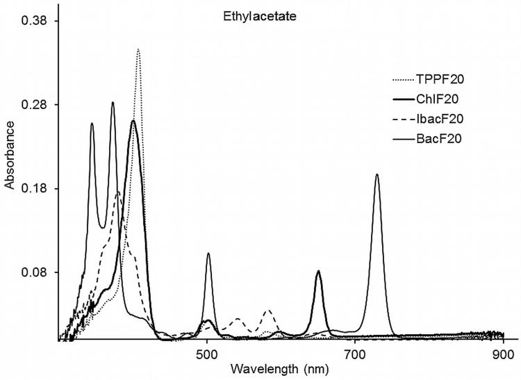
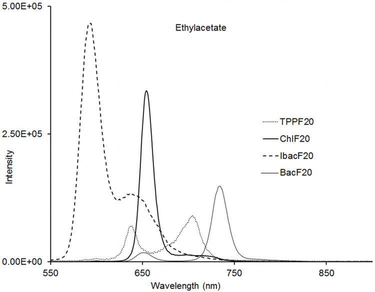
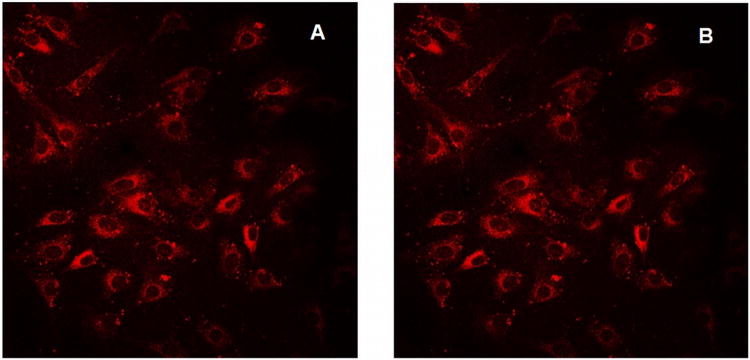
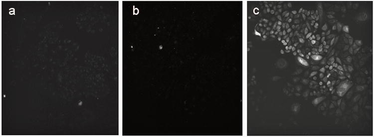
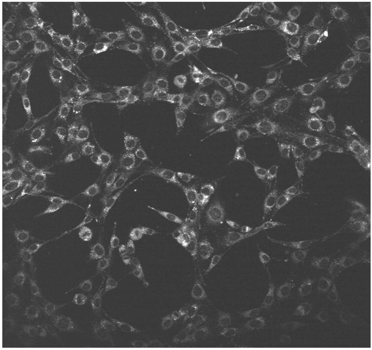
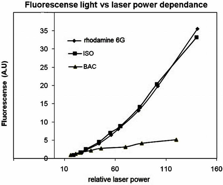
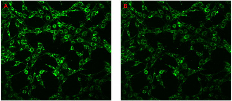
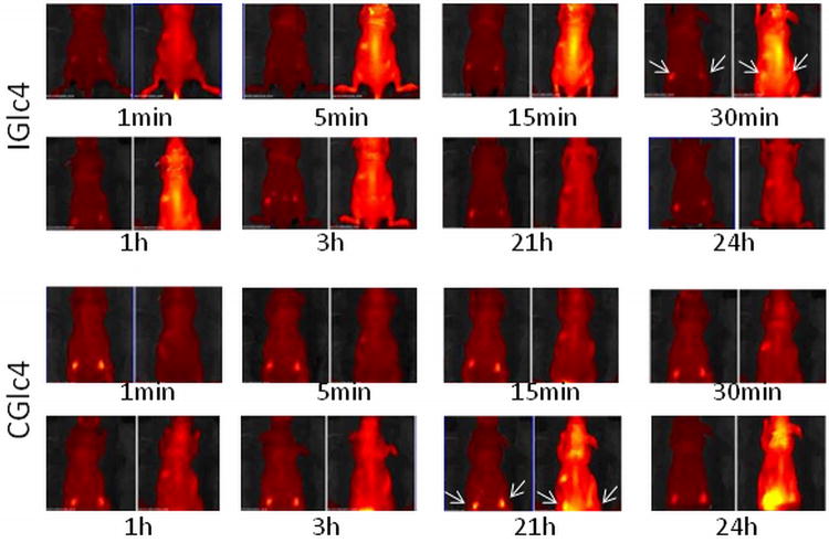
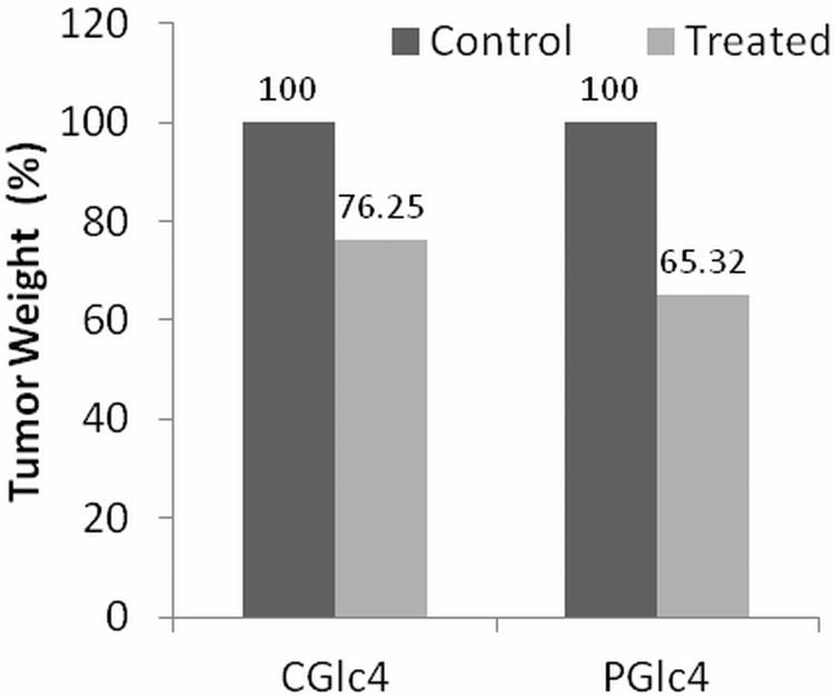
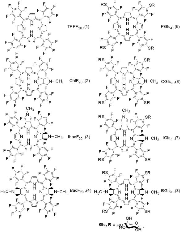
The photophysical properties of a chlorin, isobacteriochlorin and bacteriochlorin built on a core tetrapentafluorophenylporphyrin (TPPF20 ) and the nonhydrolyzable para thioglycosylated conjugates of these chromophores are presented. The photophysical characterization of these compounds was done in three different solvents to correlate with different environments in cells and tissues. Compared with TPPF20 other dyes have greater absorption in the red region of the visible spectrum and greater fluorescence quantum yields. The excited state lifetimes are from 3 to 11 ns. The radiative and nonradiative rate constants for deactivation of the excited state were estimated from the fluorescence quantum yield and excited state lifetime. The data indicate that the bacteriochlorin has strong absorption bands near 730 nm and efficiently enters the triplet manifold. The isobacteriochlorin has a 40-70% fluorescence quantum yield depending on solvent, so it may be a good fluorescent tag. The isobacteriochlorins also display enhanced two-photon absorption, thereby allowing the use of 860 nm light to excite the compound. While the two-photon cross section of 25 GM units is not large, excitation of low chromophore concentrations can induce apoptosis. The glycosylated compounds accumulate in cancer cells and a head and neck squamous carcinoma xenograft tumor model in mice. These compounds are robust to photobleaching.


Figures

Figure 1
Figure 1
UV-visible absorption spectra of compounds 1-4 in ethylacetate in 1 cm cuvettes and the concentration of each solution was 1 μM.
Figure 2
Figure 2
Emission spectra of compounds 2-4 in ethylacetate; excitation at 509 nm where the O.D was 0.014 for each compound.
Figure 3
Figure 3
K:Molv 3T3 NIH cells plated onto cover slips were incubated with CGlc4 for 24 hours, washed three times with PBS buffer, fixed with 4% paraformaldehyde, again washed three times with PBS, and mounted in Dako fluorescence mounting medium. Confocal images were taken using excitation wavelength 552 nm and emission filter was 578-700 nm. A: image after first scan, and B: image of the same sample after 25 scans. The images are as collected.
Figure 4
Figure 4
CHO cells were incubated overnight with a) 10 μM PGlc4 b) 10 μM CGlc4 c) 10 μM of a mixture of IGlc4: BGlc4 (5:1). Two-photon microscope excitation light was at 860 nm, and the detector was set for 500 nm to 670 nm. The image data was collected for 1s. The images are as collected.
Figure 5
Figure 5
K:Molv NIH 3T3 cells were incubated with 10 μM of IbacF20 overnight. Two-photon microscope excitation light was at 860 nm, and the detector was set for 500 nm to 670 nm. The image data was collected for 1 s. The image is as collected.
Figure 6
Figure 6
Dependence of light emitted versus laser power. Excitation: 860 nm. Light collected: 500 nm to 800 nm. Pink = IbacF20, blue = rhodamine 6G, yellow = BacF20.
Figure 7
Figure 7
K:Molv 3T3 NIH cells were incubated with IbacF20 for 24 hours and rinsed three times with PBS buffer, the 2-photon microscopy was done repeatedly to examine the photo stability of the compounds under these conditions. Excitation was at 860 nm, and detection between 500 nm and 670 nm. The 2-photon microscopic image after one scan (A), and the same sample after 25 scans (B). The images are as collected.
Figure 8
Figure 8
IGlc4 and CGlc4 up-take, distribution, clearance. Four-week-old, male Balb/c athymic nude mice (Nu/Nu) were obtained from Harlan Sprague Dawley, Inc. (Indianapolis, IN). Mice were housed in temperature-controlled rooms (74 ± 2°F) with a 12-hour alternating light-dark cycle. Approximately 3×10⁶ GFP-JHU-22 cells in log phase were injected subcutaneously into the mice. The up-take experiments were done after seven days after GFP-JHU-22 cell inoculation. The mice received tail-vein injections with a total volume of 100 μLof 5mg/mLIGlc4 or CGlc4.The fluorescence images were taken at the times indicated after injection. Each individual group has two images where the emission due to GFP is on the left of each time interval, and monitored using a 465-520 band pass filter. A 605 -660 nm band pass filter was used to image the tumors with CGlc4 and IGlc4.The arrows indicated the location of tumor xenografts. A Xenogren IVIS instrument (Caliper Life Sciences, Hopkinton, MA) was used to collect these images.
Figure 9
Figure 9
Decrease in tumor weight after 21 days.
Scheme 1
Scheme 1
Structure of non-glycosylated (1-4) and glycosylated (5-8) conjugates of porphyrin. The syn versus anti compounds for the bacteriochlorins and isobacteriochlorins are isolated, but the diastereomers for CGlc4 and two sets of diastereomers for IGlc4 and BGlc4 (see ref. 23) are not separated for these studies.

References

    1. Kadish K, Smith K, Guilard R. The Handbook of Porphyrin Science with Applications to Chemistry, Physics, Materials Science, Engineering, Biology and Medicine. World Scientific Publishers; Singapore: 2010.
    1. Bonnett R. Photosensitizers of the porphyrin the phthalocyanine series for phtodynamic therapy. Chem Soc Rev. 1995;24:19–33.
    1. Pandey RK. Recent advances in photodynamic therapy. J Porphyrins Phthalocyanines. 2000;4:368–373.
    1. Sternberg ED, Dolphin D, Brückner C. Porphyrin-based photosensitizers for use in photodynamic therapy. Tetrahedron. 1998;54:4151–4202.
    1. Lovell JF, Liu TWB, Chen J, Zheng G. Activatable Photosensitizers for Imaging and Therapy. Chem Rev. 2010;110:2839–2857. - PubMed

Publication types

LinkOut - more resources