Review
. 2013 Nov;25(11):1163-72.
doi: 10.1111/jne.12108.

Rapid effects of oestrogen on synaptic plasticity: interactions with actin and its signalling proteins

Affiliations
Review

Rapid effects of oestrogen on synaptic plasticity: interactions with actin and its signalling proteins

A H Babayan et al. J Neuroendocrinol. 2013 Nov.

Abstract

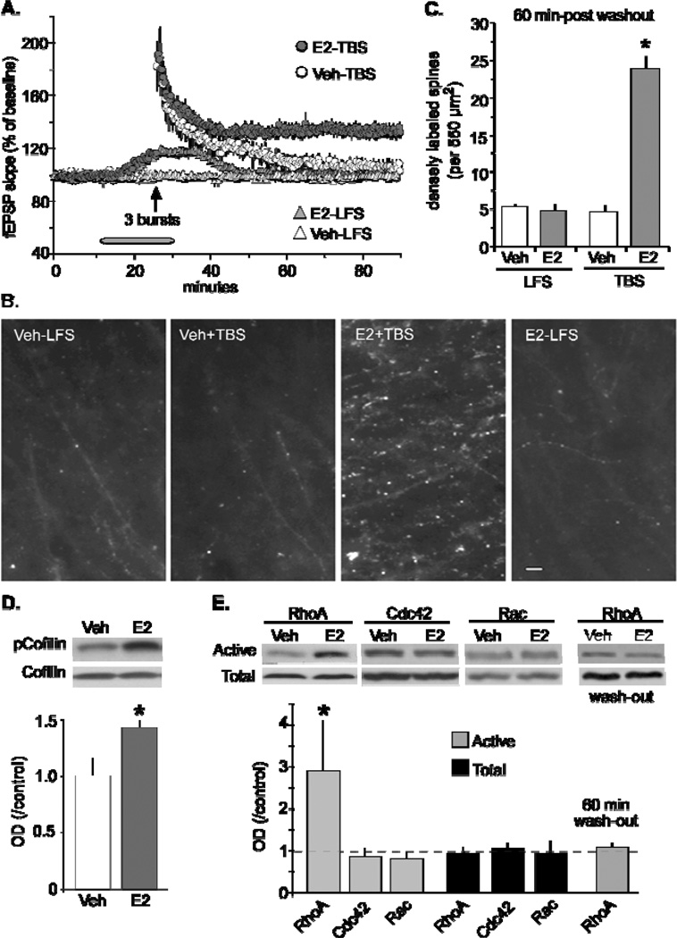
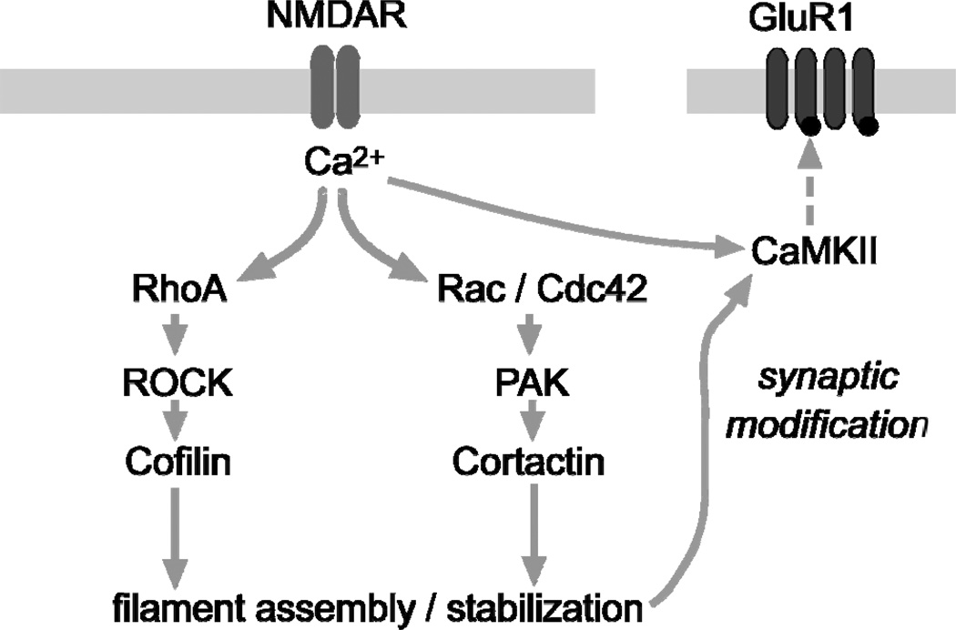
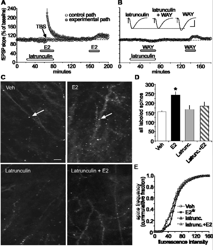
Oestrogen rapidly enhances fast excitatory postsynaptic potentials, facilitates long-term potentiation (LTP) and increases spine numbers. Each effect likely contributes to the influence of the steroid on cognition and memory. In the present review, we first describe a model for the substrates of LTP that includes an outline of the synaptic events occurring during induction, expression and consolidation. Briefly, critical signalling pathways involving the small GTPases RhoA and Rac/Cdc42 are activated by theta burst-induced calcium influx and initiate actin filament assembly via phosphorylation (inactivation) of cofilin. Reorganisation of the actin cytoskeleton changes spine and synapse morphology, resulting in increased concentrations of AMPA receptors at stimulated contacts. We then use the synaptic model to develop a specific hypothesis about how oestrogen affects both baseline transmission and plasticity. Brief infusions of 17β-oestradiol (E2 ) reversibly stimulate the RhoA, cofilin phosphorylation and actin polymerisation cascade of the LTP machinery; blocking this eliminates the effects of the steroid on transmission. We accordingly propose that E2 induces a weak form of LTP and thereby increases synaptic responses, a hypothesis that also accounts for how it markedly enhances theta burst induced potentiation. Although the effects of E2 on the cytoskeleton could be a result of the direct activation of small GTPases by oestrogen receptors on the synaptic membrane, the hormone also activates tropomyosin-related kinase B receptors for brain-derived neurotrophic factor, a neurotrophin that engages the RhoA-cofilin sequence and promotes LTP. The latter observations raise the possibility that E2 produces its effects on synaptic physiology via transactivation of neighbouring receptors that have prominent roles in the management of spine actin, synaptic physiology and plasticity.

Keywords: RhoA; cofilin; long-term potentiation; oestradiol; pTrkB; spines.


Figures

Figure 1
Figure 1
Schematic of a proposed synaptic model showing signaling cascades that regulate actin dynamics during the production of LTP. It has been shown that chemically induced NMDA receptor-dependent LTP robustly activates RhoA and Rac [53], which is consistent with evidence that Rho-GTPases are activated by Ca2+ influx through NMDA receptors (blue bars) [106]. Activity-driven activation of the RhoA>ROCK>cofilin pathway leads to rapid filament assembly, while parallel activation of Rac/Cdc42>PAK>cortactin influences later stages of LTP stabilization. The activation of these two pathways leads to several activity-dependent changes in the synapse including activation and translocation of CaMKII to the postsynaptic density that will then phosphorylate AMPARs (red bars) and initiate receptor trafficking to the synapse. The latter events increase synaptic current through AMPAR’s, and thus, increase fEPSP size.
Figure 2
Figure 2
Estrogen promotes actin polymerization in CA1 dendritic spines. (A) Latrunculin (500nM) effectively blocked both E2-induced increases in synaptic transmission in the control pathway (white circle) and theta burst-induced LTP in the experimental pathway (dark circle). Following a 1hr washout period, fEPSP responses to E2 was restored. (B) Infusions of latrunculin blocked, in a reversible manner, the increase in baseline synaptic responses produced by the selective ERβ agonist, WAY200070 (WAY; 100nM). Inset, representative traces collected during treatments with latrunculin, WAY, and both. Scale 1mV/5ms. (C) Top two panels compare phalloidin labeling in CA1 stratum radiatum in slices treated with 1nM E2 or vehicle (Scale bar = 5µm). E2 treatment increases spine-like profiles (arrows) relative to vehicle. Bottom two panels illustrate the effect of latrunculin (500nM) alone and in combination with E2-induced baseline phalloidin labeling. (D) The group mean (±SEM) number of phalloidin-labeled spines in area CA1b was significantly greater in slices treated with E2 compared to vehicle controls (*p < 0.001), and that latrunculin blocks E2-induced increases in phalloidin labeling. (E) Cumulative frequency distribution of phalloidin-labeling intensities in CA1b in control versus E2-treated slices; E2 caused a significant rightward shift in the distribution curve relative to controls (*p < 0.001). This effect was blocked by latrunculin. Modified from [17].
Figure 3
Figure 3
17β-estradial (E2) enhances theta burst-induced LTP and actin polymerization via RhoA signaling in hippocampal slices. (A) Infusions of 1 nM E2 (E2-LFS) caused a reversible increase in slope fEPSPs relative to control (vehicle) during single pulse stimulation in field CA1. The delivery of a subthreshold level of theta stimulation (upward arrow) in vehicle treated slices caused potentiation to decay back to baseline following a 60 min recording period. The same level of stimulation in the presence of E2 caused lasting potentiation. (B) At the end of the 60 min washout period, slices were processed for phalloidin labeling of field CA1 dendritic spines. Survey micrographs indicate slices that had received baseline test pulses (low-frequency stimulation; LFS) during E2 infusion (E2-LFS) did not show an increase in phalloidin positive spines above levels seen in slices treated with vehicle (Veh-LFS). In contrast, three theta bursts significantly enhanced the number of phalloidin-labeled puncta in slices infused with E2 (E2+TBS) relative to vehicle treated slices (Veh+TBS). Scale bar, 5µm. (C) The number of densely labeled spines was determined using an automatic counting program. Slices that received LFS only in the presence and absence of E2 had the same relative counts of densely labeled phalloidin after the 60 min washout period, while those that received theta bursts in the presence of E2 produced a significant increase in densely labeled spines relative to vehicle treated slices (p < 0.0001). (D) TopRepresentative Western blot images show both phosphorylated (p)-cofilin and total cofilin levels in hippocampal slices infused with 1nM E2 or vehicle. BottomOptical density measurements from blots confirm that a 20 min infusion of E2 significantly increased pCofilin relative to total cofilin (p < 0.05) in hippocampal slices from male rats. (E) TopA 20 min infusion of E2 selectively increased the activity of GTPase RhoA relative to total RhoA as assessed using the pull-down assay. BottomQuantitative analysis confirmed that E2 significantly increased activity levels of RhoA relative to related GTPases Cdc42 and Rac (p < 0.05). Following a 60 min washout of E2, activated RhoA levels were not significantly different from vehicle-treated slices. Modified from [17].

References

    1. Devi G, Hahn K, Massimi S, Zhivotovskaya E. Prevalence of memory loss complaints and other symptoms associated with the menopause transition: a community survey. Gender medicine. 2005;2:255–264. - PubMed
    1. Dumas JA, Kutz AM, Naylor MR, Johnson JV, Newhouse PA. Increased memory load-related frontal activation after estradiol treatment in postmenopausal women. Hormones and behavior. 2010;58:929–935. - PMC - PubMed
    1. Greendale GA, Huang MH, Wight RG, Seeman T, Luetters C, Avis NE, Johnston J, Karlamangla AS. Effects of the menopause transition and hormone use on cognitive performance in midlife women. Neurology. 2009;72:1850–1857. - PMC - PubMed
    1. Wegesin DJ, Stern Y. Effects of hormone replacement therapy and aging on cognition: evidence for executive dysfunction. Neuropsychology, development, and cognition. Section B, Aging, neuropsychology and cognition. 2007;14:301–328. - PubMed
    1. Loy R, Gerlach JL, McEwen BS. Autoradiographic localization of estradiol-binding neurons in the rat hippocampal formation and entorhinal cortex. Brain Res. 1988;467:245–251. - PubMed

Publication types