. 2013 Dec;43(12):1406-18.
doi: 10.1111/cea.12200.

Diesel exhaust particle exposure increases severity of allergic asthma in young mice

Affiliations

Diesel exhaust particle exposure increases severity of allergic asthma in young mice

T H Acciani et al. Clin Exp Allergy. 2013 Dec.

Abstract

Background: Epidemiologic studies have reported an association between diesel exhaust particle (DEP) exposure, allergic sensitization, and childhood wheezing, although the mechanisms remain unclear. While DEP is known to augment allergic responses in adult animal models, its effects on sensitization and asthma severity in young animals is unknown.

Objective: To examine the impact of different doses of DEP and allergen co-exposure on allergic sensitization and asthma characteristics in young mice, and whether Th17 as well as Th2 responses are induced.

Methods: Lungs of 3-week-old wild-type Balb/c mice were exposed by pharyngeal aspiration nine times over 3 weeks to DEP at 1.2 or 6.0 mg/kg body weight, house dust mite (HDM) at 0.8, 1.2 or 6.0 mg/kg of DEP in combination with HDM, or the same volume (50 μL) of 0.9% sterile saline.

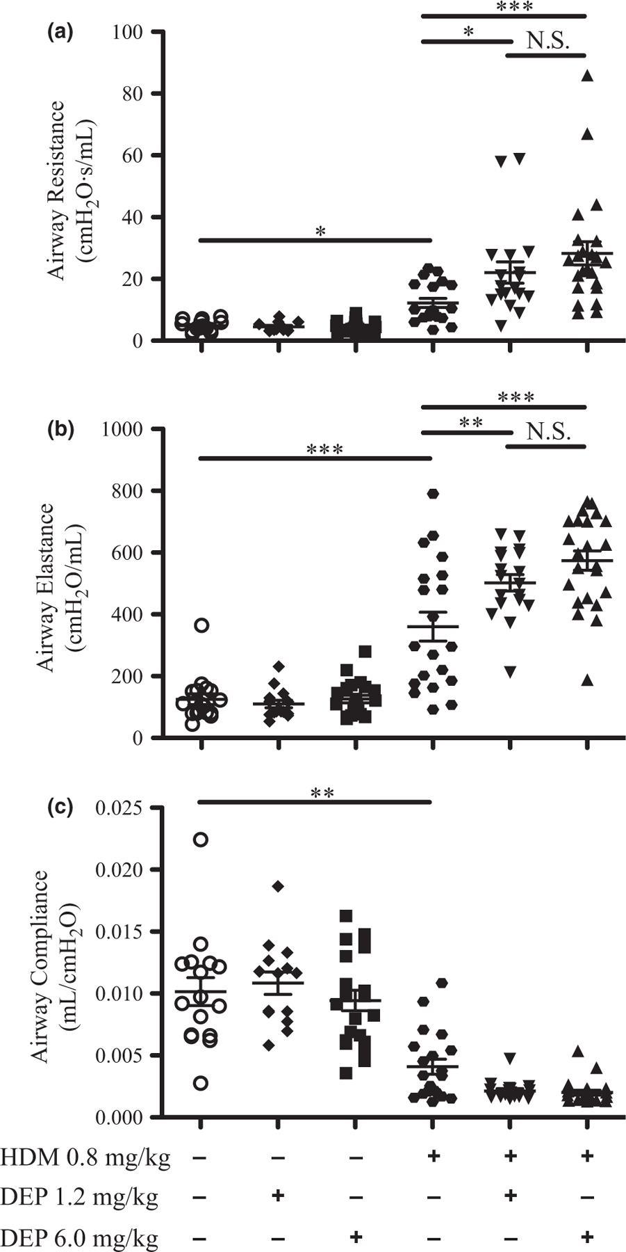
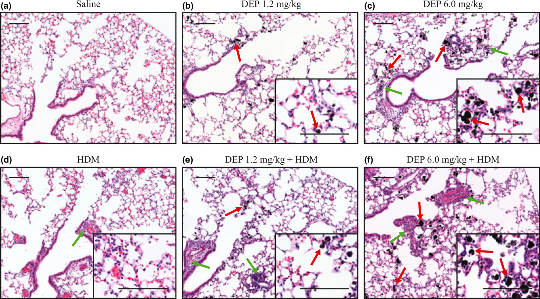
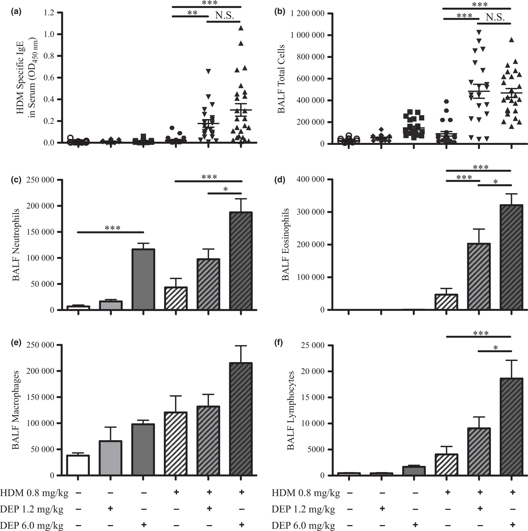
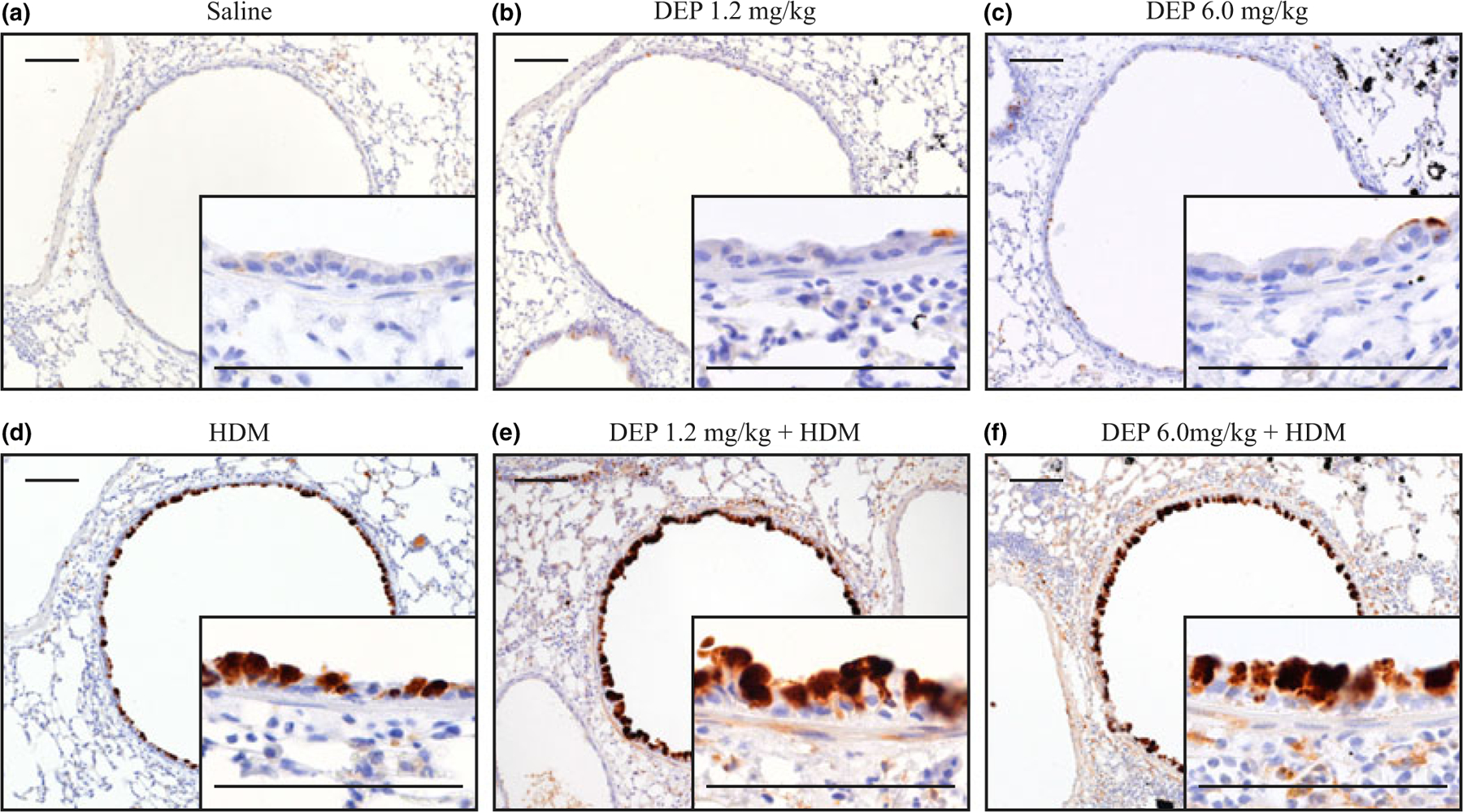
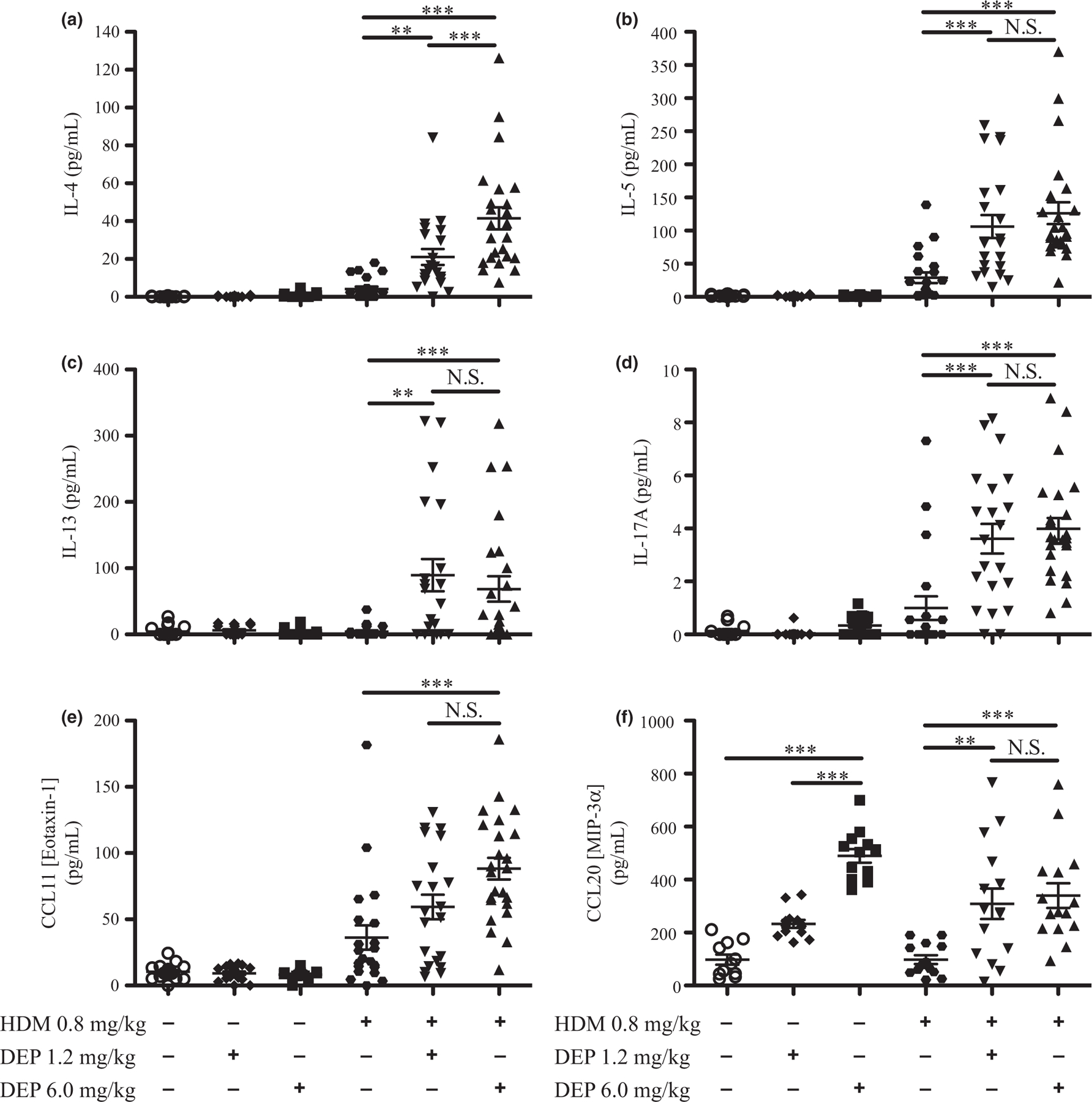
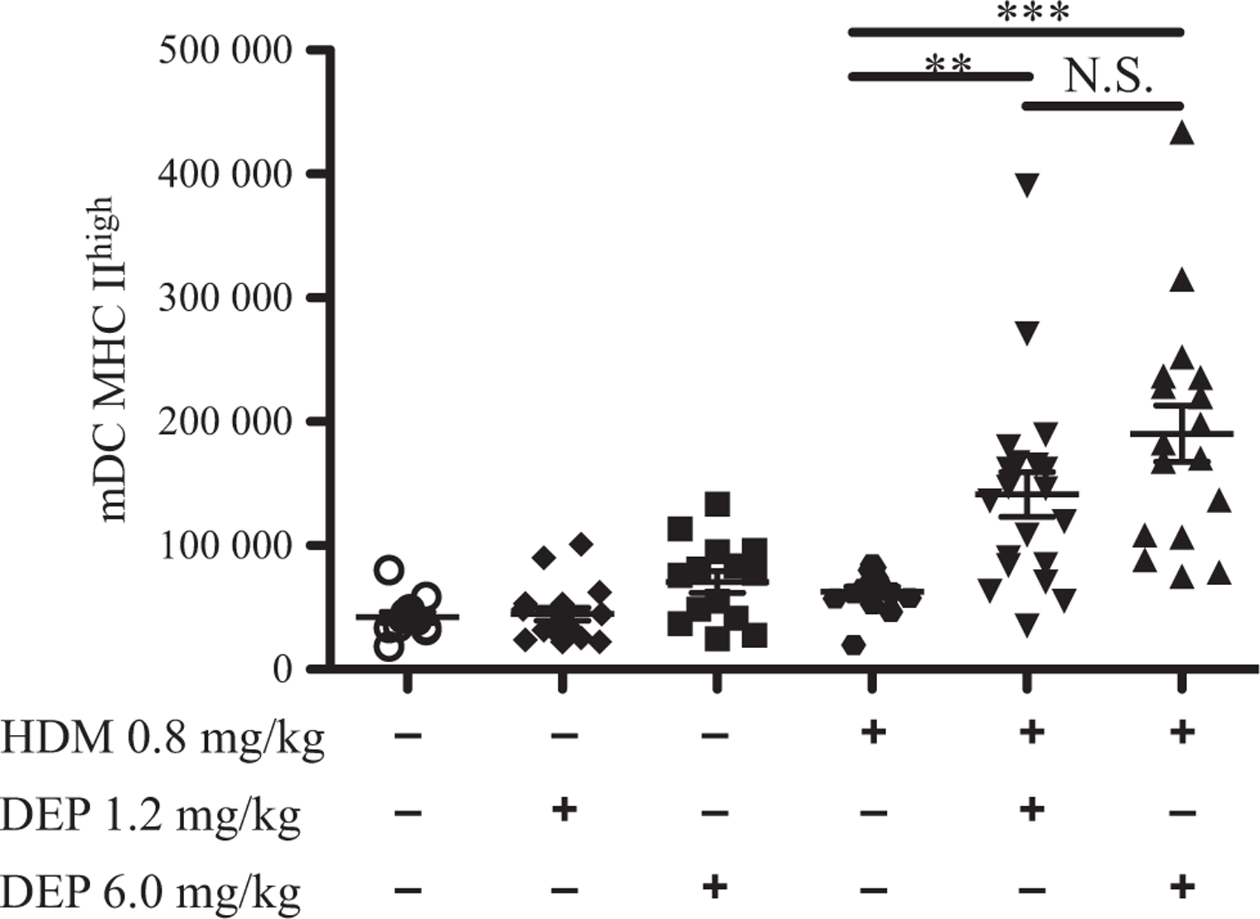
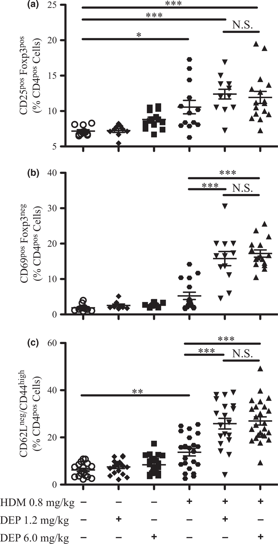
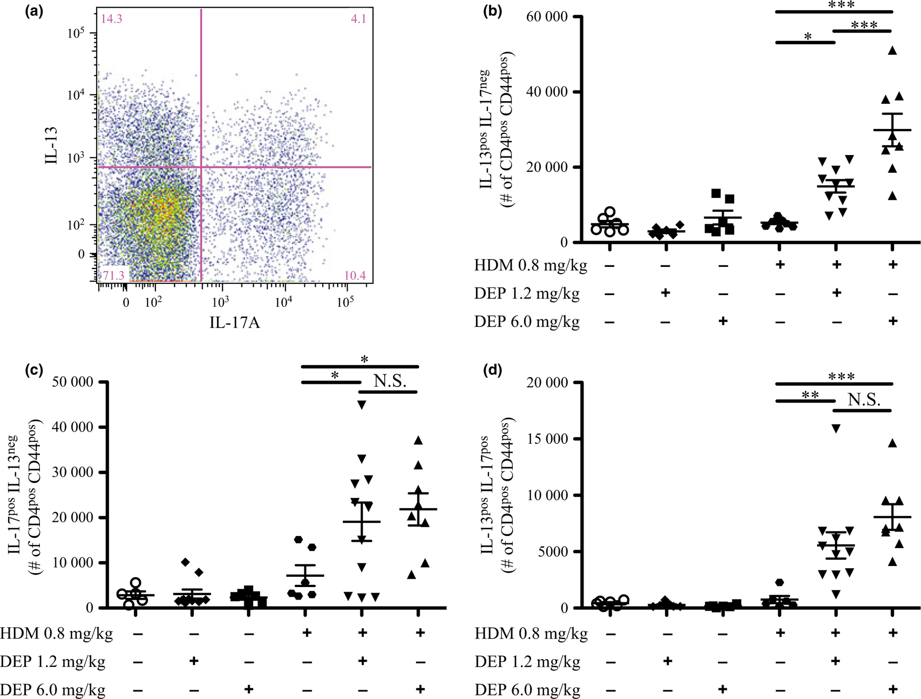
Results: In young mice, exposure to 1.2 mg/kg of DEP caused no detectable lung inflammation, but 6.0 mg/kg of DEP induced neutrophilic influx. Compared to HDM or DEP alone, mice exposed to either dose of DEP together with HDM demonstrated increased allergen-specific IgE, lung inflammation, airway hyperreactivity, goblet cell metaplasia, Th2/Th17 cytokines, dendritic cells, activated T cells, effector T cells, and IL-17(pos) and IL-13(pos) /IL-17A(pos) T effector cells.

Conclusions and clinical relevance: In young mice, co-exposure to DEP and HDM together exacerbated allergic sensitization and induced key characteristics of more severe asthma, including IL-17A, IL-17(pos) and IL-13(pos) /IL-17A(pos) T effector cells. While exposure to 1.2 mg/kg DEP alone caused no detectable changes, it did exacerbate allergic sensitization and asthma characteristics to a similar degree as a five-fold higher dose of DEP. This study demonstrates that exposure to DEP, even at a dose that alone causes no inflammation, exacerbates allergic asthma in young animals and suggests the importance of preventive measures to reduce the exposure of children to traffic related air pollution.

Keywords: T cells; Th17; Th2; air pollution; allergen; asthma; atopy; dendritic cells; diesel exhaust particle; house dust mite.


Conflict of interest statement

Conflict of interest

The authors declare no conflict of interest.

Figures

Fig. 1.
Fig. 1.
Airway hyper-reactivity is increased after DEP and allergen exposure. Airway mechanics were assessed using a flexiVent system after nebulizing PBS (baseline) and then increasing doses of methacholine (25, 50, and 100 mg/mL) into the lungs. Changes in airway resistance (a), elastance (b), and compliance (c) are shown at 100 mg/mL methacholine; dose–response curves are shown in supplemental data (Fig. S2a–c). Statistical significance was determined to be P < 0.05 (*), P < 0.01 (**), and P < 0.001 (***) or not significant if P > 0.05 (N.S.). Error bars shown represent the standard error of mean (±SEM). The number of animals per group was 15–25.
Fig. 2.
Fig. 2.
Lung histology following DEP and allergen exposure. (a–f) Haematoxylin and eosin staining of lung sections showing DEP (red arrows) and inflammation (green arrows) in the small airways and alveolar regions of the lung; scale bar = 100 μm.
Fig. 3.
Fig. 3.
HDM-specific IgE and inflammatory cells increase with DEP and allergen exposure. (a) HDM-specific IgE in serum samples from exposure groups was measured by ELISA. (b) Inflammatory cell influx was estimated by counting cells present in BALF. Changes in the number of neutrophils (c), eosinophils (d), macrophages (e), and lymphocytes (f) were assessed after differential staining; changes in percentages of cell types are shown in supplemental data (Fig. S4). Statistical significance was determined to be P < 0.05 (*), P < 0.01 (**), and P < 0.001 (***) or not significant if P > 0.05 (N.S.). Error bars shown represent the standard error of mean (±SEM). The number of animals per group was 15–25.
Fig. 4.
Fig. 4.
Goblet cell metaplasia following DEP and allergen exposure. (a–f) Immunohistochemistry for Muc5AC was performed on lung sections, positive staining for Muc5AC appears brown and DEP appears black; scale bar = 100 μm. Quantitative assessment of goblet cell metaplasia is shown by Western blot analysis of CLCA3 in supplemental data (Fig. S5a,b).
Fig. 5.
Fig. 5.
Cytokine and chemokine levels in BALF increased with DEP and allergen exposure. Cytokine and chemokine levels in BALF were measured by multiplex or ELISA (a–f). Statistical significance was determined to be P < 0.01 (**) and P < 0.001 (***) or not significant if P > 0.05 (N.S.). Error bars shown represent the standard error of mean (±SEM). The number of animals per group was 15–25.
Fig. 6.
Fig. 6.
Increased myeloid dendritic cells after DEP and allergen exposure. Flow cytometry analysis was performed on lung cells from exposure groups for activated myeloid dendritic cells. The number of myeloid dendritic cells is depicted. The gating strategy is shown in supplemental data (Fig. S8a). Statistical significance was determined to be P < 0.01 (**), and P < 0.001 (***) or not significant if P > 0.05 (N.S.). Error bars shown represent the standard error of mean (±SEM). The number of animals per group was 15–25.
Fig. 7.
Fig. 7.
Regulatory T cells, activated T cells, and effector T cells increase with DEP and allergen exposure. Flow cytometry analysis was performed on lung cells from exposure groups for CD25pos regulatory T cells (a), activated T cells (b), and effector T cells (c). The percentages of CD4pos cells are depicted. The gating strategies are shown in supplemental data for effector T cells (Fig. S8b) and regulatory T cells and activated T cells (Fig. S8c). Statistical significance was determined to be P < 0.05 (*), P < 0.01 (**), and P < 0.001 (***) or not significant if P > 0.05 (N.S.). Error bars shown represent the standard error of mean (±SEM). The number of animals per group was 15–25.
Fig. 8.
Fig. 8.
IL-13pos, IL-17Apos, and dual-positive (IL-13pos/IL-17Apos) effector T cells increase following DEP and allergen exposure. Flow cytometry analysis was performed on effector T cells from exposure groups stained for intracellular cytokines IL-13 and IL-17A, after ex vivo restimulation. A sample gating strategy (a) shows the separation of effector T cells that were only IL-13pos (b), only IL-17Apos (c), and positive for both (IL-13pos/IL-17Apos) (d). The number of each cell type is depicted. Changes in percentages of cell types are shown in supplemental data (Fig. S7a–c). The full gating strategy is also shown in supplemental data (Fig. S8d). Statistical significance was determined to be P < 0.05 (*), P < 0.01 (**), and P < 0.001 (***) or not significant if P > 0.05 (N.S.). Error bars shown represent the standard error of mean (±SEM). The number of animals per group was 6–11.

References

    1. Holgate ST. Innate and adaptive immune responses in asthma. Nat Med 2012; 18:673–83. - PubMed
    1. Simpson JL, Scott R, Boyle MJ, Gibson PG. Inflammatory subtypes in asthma: assessment and identification using induced sputum. Respirology 2006; 11:54–61. - PubMed
    1. Hastie AT, Moore WC, Meyers DA, Vestal PL, Li H, Peters SP et al. Analyses of asthma severity phenotypes and inflammatory proteins in subjects stratified by sputum granulocytes. J Allergy Clin Immunol 2010; 125:1028–36.e13. - PMC - PubMed
    1. Kim HY, DeKruyff RH, Umetsu DT. The many paths to asthma: phenotype shaped by innate and adaptive immunity. Nat Immunol 2010; 11:577–84. - PMC - PubMed
    1. Al-Ramli W, Préfontaine D, Chouiali F, Martin JG, Olivenstein R, Lemière C et al. T(H)17-associated cytokines (IL-17A and IL-17F) in severe asthma. J Allergy Clin Immunol 2009; 123:1185–7. - PubMed

Publication types

MeSH terms