. 2013 Oct 10;13(1):95.
doi: 10.1186/1475-2867-13-95.

MiRNA-497 regulates cell growth and invasion by targeting cyclin E1 in breast cancer

Affiliations

MiRNA-497 regulates cell growth and invasion by targeting cyclin E1 in breast cancer

Qifeng Luo et al. Cancer Cell Int. .

Abstract

Background: MicroRNAs are a class of endogenous single strand non-coding RNAs that are involved in many important physiological and pathological processes. The purpose of this study was to examine the expression levels of miR-497 in human breast cancer and its function in MDA-MB-231 breast cancer cells.

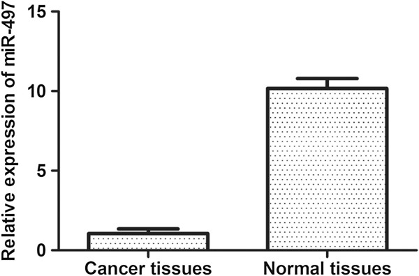
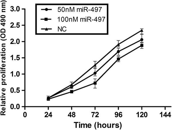
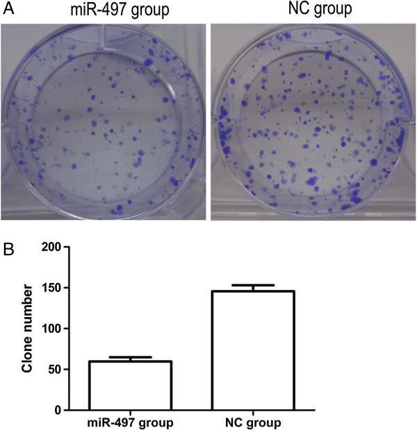
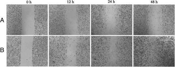
Methods: Quantitative polymerase chain reaction was used to measure the expression levels of miR-497 in 40 breast cancer specimens and adjacent normal breast tissues. MTT assays, colony formation assays, wound healing assays, transwell assays and cell cycle assays were used to explore the potential function of miR-497 in MDA-MB-231 breast cancer cells. Dual-luciferase reporter assays were performed to analyze the regulation of putative target of miR-497, and western blot assays were used to validate the dual-luciferase results.

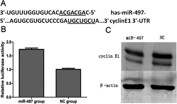
Results: The expression of miR-497 in breast cancer specimens was lower than adjacent normal tissues (P < 0.05). Overexpression of miR-497 inhibited cellular growth, suppressed cellular migration and invasion, and caused a G1 arrest. Dual-luciferase reporter assays showed that miR-497 binds the 3'-untranslated region (3'-UTR) of cyclin E1, suggesting that cyclin E1 is a direct target of miR-497. Western blot assays confirmed that overexpression of miR-497 reduced cyclin E1 protein levels.

Conclusions: MiR-497 may act as a tumor suppressor gene in breast cancer. Inhibited cellular growth, suppressed cellular migration and invasion, and G1 cell cycle arrest were observed upon overexpression of miR-497 in cells, possibly by targeting cyclin E1. These results indicate miR-497 could be considered a therapeutic target for the development of treatment for breast cancer.


Figures

Figure 1
Figure 1
MiR-497 levels are significantly decreased in breast cancer specimens. The graph represents the 2-ΔΔCt values ± SEM, *P < 0.05.
Figure 2
Figure 2
MiR-497 expression inhibits MDA-MB-231 proliferation. MTT cell proliferation assays were performed with miR-497 and NC expressing cells. The proliferation of MDA-MB-231 cells transfected with miR-497 was inhibited in a dose- and time-dependent manner compared with the NC group. The graph represents OD Values ± SEM, *P < 0.05.
Figure 3
Figure 3
Overexpression of miR-497 inhibited colony formation ability of MDA-MB-231 cells. (A) Colony formation assays showed Crystal Violet staining of the miR-497-transfected group and NC-transfected group. (B) Graph indicates colony numbers of each experimental transfected group (60 ± 2 miR-497 group vs. 145 ± 2 NC group, *P < 0.05).
Figure 4
Figure 4
Overexpressing of miR-497 in MDA-MB-231 cells showed impaired migration in wound healing assays. (A) Image showed the gap of the scratched region of the miR-497-expressing MDA-MB-231 cells. (B) Image showed the region of the NC group cells.
Figure 5
Figure 5
MiR-497 overexpression affects invasion ability of MDA-MB-231 cells. (A) Crystal Violet-stained invaded cells from transwell experiments. Images were obtained on an inverted microscope with × 200 magnification. (B) Invasion rates were determined by counting the number of cells invading through matrigel, *P < 0.05.
Figure 6
Figure 6
Overexpression of miR-497 in MDA-MB-231 cells caused G1 phase accumulation. MDA-MB-231 cells transfected with miR-497 or NC mimics were analyzed by flow cytometry. The respective proportion of G1 phase, S phase and G2 phase cells of miR-497 and NC groups are indicated.
Figure 7
Figure 7
miR-497 targeted cyclin E1. (A) The miR-497 binding site in cyclin E1 3′-UTR, located 247–254 bp upstream of the cyclin E1 3′-UTR. (B) The relative luciferase of activity in miR-497 group and NC group (*P < 0.05). (C) Western blot analysis of cyclin E1 protein levels in miR-497-overexpressing and NC cells.

References

    1. Bartel DP. MicroRNAs: genomics, biogenesis, mechanism, and function. Cell. 2004;116(2):281–297. doi: 10.1016/S0092-8674(04)00045-5. - DOI - PubMed
    1. Meister G, Landthaler M, Dorsett Y, Tuschl T. Sequence-specific inhibition of microRNA- and siRNA-induced RNA silencing. RNA. 2004;10(3):544–550. doi: 10.1261/rna.5235104. - DOI - PMC - PubMed
    1. Esquela-Kerscher A, Slack FJ. Oncomirs - microRNAs with a role in cancer. Nature reviews Cancer. 2006;6(4):259–269. - PubMed
    1. Ambros V. MicroRNA pathways in flies and worms: growth, death, fat, stress, and timing. Cell. 2003;113(6):673–676. doi: 10.1016/S0092-8674(03)00428-8. - DOI - PubMed
    1. Carlsson J, Davidsson S, Helenius G, Karlsson M, Lubovac Z, Andrén O, Olsson B, Klinga-Levan K. A miRNA expression signature that separates between normal and malignant prostate tissues. Cancer Cell Int. 2011;11(1):14. doi: 10.1186/1475-2867-11-14. - DOI - PMC - PubMed

LinkOut - more resources