. 2013 Nov;2(11):862-70.
doi: 10.5966/sctm.2013-0080. Epub 2013 Oct 10.

Efficient and rapid derivation of primitive neural stem cells and generation of brain subtype neurons from human pluripotent stem cells

Affiliations

Efficient and rapid derivation of primitive neural stem cells and generation of brain subtype neurons from human pluripotent stem cells

Yiping Yan et al. Stem Cells Transl Med. 2013 Nov.

Erratum in

  • Stem Cells Transl Med. 2013 Dec;2(12):1022

Abstract

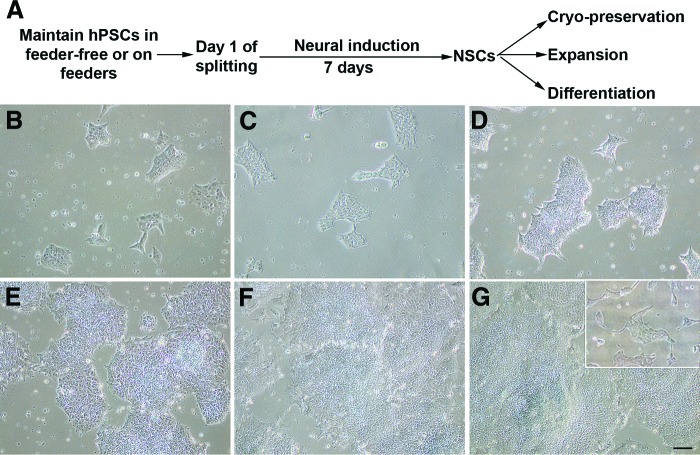
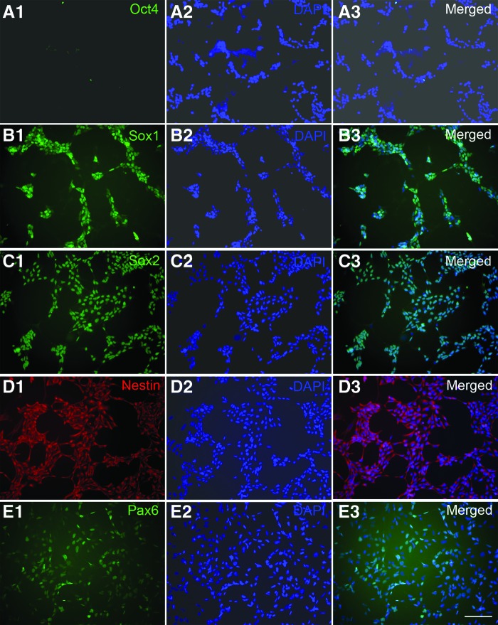
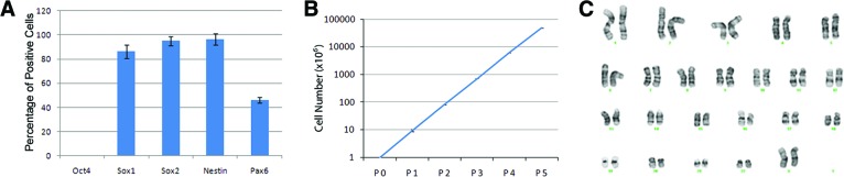
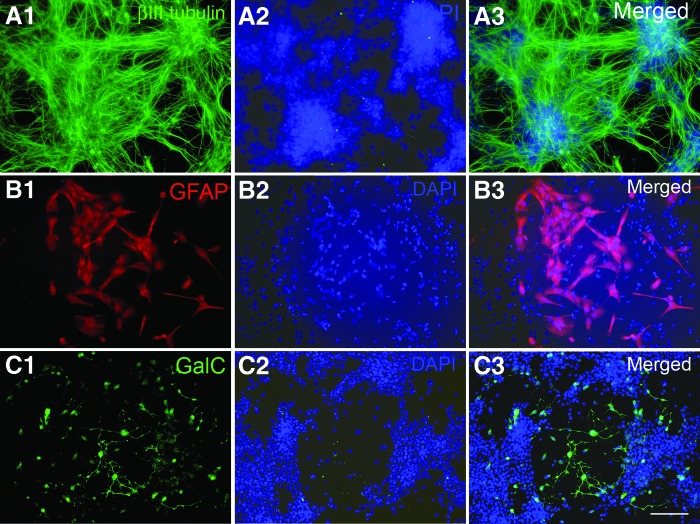
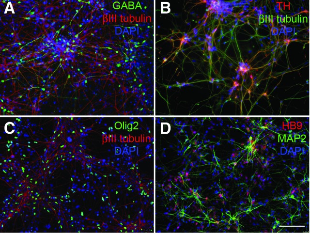
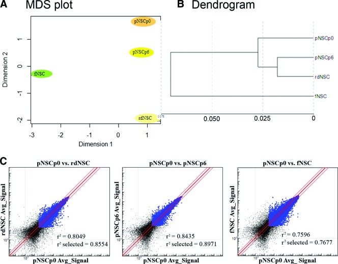
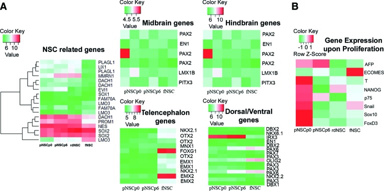
Human pluripotent stem cells (hPSCs), including human embryonic stem cells and human induced pluripotent stem cells, are unique cell sources for disease modeling, drug discovery screens, and cell therapy applications. The first step in producing neural lineages from hPSCs is the generation of neural stem cells (NSCs). Current methods of NSC derivation involve the time-consuming, labor-intensive steps of an embryoid body generation or coculture with stromal cell lines that result in low-efficiency derivation of NSCs. In this study, we report a highly efficient serum-free pluripotent stem cell neural induction medium that can induce hPSCs into primitive NSCs (pNSCs) in 7 days, obviating the need for time-consuming, laborious embryoid body generation or rosette picking. The pNSCs expressed the neural stem cell markers Pax6, Sox1, Sox2, and Nestin; were negative for Oct4; could be expanded for multiple passages; and could be differentiated into neurons, astrocytes, and oligodendrocytes, in addition to the brain region-specific neuronal subtypes GABAergic, dopaminergic, and motor neurons. Global gene expression of the transcripts of pNSCs was comparable to that of rosette-derived and human fetal-derived NSCs. This work demonstrates an efficient method to generate expandable pNSCs, which can be further differentiated into central nervous system neurons and glia with temporal, spatial, and positional cues of brain regional heterogeneity. This method of pNSC derivation sets the stage for the scalable production of clinically relevant neural cells for cell therapy applications in good manufacturing practice conditions.

Keywords: Astrocytes; Cell culture; Nestin; Neural differentiation; Neural induction; Neural stem cell; Neuron; Oligodendrocytes.


Figures

Figure 1.
Figure 1.
The morphology of cells during neural induction. (A): Workflow of NSC derivation from hPSCs. (B): hPSCs cultured in feeder-free conditions at day 1 of splitting with 10%–15% of confluence. (C–G): The morphology of cells at days 1 (C), 2 (D), 4 (E), 6 (F), and 7 (G) after neural induction. Inset in (G) shows passage 0 NSCs at day 1 of replating after dissociation. Scale bar = 100 μm. Abbreviations: hPSC, human pluripotent stem cell; NSC, neural stem cell.
Figure 2.
Figure 2.
Expression of pluripotent and neural markers of neural stem cells (NSCs). (A–E): Passage 0 NSCs derived from H9 embryonic stem cells were dissociated and plated for staining with antibodies against the pluripotent marker Oct4 (A1–A3) and the neural markers Sox1 (B1–B3), Sox2 (C1–C3), Nestin (D1–D3), and Pax6 (E1–E3). Cell nuclei were stained with DAPI (blue). Scale bar = 100 μm. Abbreviation: DAPI, 4′,6-diamidino-2-phenylindole.
Figure 3.
Figure 3.
Quantification of neural stem cell (NSC) marker expression, expansion, and karyotyping. (A): The percentages of Oct4-, Sox1-, Sox2-, Nestin-, and Pax6-positive cells were counted over total cells in P0 H9 embryonic stem cell (ESC)-derived NSCs. (B): The dynamics of NSC expansion with an 8–10-fold increase in cell number of each passage. (C): No gross chromosomal aberrations were observed by karyotyping of P5 H9 embryonic stem cell-derived NSCs. Abbreviation: P, passage.
Figure 4.
Figure 4.
Triple lineage differentiation of expanded neural stem cells derived from H9 embryonic stem cells. (A–C): Differentiated cells were stained with antibodies against the neuronal marker βIII tubulin (A1–A3), the astrocyte marker GFAP (B1–B3), and the oligodendrocyte marker GalC (C1–C3). Cell nuclei were stained with DAPI (blue). Scale bar = 100 μm. Abbreviations: DAPI, 4′,6-diamidino-2-phenylindole; GalC, galactosylceramidase; GFAP, glial fibrillary acidic protein.
Figure 5.
Figure 5.
Subtypes of neuronal differentiation of expanded H9 embryonic stem cell-derived neural stem cells. Differentiated cells were stained with antibodies against the neuronal markers βIII tubulin (A–C) or MAP2 (D). Region-specific neuronal subtypes were evaluated by staining with antibodies against the GABAergic marker GABA (A), the dopaminergic marker TH (B), and the motor neuron markers Olig2 (C) and HB9 (D). Cell nuclei were stained with DAPI (blue). Scale bar = 100 μm. Abbreviations: DAPI, 4′,6-diamidino-2-phenylindole; MAP2, microtubule-associated protein 2; TH, tyrosine hydroxylase.
Figure 6.
Figure 6.
MDS dendrogram and scatter plot. (A, B): MDS plot and dendrogram to show clustering among examined neural stem cell samples. Pluripotent stem cell-derived neural stem cells (NSCs) were clustered together compared with fetal-derived NSCs and rdNSCs. (C): Scatter plots. The population similarity was shown in scatter plots. Correlation coefficients of whole genes (r2) and of genes expressed by both samples (r2 select) were calculated using GenomeStudio software. Abbreviations: Avg., average; fNSC, fetal cortex-derived neural stem cell; MDS, multidimensional scaling; pNSCp0, NSCs on differentiation day 7 in neural induction medium; pNSCp6, pNSCp0 passaged six times; rdNSC, rosette-derived neural stem cell, >28 passages.
Figure 7.
Figure 7.
Heat map. (A): Expression of NSC-related genes and default regional identity of the sample population shown by a heat map generated by the R software. NSCs from pluripotent stem cell (PSC) origin expressed most of the NSC-related genes that are also expressed by fNSCs. Primitive NSCs expressed genes related to ventral hindbrain as in default. The gene expression data are log2 transformed. (B): Expression of select non-neural genes and identity of the sample population shown by heat map. NSCs from PSCs at passage 0 (P0) (pNSCp0) and expanded NSCs to P6 (pNSCp6), rdNSCs, and human fNSCs are included for comparison. The gene expression data are normalized (i.e., all genes have a mean of 0 and an SD of 1). Abbreviations: fNSC, fetal cortex-derived neural stem cell; NSC, neural stem cell; pNSCp0, NSCs on differentiation day 7 in neural induction medium; pNSCp6, pNSCp0 passaged six times; rdNSC, rosette-derived neural stem cell.

References

    1. Chambers SM, Fasano CA, Papapetrou EP, et al. Highly efficient neural conversion of human ES and iPS cells by dual inhibition of SMAD signaling. Nat Biotechnol. 2009;27:275–380. - PMC - PubMed
    1. Hitoshi S, Seaberg RM, Koscik C, et al. Primitive neural stem cells from the mammalian epiblast differentiate to definitive neural stem cells under the control of Notch signaling. Genes Dev. 2004;18:1806–1811. - PMC - PubMed
    1. Viti J, Feathers A, Phillips J, et al. Epidermal growth factor receptors control competence to interpret leukemia inhibitory factor as an astrocyte inducer in developing cortex. J Neurosci. 2003;23:3385–3393. - PMC - PubMed
    1. Kalyani AJ, Mujtaba T, Rao MS. Expression of EGF receptor and FGF receptor isoforms during neuroepithelial stem cell differentiation. J Neurobiol. 1999;38:207–224. - PubMed
    1. Kalyani A, Hobson K, Rao MS. Neuroepithelial stem cells from the embryonic spinal cord: Isolation, characterization, and clonal analysis. Dev Biol. 1997;186:202–223. - PubMed

Substances