. 2014 Feb 15;23(4):1013-24.
doi: 10.1093/hmg/ddt497. Epub 2013 Oct 9.

Ataxia and hypogonadism caused by the loss of ubiquitin ligase activity of the U box protein CHIP

Affiliations

Ataxia and hypogonadism caused by the loss of ubiquitin ligase activity of the U box protein CHIP

Chang-He Shi et al. Hum Mol Genet. .

Abstract

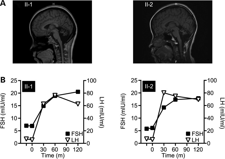
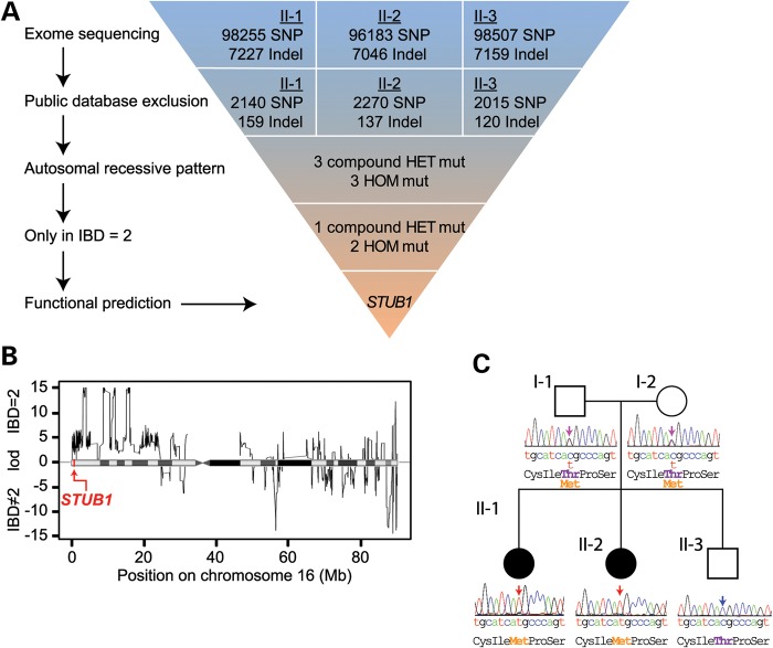
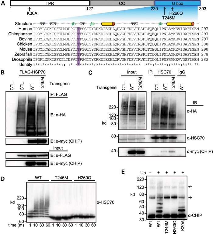
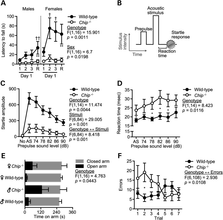
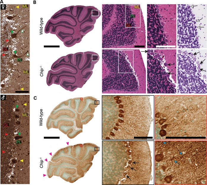
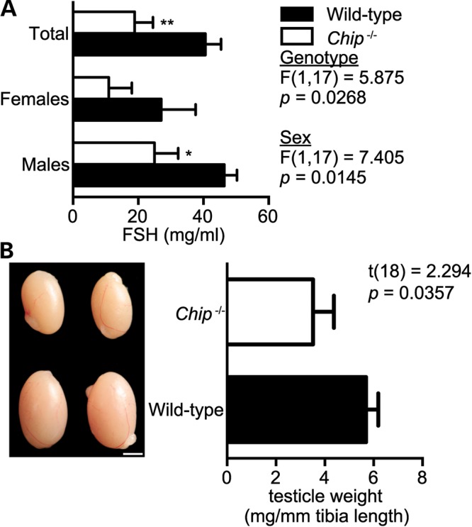
Gordon Holmes syndrome (GHS) is a rare Mendelian neurodegenerative disorder characterized by ataxia and hypogonadism. Recently, it was suggested that disordered ubiquitination underlies GHS though the discovery of exome mutations in the E3 ligase RNF216 and deubiquitinase OTUD4. We performed exome sequencing in a family with two of three siblings afflicted with ataxia and hypogonadism and identified a homozygous mutation in STUB1 (NM_005861) c.737C→T, p.Thr246Met, a gene that encodes the protein CHIP (C-terminus of HSC70-interacting protein). CHIP plays a central role in regulating protein quality control, in part through its ability to function as an E3 ligase. Loss of CHIP function has long been associated with protein misfolding and aggregation in several genetic mouse models of neurodegenerative disorders; however, a role for CHIP in human neurological disease has yet to be identified. Introduction of the Thr246Met mutation into CHIP results in a loss of ubiquitin ligase activity measured directly using recombinant proteins as well as in cell culture models. Loss of CHIP function in mice resulted in behavioral and reproductive impairments that mimic human ataxia and hypogonadism. We conclude that GHS can be caused by a loss-of-function mutation in CHIP. Our findings further highlight the role of disordered ubiquitination and protein quality control in the pathogenesis of neurodegenerative disease and demonstrate the utility of combining whole-exome sequencing with molecular analyses and animal models to define causal disease polymorphisms.


Figures

Figure 1.
Figure 1.
Clinical manifestations in patients presenting with ataxia and hypogonadism. (A) MRI scans revealed remarkable cerebellum atrophy of Patients II-1 (left) and II-2 (right). (B) GnRH stimulation tests measured the response in circulating FSH and LH serum levels to exogenous GnRH administration (at time = 0) in Patients II-1 (left) and II-2 (right).
Figure 2.
Figure 2.
Exome sequencing identifies a p.Thr246Met mutation in the GHS family. (A) Schematic representation of our exome data-filtering approach to identify mutations with recessive inheritance patterns in the family. (B) Posterior probabilities of IBD = 2 classification. The logarithmic ratio (LOD) of the posterior probabilities of being IBD = 2 versus IBD ≠ 2 are plotted for all classified variant positions on chromosome 16. A disease-causing mutation in the STUB1 gene was identified in an IBD = 2 region of high posterior probability, indicated by the red arrow. (C) A pedigree of the family indicating the unaffected (open symbols) and affected (filled symbols) members. Sanger sequencing confirmed the cosegregation of the c.737C→T resulting in p.Thr246Met mutation in STUB1 within the family.
Figure 3.
Figure 3.
The T246M substitution mutation in CHIP abolishes ubiquitin ligase activity. (A) CHIP comprises three functional domains: TPR, coiled-coil (CC) and U box. The arrows indicate the location and identity of point mutations used to measure the functional parameters of CHIP (top). The structural features of the U box include alpha helices (cylinders), beta strands (arrows), beta turns (TT) and alpha turns (TTT). Sequence alignment demonstrates the evolutionary conservation of the T246 in the U box domain of the CHIP protein across the indicated species. Conservation of residues are labeled as fully conserved (*), strongly similar (:) or non-similar (). (B) COS-7 cells were co-transfected with the indicated vectors (transgenes, CTL = pcDNA3, WT = pcDNA3-CHIP, T246M = pcDNA3-CHIP-T246M) in addition to HA-tagged ubiquitin. HSP70 was immunoprecipitated (IP) with FLAG beads and the resulting precipitants and inputs were immunoblotted (IB) with the indicated antibodies. (C) COS-7 cells were co-transfected with the indicated transgenes in addition to HA-tagged ubiquitin and immunoprecipitated with either an HSC70 antibody or rat IgG. The inputs and resulting precipitants (IP) were immunoblotted with the indicated antibodies. Approximate molecular weights in kDa are also provided. (D and E) Cell-free ubiquitination reactions containing recombinant HSC70 and the indicated CHIP proteins resolved via SDS–PAGE and immunoblotted for an antibody recognizing HSC70 (D) or CHIP (E). Ubiquitin (Ub) was excluded in Lane 1 (E) to demonstrate the autoubiquitination of CHIP, arrows indicate the incomplete reduction of CHIP oligomers.
Figure 4.
Figure 4.
Chip−/− mice have extreme ataxia and other selective motoric and cognitive impairments. (A) Latency to fall from an accelerating rotarod represented by the mean ± SEM for either Chip−/− or wild-type mice (n = 5 per genotype per gender). The first three trials were given on the first day of rotarod testing. Retest (R) indicates the highest latency across two trials given 48 h after the day one trials: **P < 0.01 comparing Chip−/− versus wild-type mice at the retest; P < 0.05 and ††P < 0.01 comparing the indicated time point with first trial within the genotype. (B) The acoustic startle response comprises a prepulse followed by an acoustic stimulus (AS, 120 dB). Both the reaction time to the AS and the magnitude of the response were measured. (C and D) Amplitude and reaction time of the startle response following AS are represented by the mean ± SEM for each genotype (n = 6 and 10 for Chip−/− and wild-type mice, respectively). Trials included no stimulus trials (No) and AS alone trials: P < 0.05 comparing Chip−/− versus wild-type mice across all stimulus conditions shown in (C) or as indicated by * in (D). (E and F) Time on the open and closed arms of an EPM (E) and the number of errors (incorrect holes explored) before finding the target hole on the Barnes maze (F) represented by the mean ± SEM for each genotype (n = 10): *P < 0.05 comparing Chip−/− versus wild-type mice.
Figure 5.
Figure 5.
CHIP expression in human cerebellum and the loss of Purkinje cells in Chip−/− mice. (A) Immunohistochemistry of CHIP expression in adult human cerebellum from a healthy female (♀) and male (♂) with the major regions of the cerebellum identified: molecular layer (ML), Purkinje cell layer (PL) and the granular layer (GL). The colored arrows highlight intense CHIP immunoreactivity throughout the cerebellum including increased reactivity in Purkinje cells, both in the cell body (downward arrows) and dendrites (upward arrows). Scale bar represents 100 μm. (B) Representative whole cerebellar sagittal sections from wild-type and Chip−/− cerebellums (left) with the major regions labeled at higher power (middle) as shown in (A) stained with either hematoxylin and eosin (left, middle) or cresyl violet (right). The open arrows identify normal Purkinje cells in wild-type mice, whereas the closed arrows identify the pyknotic nuclei in Purkinje cells in Chip−/− cerebellums. (C) Representative whole cerebellar sagittal sections from wild-type and Chip−/− cerebellums immunostained for calbindin (left). Magenta arrowheads (left) indicate regions with no calbindin immunoreactivity and the black and red boxes correspond to the higher power images (middle and right, respectively). The closed arrows identify the pyknotic nuclei present in Purkinje cells (middle) and the cyan arrows identify swollen dendrites in Chip−/− cerebellums. (B and C) Scale bars for whole cerebellum and higher power images are 1 mm and 100μm, respectively.
Figure 6.
Figure 6.
Hypogonadism in Chip−/− mice. (A) Serum levels of FSH in wild-type and Chip−/− mice represented by the mean ± SEM for each genotype (n = 10): *P < 0.05; **P < 0.01 comparing Chip−/− versus wild-type mice. (B) Representative pictographs of testes and testicle weights from wild-type and Chip−/− mice. Scale bar represents 20 mm. Weights are represented in mg of testicle per mm of tibia length to control for animal size and represented by the mean ± SEM for each genotype (n = 10 and 8, wild-type and Chip−/− mice, respectively).

Similar articles

Cited by

References

    1. Holmes G. A form of familial degeneration of the cerebellum. Brain. 1908;30:466–489.
    1. Margolin D.H., Kousi M., Chan Y.M., Lim E.T., Schmahmann J.D., Hadjivassiliou M., Hall J.E., Adam I., Dwyer A., Plummer L., et al. Ataxia, dementia, and hypogonadotropism caused by disordered ubiquitination. N. Engl. J. Med. 2013;368:1992–2003. - PMC - PubMed
    1. Ballinger C.A., Connell P., Wu Y., Hu Z., Thompson L.J., Yin L.Y., Patterson C. Identification of CHIP, a novel tetratricopeptide repeat-containing protein that interacts with heat shock proteins and negatively regulates chaperone functions. Mol. Cell. Biol. 1999;19:4535–4545. - PMC - PubMed
    1. Connell P., Ballinger C.A., Jiang J., Wu Y., Thompson L.J., Hohfeld J., Patterson C. The co-chaperone CHIP regulates protein triage decisions mediated by heat-shock proteins. Nat. Cell Biol. 2001;3:93–96. - PubMed
    1. Jiang J., Ballinger C.A., Wu Y., Dai Q., Cyr D.M., Hohfeld J., Patterson C. CHIP is a U-box-dependent E3 ubiquitin ligase: identification of Hsc70 as a target for ubiquitylation. J. Biol. Chem. 2001;276:42938–42944. - PubMed

Publication types

MeSH terms

Substances

Supplementary concepts