

Autophagy prevents irradiation injury and maintains stemness through decreasing ROS generation in mesenchymal stem cells
- PMID: 24113178
- PMCID: PMC3824648
- DOI: 10.1038/cddis.2013.338
Autophagy prevents irradiation injury and maintains stemness through decreasing ROS generation in mesenchymal stem cells
Abstract
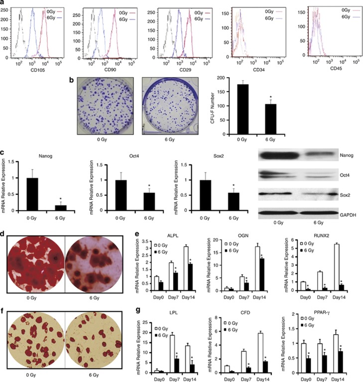
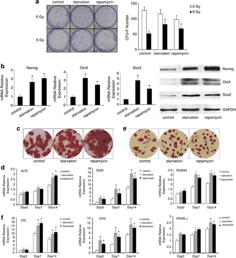
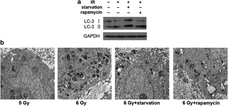
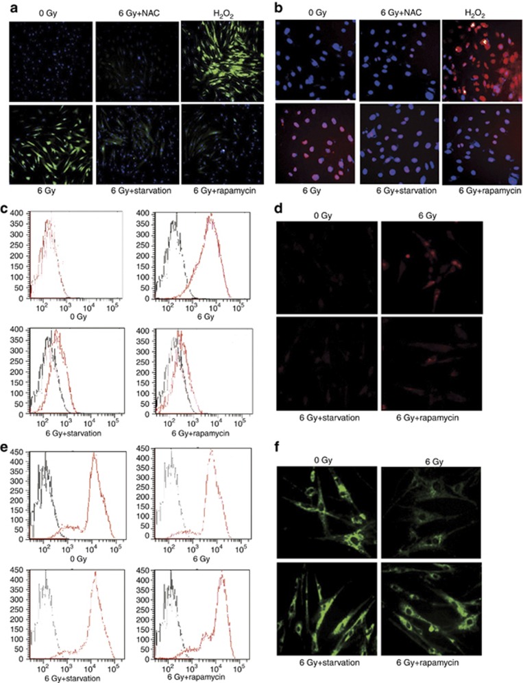
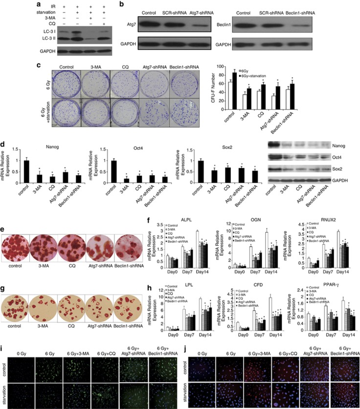
Stem cells were characterized by their stemness: self-renewal and pluripotency. Mesenchymal stem cells (MSCs) are a unique type of adult stem cells that have been proven to be involved in tissue repair, immunoloregulation and tumorigenesis. Irradiation is a well-known factor that leads to functional obstacle in stem cells. However, the mechanism of stemness maintenance in human MSCs exposed to irradiation remains unknown. We demonstrated that irradiation could induce reactive oxygen species (ROS) accumulation that resulted in DNA damage and stemness injury in MSCs. Autophagy induced by starvation or rapamycin can reduce ROS accumulation-associated DNA damage and maintain stemness in MSCs. Further, inhibition of autophagy leads to augment of ROS accumulation and DNA damage, which results in the loss of stemness in MSCs. Our results indicate that autophagy may have an important role in protecting stemness of MSCs from irradiation injury.
Figures
Similar articles
-
Autophagy Prevents Oxidative Stress-Induced Loss of Self-Renewal Capacity and Stemness in Human Tendon Stem Cells by Reducing ROS Accumulation.Cell Physiol Biochem. 2016;39(6):2227-2238. doi: 10.1159/000447916. Epub 2016 Nov 7. Cell Physiol Biochem. 2016. PMID: 27832632
-
Regulatory roles of miR-22/Redd1-mediated mitochondrial ROS and cellular autophagy in ionizing radiation-induced BMSC injury.Cell Death Dis. 2019 Mar 7;10(3):227. doi: 10.1038/s41419-019-1373-z. Cell Death Dis. 2019. PMID: 30846680 Free PMC article.
-
Rspo1-LGR4 axis in BMSCs protects bone against radiation-induced injury through the mTOR-dependent autophagy pathway.J Cell Physiol. 2021 Jun;236(6):4273-4289. doi: 10.1002/jcp.30051. Epub 2021 Jan 16. J Cell Physiol. 2021. PMID: 33452710
-
Modulating autophagy in mesenchymal stem cells effectively protects against hypoxia- or ischemia-induced injury.Stem Cell Res Ther. 2019 Apr 17;10(1):120. doi: 10.1186/s13287-019-1225-x. Stem Cell Res Ther. 2019. PMID: 30995935 Free PMC article. Review.
-
Regulation of the mitochondrial reactive oxygen species: Strategies to control mesenchymal stem cell fates ex vivo and in vivo.J Cell Mol Med. 2018 Nov;22(11):5196-5207. doi: 10.1111/jcmm.13835. Epub 2018 Aug 30. J Cell Mol Med. 2018. PMID: 30160351 Free PMC article. Review.
Cited by
-
Low dose radiation induced senescence of human mesenchymal stromal cells and impaired the autophagy process.Oncotarget. 2015 Apr 10;6(10):8155-66. doi: 10.18632/oncotarget.2692. Oncotarget. 2015. PMID: 25544750 Free PMC article.
-
Surface-Selective Preferential Production of Reactive Oxygen Species on Piezoelectric Ceramics for Bacterial Killing.ACS Appl Mater Interfaces. 2016 Sep 21;8(37):24306-9. doi: 10.1021/acsami.6b07440. Epub 2016 Sep 8. ACS Appl Mater Interfaces. 2016. PMID: 27599911 Free PMC article.
-
Radiation Induces Apoptosis and Osteogenic Impairment through miR-22-Mediated Intracellular Oxidative Stress in Bone Marrow Mesenchymal Stem Cells.Stem Cells Int. 2018 Aug 12;2018:5845402. doi: 10.1155/2018/5845402. eCollection 2018. Stem Cells Int. 2018. PMID: 30158985 Free PMC article.
-
Kynurenine inhibits autophagy and promotes senescence in aged bone marrow mesenchymal stem cells through the aryl hydrocarbon receptor pathway.Exp Gerontol. 2020 Feb;130:110805. doi: 10.1016/j.exger.2019.110805. Epub 2019 Dec 5. Exp Gerontol. 2020. PMID: 31812582 Free PMC article.
-
Radiation Responses of Human Mesenchymal Stem Cells Derived From Different Sources.Dose Response. 2019 Dec 9;17(4):1559325819893210. doi: 10.1177/1559325819893210. eCollection 2019 Oct-Dec. Dose Response. 2019. PMID: 31839760 Free PMC article.
References
-
- Prockop DJ. Marrow stromal cells as stem cells for nonhematopoietic tissues. Science. 1997;276:71–74. - PubMed
-
- Granero-Molto F, Weis JA, Longobardi L, Spagnoli A. Role of mesenchymal stem cells in regenerative medicine: application to bone and cartilage repair. Expert Opin Biol Ther. 2008;8:255–268. - PubMed
-
- Dezawa M, Ishikawa H, Itokazu Y, Yoshihara T, Hoshino M, Takeda S, et al. Bone marrow stromal cells generate muscle cells and repair muscle degeneration. Science. 2005;309:314–317. - PubMed
Publication types
MeSH terms
Substances
LinkOut - more resources
Full Text Sources
Other Literature Sources
