. 2013 Oct 10;4(10):e847.
doi: 10.1038/cddis.2013.375.

The AKT1/NF-kappaB/Notch1/PTEN axis has an important role in chemoresistance of gastric cancer cells

Affiliations

The AKT1/NF-kappaB/Notch1/PTEN axis has an important role in chemoresistance of gastric cancer cells

W Zhou et al. Cell Death Dis. .

Abstract

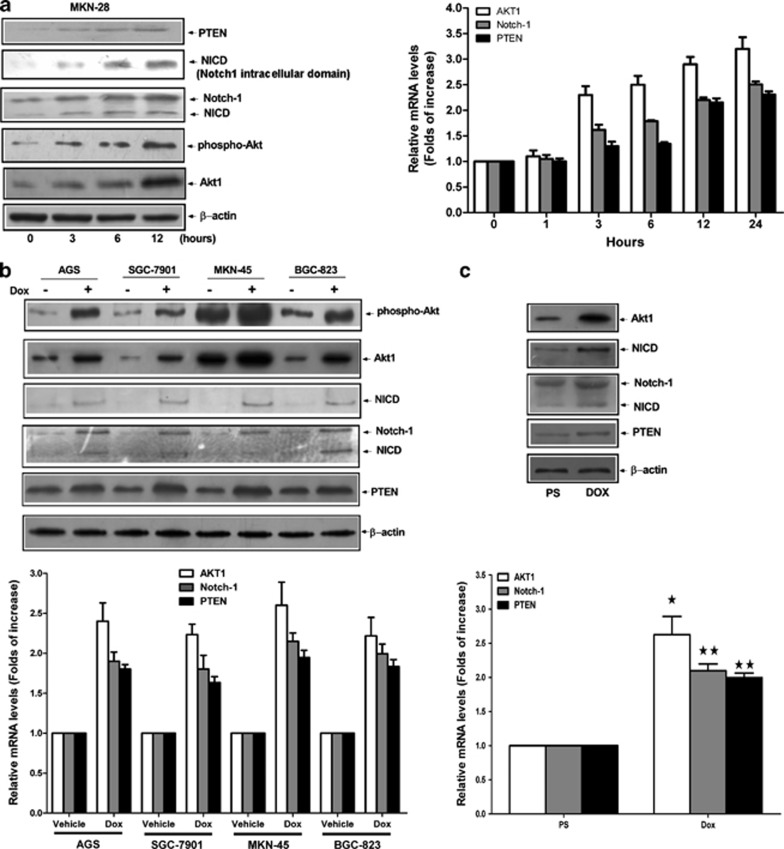
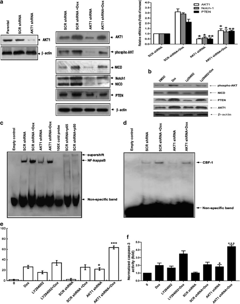
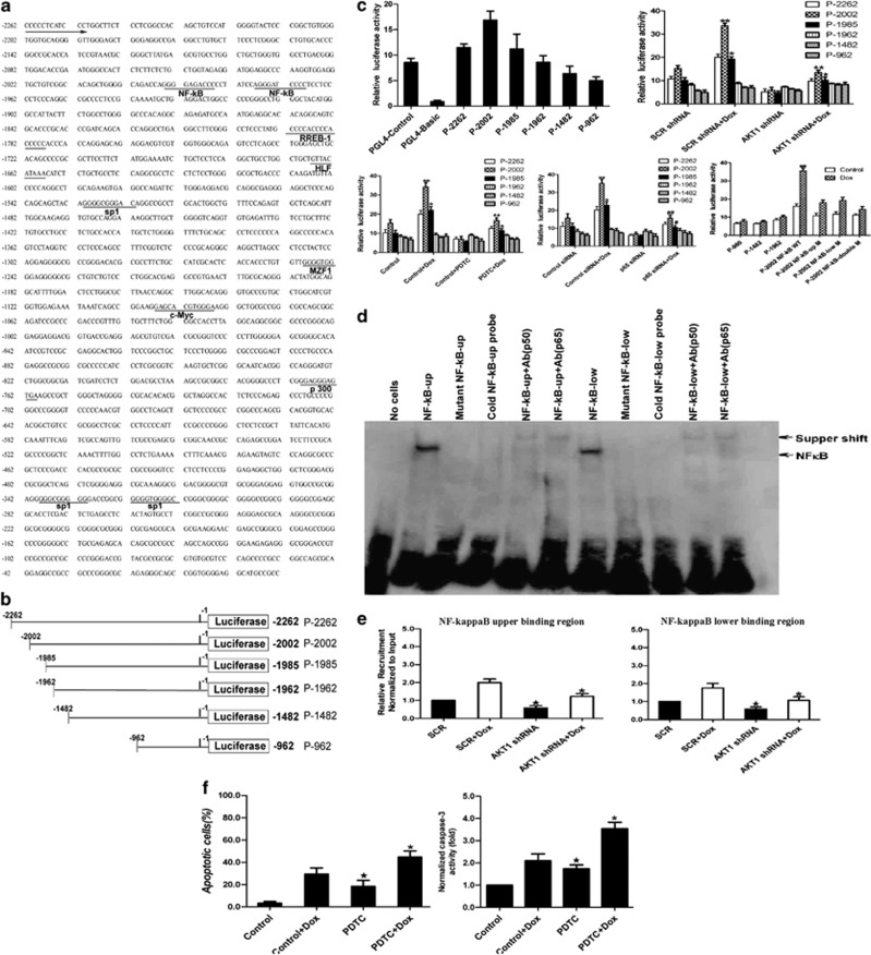
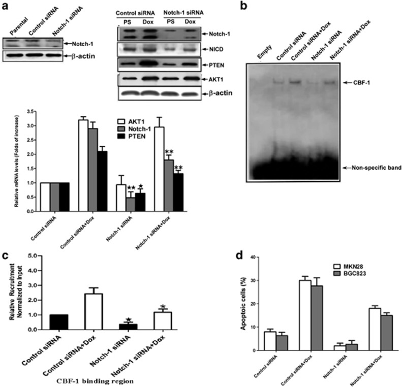
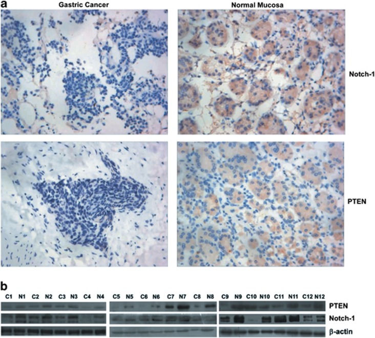
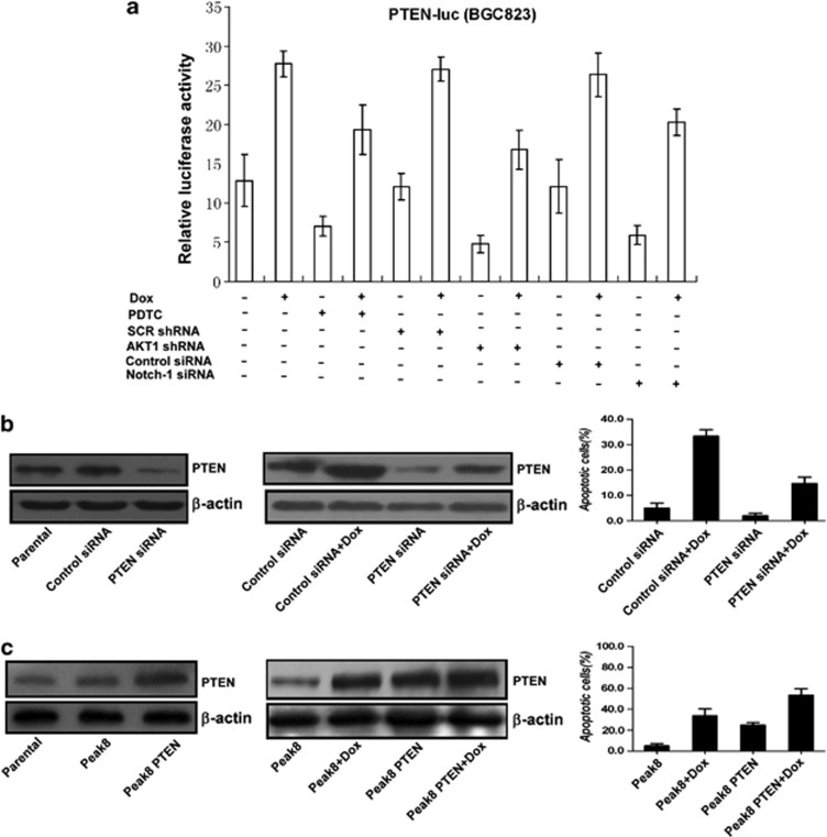
The inherent resistance of tumors to DNA damage often limits the efficacy of chemotherapy. The aim of this work is to explore the potential mechanism for development of chemoresistance in gastric cancer. Our data revealed that AKT1 mRNA and protein expression were induced by doxorubicin (a chemotherapeutic agent); the doxorubicin-induced AKT1 expression and activation increased the binding of NF-kappaB on Notch1 DNA promoter and then promoted the Notch1 transcription and expression; enhanced expression of Notch1 further upregulated PTEN expression through CBF-1 binding to PTEN DNA promoter; and inhibition of AKT1 expression and activity sensitized the gastric cancer cell to doxorubicin treatment in cultured gastric cancer cell lines and xenograft nude mice gastric cancer model. Furthermore, our data demonstrated that both Notch1 and PTEN were absent or minimally expressed in gastric cancer tissue but abundant in paired normal gastric mucosa, and the expression of Notch1 correlated with that of PTEN. Together, these novel results suggested that a novel AKT1/NF-kappaB/Notch1/PTEN axis has an important role in the development of chemoresistance in gastric cancer. Notch1 has an anti-cancer role in gastric cancer.


Figures

Figure 1
Figure 1
Elevated expression of AKT1, Notch-1, and PTEN in human gastric cancer cells after treatment of doxorubicin. (a) The MKN-28 cells were incubated with 3 μM doxorubicin for various time courses, and the protein and mRNA levels of AKT1, Notch1, and PTEN were determined by immunoblotting and real-time RT-PCR, respectively. Values are the mean±S.D. from three different experiments. (b) The protein and mRNA levels of AKT1, Notch1, and PTEN in the indicated human gastric cancer cell lines after 12 h treatment of doxorubicin (3 μM) were determined by immunoblotting and real-time RT-PCR, respectively. Values are the mean±S.D. from three different experiments. (c) Nude mice bearing MKN-28 xenograft tumor ( × 100 mm3) were intraperitoneally administered with physiological saline (PS) and 8 mg/kg doxorubicin, respectively, once for 4 days. All nude mice were killed at day 21 and the protein and mRNA levels of AKT1, Notch-1, and PTEN in xenograft tumors were determined by immunoblotting and real-time RT-PCR, respectively. Values are the mean±S.D. from three different experiments. Statistical significance is compared with PS group. P<0.05, ★★P<0.01. In all the above experiments, immunoblotting results of β-actin were used to show equal loading
Figure 2
Figure 2
AKT1 regulated Notch-1 and PTEN expression through NFκB and had an anti-apoptotic role in gastric cancer cells. (a) Lysate from MKN-28 cells (parental), MKN-28 cells expressing lentiviral scramble shRNA (SCR shRNA), or lentiviral AKT1 shRNA were analyzed by anti-AKT1 blotting. The MKN-28 cells expressing lentiviral SCR shRNA or lentiviral AKT1 shRNA were treated with PS or 3 μM doxorubicin for 12 h, respectively. The protein and mRNA levels of AKT1, Notch1 and PTEN were detected by immunoblotting and real-time RT-PCR. Immunoblotting results of β-actin were used to show equal loading. Values are the mean±S.D. from three different experiments. P<0.05, ★★P<0.01 versus SCR shRNA group; *P<0.05, **P<0.01 versus SCR shRNA+Dox group. (b) Effect of doxorunicin and LY294002 (10 μM) on expression of AKT1, p-AKT (473), NICD, and PTEN. Immunoblotting results of β-actin were used to show equal loading. (c) The MKN-28 cells expressing lentiviral SCR shRNA or lentiviral AKT1 shRNA were treated with PS or 3 μM doxorubicin for 12 h, respectively. The DNA binding capability of NFκB was detected by EMSA analysis. (d) The MKN-28 cells and MKN-28 cells expressing lentiviral SCR shRNA or lentiviral AKT1 shRNA were treated with PS or 3 μM doxorubicin for 12 h, respectively. The DNA binding capability of CBF-1 on PTEN minimal promoter was detected by EMSA analysis. (e) MKN-28 cells' preincubation with DMSO or 10 μM LY294002 and MKN-28 cells expressing lentiviral SCR shRNA or lentiviral AKT1 shRNA were treated with PS or 3 μM doxorubicin for 12 h, respectively. Cell apoptosis was determined by flow cytometry. P<0.05, versus SCR shRNA group. ***P<0.01 versus SCR shRNA+Dox group. (f) Analysis of relative caspase-3 activity in cells. Normalized caspase-3 activity. All experiments were performed in triplicate. P<0.05, versus SCR shRNA group. ***P<0.01 versus SCR shRNA+Dox group
Figure 3
Figure 3
p65/NFκB bound to the Notch1 promoter and regulated the transcription of Notch1 gene. (a) The human Notch1 promoter sequence. (b) The schematic diagram of constructs with different 5′ upstream deletions. (c) Luciferase activity of the deleted constructs in MKN-28 cells. After transfection of reporter gene, cells were treated by doxorubicin (3 μM, 12 h) with or without pretreatment with PDTC or infection of Akt1 shRNA lentivirus and p65 siRNA. All transfection experiments were repeated three times, and luciferase activity was normalized to the Renilla minimal reporter. Mean data±S.D. were results from three different experiments. *P<0.05 **P<0.01, cells with DOX treatment versus same cells without DOX treatment; P<0.05, ★★P<0.01, PDTC+DOX group or AKT1 shRNA+DOX group versus DOX group or SCR shRNA+DOX group; #P<0.05, ##P<0.01, p65 siRNA+DOX group versus Control siRNA+DOX group. (d) EMSA analysis for DNA binding activity of the upper and lower NF-κB sites (NF-κB up and NF-κB low) in the Notch1 promoter of MKN-28 cells. Excess cold consensus NF-κB probe and anti- NF-κB (p50 and p65) were added 10 min before binding reaction. Arrows indicate NF-κB specific binding or supershift bands. (e) Recruitment of NF-κB to the Notch1 promoter. Data were presented as the relative amount of immunoprecipitated DNA normalized to input as measured by quantitative real-time PCR. The data for SCR samples were arbitrarily set as 1. P<0.05, versus SCR shRNA group; *P<0.05, versus SCR shRNA+Dox group. (f) Effect of PDTC (50 μM) pretreatment (2 h) on doxorubicin (3 μM) induced apoptosis and caspase-3 activity. P<0.05, versus Control group; *P<0.05, versus Control+Dox group. All experiments were performed in triplicate
Figure 4
Figure 4
Notch1 regulated PTEN expression through CBF-1 and had a pro-apoptotic role in gastric cancer cells. (a) Lysate from MKN-28 cells (parental), MKN-28 cells transfected with control siRNA or Notch1 siRNA were analyzed by anti-Notch1 blotting. The MKN-28 cells transfected with control siRNA or Notch-1 siRNA were treated with PS or 3 μM doxorubicin for 12 h, respectively. The protein and mRNA levels of AKT1, Notch1 and PTEN were detected by immunoblotting and real-time RT-PCR. Immunoblotting results of β-actin were used to show equal loading. Values are the mean±S.D. from three different experiments. P<0.05, ★★P<0.01, versus Control siRNA group; **P<0.01 versus Control siRNA+Dox group. (b and c) The MKN-28 cells and MKN-28 cells transfected with control siRNA or Notch1 siRNA were treated with PS or 3 μM doxorubicin for 12 h, respectively. The DNA binding capability of CBF-1 on PTEN minimal promoter was detected by EMSA and ChIP analysis. P<0.05, versus Control siRNA group; *P<0.05 versus Control siRNA+Dox group. (d) BGC-823 cells and MKN-28 cells were transfected with control siRNA or Notch1 siRNA and then were treated with PS or 3 μM doxorubicin for 12 h, respectively. Cell apoptosis were determined by flow cytometry. Values are the mean±S.D. from experiments in triplicate
Figure 5
Figure 5
Lack of Notch1 and PTEN expression in gastric cancer tissues. (a) Immunohistochemical analysis of Notch1 and PTEN expression in gastric cancer and paired normal mucosa. ( × 400 magnification). (b) The expression of Notch1 and PTEN in representative paired tissues from gastric cancer and adjacent normal mucosa detected by immunoblotting. N, normal mucosa; C, cancer. Immunoblotting result of β-actin is used to show equal loading
Figure 6
Figure 6
Doxorubicin-induced PTEN expression had a pro-apoptotic function in gastric cancer cells. (a) PTEN transactivation was mediated by NFκB, AKT1, and Notch1. MKN-28 cells were transfected for 9 h with the PTEN-luc reporter construct. Some cells were transfected together with control or Notch1 siRNA. Twenty-four hours later, cells were either left or pretreated with 10 μM PDTC for 1 h, left untreated or treated with 3 μM doxorubicin for an additional 12-h period, after which the luciferase activity was determined. The MKN-28 cells expressing lentiviral SCR shRNA or lentiviral AKT1 shRNA were also transfected with PTEN-luc reporter construct followed by treatment with or without 3 μM doxorubicin, after which the luciferase activity was determined. (b) Knockdown of PTEN by siRNA inhibited the basal and doxorubicin-induced cell apoptosis in gastric cancer cells. (c) Upregulation of PTEN expression by wild-type PTEN plasmid transfection promoted cell apoptosis in gastric cancer cells

Similar articles

Cited by

References

    1. Basu B, Yap TA, Molife LR, de Bono JS. Targeting the DNA damage response in oncology: past, present and future perspectives. Curr Opin Oncol. 2012;24:316–324. - PubMed
    1. Koo DH, Lee JL, Kim TW, Chang HM, Ryu MH, Yook Jh, et al. Adjuvant chemotherapy with 5-fluorouracil, doxorubicin and mitomycin-C (FAM) for 6 months after curative resection of gastric carcinoma. Eur J Surg Oncol. 2007;33:843–848. - PubMed
    1. Kulke MH, Wu B, Clark JW, Enzinger PC, Lynch TJ, Vincitore M, et al. A phase II study of doxorubicin, cisplatin, and 5-fluorouracil in patients with advanced adenocarcinoma of the stomach or esophagus. Cancer Invest. 2006;24:229–234. - PubMed
    1. Galluzzi L, Larochette N, Zamzami N, Kroemer G. Mitochondria as therapeutic targets for cancer chemotherapy. Oncogene. 2006;25:4812–4830. - PubMed
    1. Fulda S, Debatin KM. Extrinsic versus intrinsic apoptosis pathways in anticancer chemotherapy. Oncogene. 2006;25:4798–4811. - PubMed

Publication types

MeSH terms