. 2013 Oct 10;4(10):e854.
doi: 10.1038/cddis.2013.370.

Down-modulation of nucleoporin RanBP2/Nup358 impaired chromosomal alignment and induced mitotic catastrophe

Affiliations

Down-modulation of nucleoporin RanBP2/Nup358 impaired chromosomal alignment and induced mitotic catastrophe

C Hashizume et al. Cell Death Dis. .

Abstract

Chromosomal missegregation is a common feature of many human tumors. Recent studies have indicated a link between nucleoporin RanBP2/Nup358 and chromosomal segregation during mitosis; however, the molecular details have yet to be fully established. Observed through live cell imaging and flow cytometry, here we show that RNA interference-mediated knockdown of RanBP2 induced G2/M phase arrest, metaphase catastrophe and mitotic cell death. Furthermore, RanBP2 down-modulation disrupted importin/karyopherin β1 as well as the expression and localization of the Ran GTPase activating protein 1. We found that N-terminal of RanBP2 interacted with the N-terminal of importin β1. Moreover, at least a portion of RanBP2 partially localizes at the centrosome during mitosis. Notably, we also found that GTPase Ran is also involved in the regulation of RanBP2-importin β1 interaction. Overall, our results suggest that mitotic arrest and the following cell death were caused by depletion of RanBP2. Our findings point to a crucial role for RanBP2 in proper mitotic progression and faithful chromosomal segregation.


Figures

Figure 1
Figure 1
RanBP2 is associated with importin β1 during mitosis. (a) Confocal microscopic images of HeLa cells at different mitotic stages, stained with anti-RanBP2 (green) and anti-α-tubulin (red) antibodies. Chromatin was stained with 4',6-diamidino-2-phenylindole (DAPI; blue). Bars=5 μm. In lanes marked ‘Input,' 20 μl of the 500 μl of extract used per IP were analyzed directly. (b) Asynchronized (upper panel) and synchronized mitotic (lower panel) HeLa cells (G2/M) were prepared via double thymidine block and confirmed by flow cytometry. (c) IP of mitotic HeLa cell extracts incubated with anti-RanBP2 or nonspecific rabbit antibodies (IgG) were analyzed by IB with anti-Imp-β1, anti-RanGAP1, anti-TopoIIα and anti-Lamin A/C antibodies. (d) IP of mitotic HeLa cell extracts incubated with anti-Imp-β1 or nonspecific rabbit antibodies (IgG) were analyzed by IB with the indicated antibodies. (e) Confocal microscopic images of HeLa cells at different mitotic stages, stained with anti-RanBP2 (green) and anti-Imp-β1 (red) antibodies. Chromatin was stained with DAPI (blue). Bars=5 μm
Figure 2
Figure 2
Importin β1 siRNA treatment reduces RanBP2 and causes abnormal chromosomal congression and defective mitosis. (a) Schedule of fixing or collecting mitotic HeLa cells after siRNA Imp-β1 depletion. (b) Lysates of mitotic HeLa cells transfected with control or Imp-β1 siRNAs were analyzed 72 h later by IB with anti-Imp-β1 antibody. The same membrane was stripped and reprobed with anti-β-actin and anti-α-tubulin (as loading control). (c) Effects of Imp-β1 depletion on the protein levels of RanBP2-associated proteins. The same membranes as in (b) were analyzed by IB with the indicated antibodies. RanBP2 was significantly reduced in the Imp-β1-depleted lysates, whereas TopoIIα, Pom121 and Lamin A/C were not affected. (d) Quantification (relative percentage) of mitotic chromosomal defect phenotypes for the indicated siRNA and/or plasmid. Values are based on three independent experiments, counting 100 mitotic cells in each experiment. Mean values±S.D. (error bars) are shown. All cells were treated with double thymidine block, stained with 4',6-diamidino-2-phenylindole (DAPI) and visualized by confocal microscopy. (e) Confocal microscopic images of mitotic HeLa cells transfected with control or Imp-β1 siRNA and analyzed 72 h post-transfection; the cells were stained with anti-RanBP2 (green) and anti-Imp-β1 (red) antibodies and DAPI (blue). (fh) Representative confocal microscopic images were stained with various antibodies as indicated. White boxes show the enlarged region. Bar=5 μm
Figure 3
Figure 3
RanBP2 siRNA treatment reduces importin β1 and causes abnormal chromosomal congression and defective mitosis. (a) Lysates of mitotic HeLa cells transfected with control or RanBP2 siRNAs were analyzed 72 h post transfection by IB with anti-RanBP2 antibody. The same membrane was stripped and reprobed with anti-β-actin (as a loading control). (b) Effects of RanBP2 depletion on the protein levels of RanBP2-associated proteins. The same membranes as in panel (a) were analyzed by IB with the indicated antibodies. (c and d) Confocal microscopic images of mitotic HeLa cells transfected with control or RanBP2 siRNAs. At 72 h post transfection, the cells were stained with RanBP2 (green) antibody and anti-RanGAP1 (red) or Imp-β1 (red) antibodies and 4',6-diamidino-2-phenylindole (DAPI; blue). Bar=5 μm. (e) Quantification (relative percentage) of mitotic chromosomal defect phenotypes in cells transfected with control siRNA or RanBP2 siRNA. Values are based on three independent experiments, counting 100 mitotic cells in each experiment. Mean values±S.D. (error bars) are shown. All cells were treated with double thymidine block, stained with DAPI and visualized by confocal microscopy
Figure 4
Figure 4
RanBP2 down-modulation induces G2/M phase arrest, metaphase catastrophe and cell death. (ac) Time-lapse microscopy of HeLa cells stably expressing histone H2B-GFP. At 72 h post transfection with (a) control or (b and c) RanBP2 siRNAs, the cells were processed for time-lapse microscopic analysis. Images were acquired every 3 min. Arrow indicates a fragmented chromosome. White arrowhead indicates an apoptotic bleb. (d) siRNAs against RanBP2 and control were transfected, respectively. Cell images were acquired using a phase-contrast microscope 24 or 48 h after siRNA transfection. White arrowhead indicates an apoptotic cell. (e) Cell death induced by knockdown of RanBP2. HeLa cells 48 h after siRNA transfection were stained with FITC-labeled Annexin V and PI. (f) Annexin V and PI-positive HeLa cells 48 h after siRNA transfection. HeLa cells randomly selected (n=300) were counted. Values are an average of three independent experiments. Error bars represent S.D. (g) At 48 h post transfection with control or RanBP2 siRNAs, HeLa cells were examined for their cell-cycle profiles by flow cytometry. (h) Percentage of cell-cycle phases in cells from panel (g). Asterisks indicates the significant P values (*P<0.05; **P<0.01)
Figure 5
Figure 5
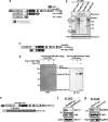
Ran binding is important for the interaction between the N-terminal of RanBP2 and importin β1. (a) Scheme of full-length RanBP2 and two C-terminal deletion mutants with GFP tags. Numbers on the right refer to amino acids. LRD, leucine-rich domain; Zinc, zinc finger motifs; E3, SUMO E3 ligase domain; CY, cyclophilin homology domain. (b) HeLa cells were transfected with a GFP, GFP-RanBP2-FL, RanBP2-N2 or RanBP2-N1 expression plasmid. After double thymidine block, lysates prepared from the cells were immunoprecipitated with anti-GFP antibody and then analyzed by IB with anti-Imp-β1 or anti-GFP antibodies. Asterisks indicate the GFP fusion proteins. Numbers indicate molecular mass markers in kilodaltons. HC, IgG heavy chain; LC, IgG light chain. In lanes marked ‘Input,' 20 μl of the 500 μl of extract used per IP were analyzed directly. (c) Scheme of full-length RanBP2 and the N- and C-terminal deletion mutant (M1) with the GST tag. Numbers on the right refer to amino acids. (d) In vitro expressed and purified GST-RanBP2-M1, Imp-β1-N2-His and His-Ran-GTP were mixed and pulled-down with glutathione-Sepharose beads. Pulled-down lysates were separated by SDS-PAGE and then either stained with Coomassie brilliant blue (CBB) or subjected to IB with anti-6 × His antibody. Numbers indicate molecular mass markers in kilodaltons. (e) Scheme of GFP-tagged full-length RanBP2 carrying the W1211R/K1212M mutation and FLAG-tagged Imp-β1-FL carrying the I178D/Y255A mutation. (f and g) HeLa cells were transfected with a GFP-RanBP2-FL (wt), GFP-RanBP2-FL_W1211R/K1212M (mt), FLAG-Imp-β1-FL (wt) or FLAG-Imp-β1-FL_ I178D/Y255A (mt) expression plasmid. After double thymidine block, lysates prepared from the cells were immunoprecipitated with anti-GFP or anti-FLAG antibody and then analyzed by IB with anti-Imp-β1, anti-GFP, anti-RanBP2 or anti-FLAG antibodies. In lanes marked ‘Input,' 20 μl of the 500 μl of extract used per IP were analyzed directly
Figure 6
Figure 6
N-terminal of importin β1 interacts with RanBP2. (a) Scheme of full-length Imp-β1 and four mutants with 6 × His tags. Numbers on the right refer to amino acids (left). An illustration of an interaction model between importin β1 and RanBP2 (right). (b) In vitro expressed and purified 6 × His tagged Imp-β1 fragments were mixed with mitotic HeLa lysates and pulled-down with Ni-NTA beads. Pulled-down lysates were analyzed by IB with anti-RanBP2 or anti-6 × His antibodies. Asterisks indicate the 6 × His fusion proteins. Numbers indicate molecular mass markers in kilodaltons. In lanes marked ‘Input,' 20 μl of the 500 μl of lysate used per pull-down were analyzed directly. (c) Quantification (relative percentage) of mitotic chromosomal defect phenotypes in FLAG-net-vector, FLAG-Imp-β1-FL, FLAG-Imp-β1-N1 and FLAG-Imp-β1-N2 transfected cells. Values are based on three independent experiments, counting 100 mitotic cells in each experiment. Mean values±S.D. (error bars) are shown. All cells were treated with double thymidine block, stained with DAPI (4',6-diamidino-2-phenylindole) and visualized by confocal microscopy
Figure 7
Figure 7
Model for RanBP2 in proper mitotic progression and faithful chromosomal segregation. Absence of RanBP2 during mitosis caused abnormal mitotic spindles, supernumerary centrosomes, impaired chromosomal alignment, induced mitotic catastrophe and cell death

References

    1. Nakano H, Wang W, Hashizume C, Funasaka T, Sato H, Wong RW. Unexpected role of nucleoporins in coordination of cell cycle progression. Cell Cycle. 2011;10:425–433. - PubMed
    1. Guttinger S, Laurell E, Kutay U. Orchestrating nuclear envelope disassembly and reassembly during mitosis. Nat Rev Mol Cell Biol. 2009;10:178–191. - PubMed
    1. Raices M, D'Angelo MA. Nuclear pore complex composition: a new regulator of tissue-specific and developmental functions. Nat Rev Mol Cell Biol. 2012;13:687–699. - PubMed
    1. Bukata L, Parker SL, D'Angelo MA. Nuclear pore complexes in the maintenance of genome integrity. Curr Opin Cell Biol. 2013;25:378–386. - PubMed
    1. Wong RW. Interaction between Rae1 and cohesin subunit SMC1 is required for proper spindle formation. Cell Cycle. 2010;9:198–200. - PubMed

Publication types

MeSH terms