

Role of hepatic progenitor cells in nonalcoholic fatty liver disease development: cellular cross-talks and molecular networks
- PMID: 24113587
- PMCID: PMC3821605
- DOI: 10.3390/ijms141020112
Role of hepatic progenitor cells in nonalcoholic fatty liver disease development: cellular cross-talks and molecular networks
Abstract
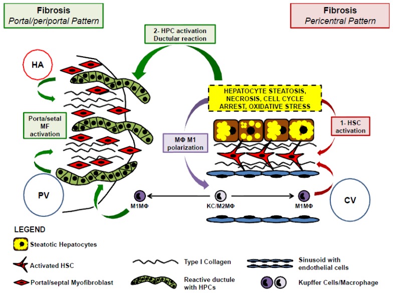
Nonalcoholic fatty liver disease (NAFLD) includes a spectrum of diseases ranging from simple fatty liver to nonalcoholic steatohepatitis, (NASH) which may progress to cirrhosis and hepatocellular carcinoma. NASH has been independently correlated with atherosclerosis progression and cardiovascular risk. NASH development is characterized by intricate interactions between resident and recruited cells that enable liver damage progression. The increasing general agreement is that the cross-talk between hepatocytes, hepatic stellate cells (HSCs) and macrophages in NAFLD has a main role in the derangement of lipid homeostasis, insulin resistance, danger recognition, immune tolerance response and fibrogenesis. Moreover, several evidences have suggested that hepatic stem/progenitor cell (HPCs) activation is a component of the adaptive response of the liver to oxidative stress in NAFLD. HPC activation determines the appearance of a ductular reaction. In NASH, ductular reaction is independently correlated with progressive portal fibrosis raising the possibility of a periportal fibrogenetic pathway for fibrogenesis that is parallel to the deposition of subsinusoidal collagen in zone 3 by HSCs. Recent evidences indicated that adipokines, a class of circulating factors, have a key role in the cross-talk among HSCs, HPCs and liver macrophages. This review will be focused on cellular cross-talk and the relative molecular networks which are at the base of NASH progression and fibrosis.
Figures

References
-
- Angulo P. Nonalcoholic fatty liver disease. N. Engl. J. Med. 2002;346:1221–1231. - PubMed
-
- Gaudio E., Nobili V., Franchitto A., Onori P., Carpino G. Nonalcoholic fatty liver disease and atherosclerosis. Intern. Emerg. Med. 2012;7:S297–S305. - PubMed
-
- Alisi A., Locatelli M., Nobili V. Nonalcoholic fatty liver disease in children. Curr. Opin. Clin. Nutr. Metab. Care. 2010;13:397–402. - PubMed
-
- Bieghs V., Rensen P.C., Hofker M.H., Shiri-Sverdlov R. NASH and atherosclerosis are two aspects of a shared disease: Central role for macrophages. Atherosclerosis. 2012;220:287–293. - PubMed
-
- Sookoian S., Gianotti T.F., Rosselli M.S., Burgueno A.L., Castano G.O., Pirola C.J. Liver transcriptional profile of atherosclerosis-related genes in human nonalcoholic fatty liver disease. Atherosclerosis. 2011;218:378–385. - PubMed
Publication types
MeSH terms
LinkOut - more resources
Full Text Sources
Other Literature Sources
Medical
