. 2013 Dec;126(6):809-27.
doi: 10.1007/s00401-013-1188-4. Epub 2013 Oct 11.

LRRK2 phosphorylates novel tau epitopes and promotes tauopathy

Affiliations

LRRK2 phosphorylates novel tau epitopes and promotes tauopathy

Rachel M Bailey et al. Acta Neuropathol. 2013 Dec.

Abstract

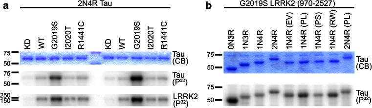
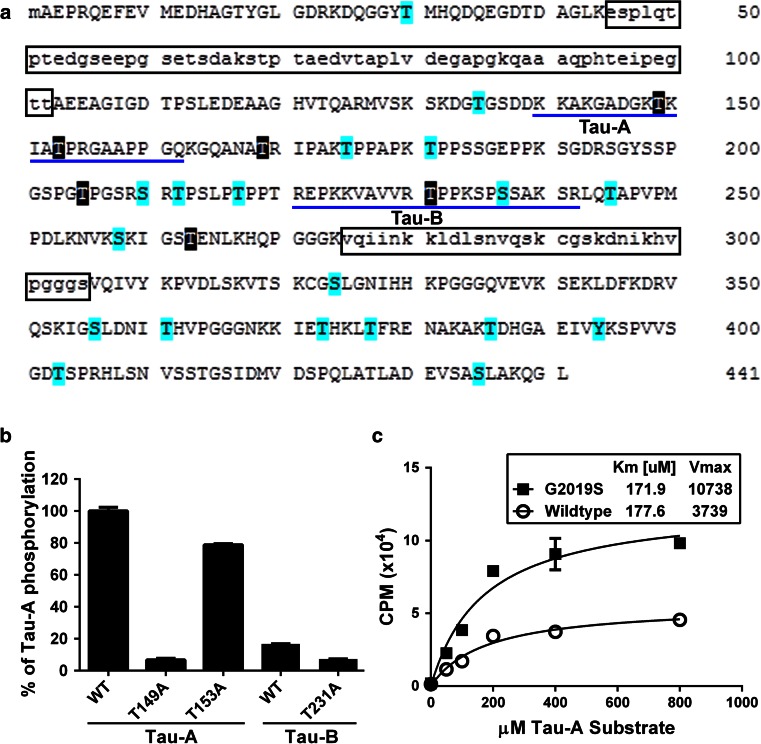
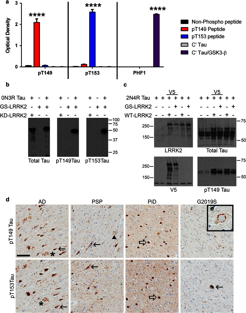
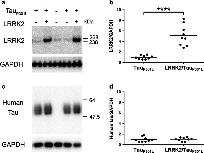
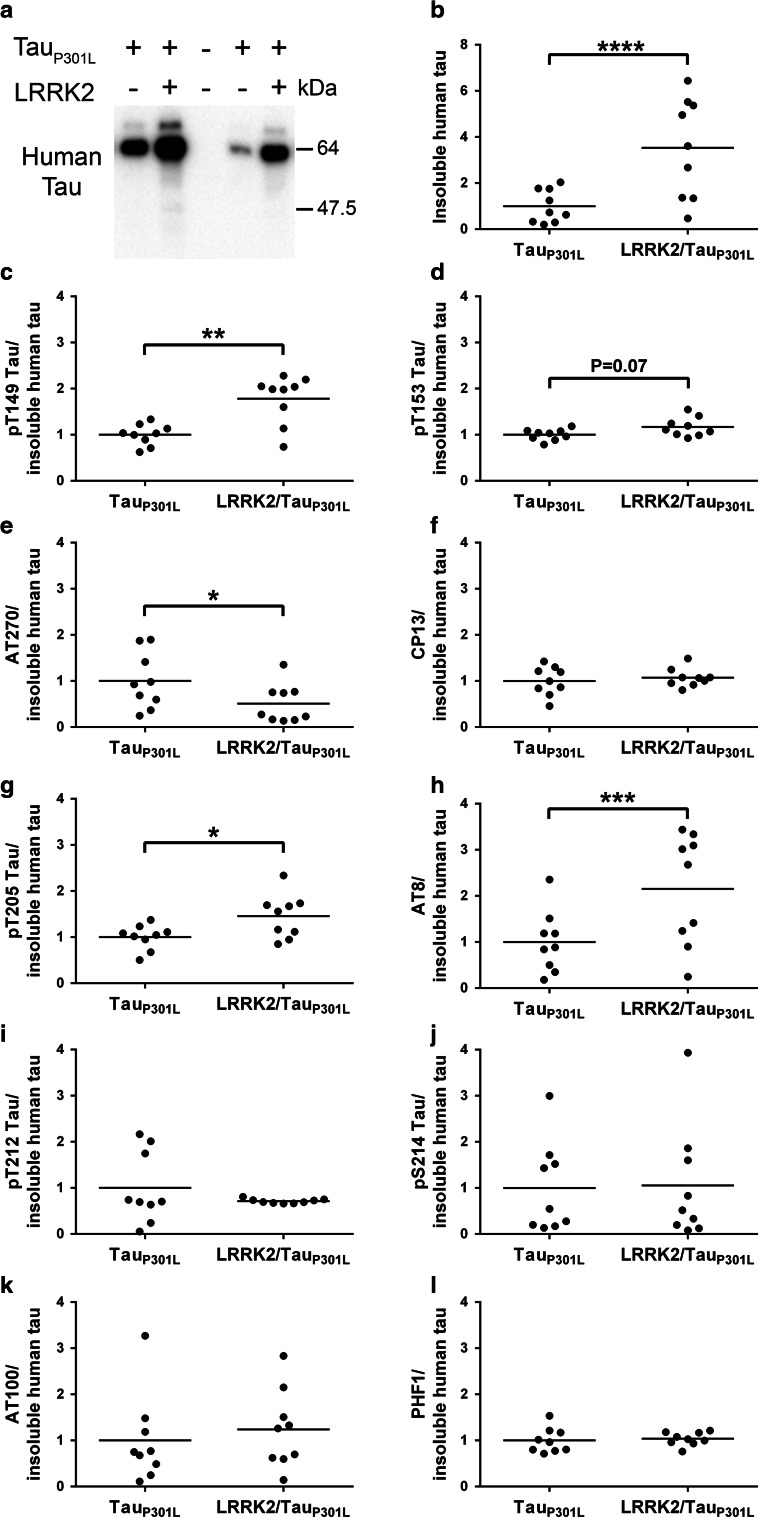
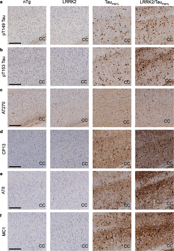
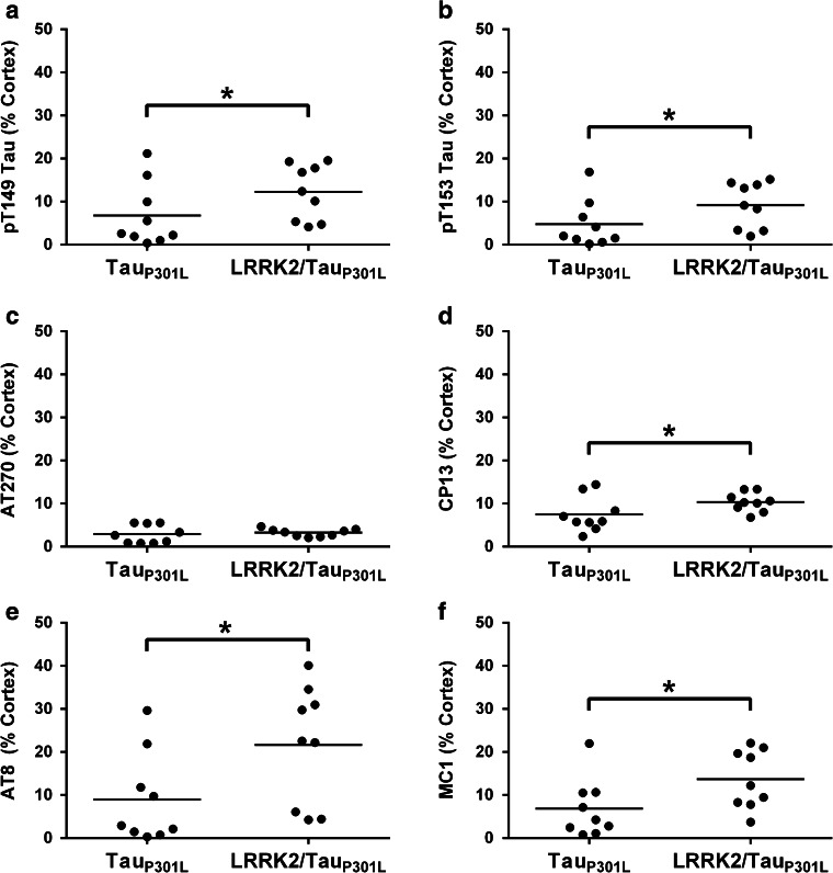
Mutations in the gene encoding leucine-rich repeat kinase 2 (LRRK2) are the most frequent cause of familial Parkinson's disease (PD). The neuropathology of LRRK2-related PD is heterogeneous and can include aberrant tau phosphorylation or neurofibrillary tau pathology. Recently, LRRK2 has been shown to phosphorylate tau in vitro; however, the major epitopes phosphorylated by LRRK2 and the physiological or pathogenic consequences of these modifications in vivo are unknown. Using mass spectrometry, we identified multiple sites on recombinant tau that are phosphorylated by LRRK2 in vitro, including pT149 and pT153, which are phospho-epitopes that to date have been largely unexplored. Importantly, we demonstrate that expression of transgenic LRRK2 in a mouse model of tauopathy increased the aggregation of insoluble tau and its phosphorylation at T149, T153, T205, and S199/S202/T205 epitopes. These findings indicate that tau can be a LRRK2 substrate and that this interaction can enhance salient features of human disease.


Figures

Fig. 1
Fig. 1
Wild-type and mutant LRRK2 phosphorylates tau in vitro. a Wild-type (WT) and mutant LRRK2 proteins directly phosphorylate 2N4R tau in vitro. Recombinant human tau (2N4R) is phosphorylated by recombinant WT, G2019S, I2020T, and R1441C human GST-LRRK2 (970–2,527), but not the kinase dead (KD) D1994K mutant as shown in duplicate by autoradiography (P32). Kinase activity of each LRRK2 construct was validated by LRRK2 auto-phosphorylation. b G2019S LRRK2 directly phosphorylates multiple tau isoforms with and without FTDP-17t mutations. Recombinant GST-G2019S LRRK2 (970–2,527) phosphorylates tau, regardless of tau isoform or mutation as shown by autoradiography (P32). In both a and b, Coomassie Blue (CB) shows equal loading of tau. Tau mutation abbreviations: EV E342V, PL P301L, PS P301S, RW R406W
Fig. 2
Fig. 2
LRRK2 phosphorylates multiple tau epitopes in vitro. a Highlighted amino acids indicate residues of 0N3R tau phosphorylated by GST-G2019S LRRK2 (970–2,527), but not by kinase dead GST-LRRK2 (970–2,527) as identified as putative sites by mass spectrometry. We obtained coverage of ~99 % of the 0N3R construct as indicated by capital letters. Major phosphorylation sites are highlighted in black. The entire amino acid sequence for the longest human tau isoform, 2N4R, is denoted and the tau domains, encoded by the alternatively spliced exons 2, 3, and 10 that were not included in the 0N3R tau construct are boxed. b We compared GST-G2019S LRRK2 (970–2,527) phosphorylation using synthetic peptides (Tau-A and Tau-B) as indicated by blue underlines in a. The degree of G2019S LRRK2-directed phosphorylation of WT Tau-A (T149, T153) and Tau-B (T231) peptides was compared to Tau-A and Tau-B with individual alanine mutants (T149A, T153A, T231A), which blocked phosphorylation at each residue, to determine the relative substrate potential of each epitope. c Kinetics of GST-WT and -G2019S LRRK2 (970–2,527) phosphorylation of Tau-A peptide. The concentration of Tau-A substrate was varied as indicated while the concentration of ATP was constant at 200 μM
Fig. 3
Fig. 3
Novel pT149 and pT153 tau antibodies recognize human tau pathology. a ELISA of phosphorylated and non-phosphorylated tau peptides with antibodies to pT149, pT153 and the PHF1 antibody, as a well-characterized control. The pT149 antibody reacted strongly with tau peptides phosphorylated at T149 while the pT153 antibody had the greatest interaction with tau phosphorylated at T153. Neither pT149 nor pT153 tau antibodies reacted with non-phosphorylated tau nor with the C-terminal half of 3R tau (C′ Tau) phosphorylated by GSK-3β. The pT153 antibody had low cross reactivity with tau phosphorylated at T149 as compared to non-phosphorylated C′ Tau, but not the other control peptides. ****P < 0.0001 [two-way ANOVA (antibody × peptide) with post hoc Bonferroni multiple comparisons test] (b) 0N3R tau was subjected to in vitro phosphorylation by either GST-G2019S (GS) or kinase dead (KD) LRRK2 (970–2,527) and analyzed via western blotting with 17025 (total tau), pT149 and pT153 antibodies. pT149 and pT153 antibodies recognize tau phosphorylated in vitro by G2019S LRRK2, but not tau incubated with the KD LRRK2. c HEK 293T cells were co-transfected with expression plasmids for 2N4R tau and pcDNA3.1, WT LRRK2-V5, GS LRRK2-V5, WT LRRK2 or GS LRRK2 and cell lysates were analyzed via western blot with LRRK2 (1182), V5, 17025 (total tau) and pT149 tau antibodies. Our novel pT149 antibody specifically detects tau phosphorylated by full-length WT and GS LRRK2 in cell culture. d In human tauopathies, pT149 (top panels) and pT153 (bottom panels) tau antibodies recognized neurofibrillary tangles (filled arrows) and neuritic tau pathology (asterisk) associated with plaques in Alzheimer’s disease (AD, left panels), neurofibrillary tangles (filled arrows) and tufted astrocytes (arrowhead) in progressive supranuclear palsy (PSP, middle-left panels), Pick bodies (open arrows) in Pick’s disease (PiD, middle-right panels), and recognized neurofibrillary tangles (filled arrow) in G2019S LRRK2 patients (G2019S, right panels). e The pT149 antibody stains tau pathology surrounding a Lewy body in the midbrain. Scale bar, 100 μm for panel d. Panel (e) is 50 μm × 50 μm
Fig. 4
Fig. 4
Tau expression in TauP301L transgenic mice is unaffected by addition of transgenic LRRK2 expression in TauP301L mice. The steady state amounts of LRRK2, human tau and GAPDH (loading control) were estimated by western blot analyses of the soluble fractions of whole brain lysates from TauP301L and LRRK2/TauP301L mice. a Representative western blot of LRRK2 and GAPDH with a non-transgenic littermate shown as a negative control. b Densitometric quantification of LRRK2 levels normalized to GAPDH. c Representative western blot of soluble human tau and GAPDH. d Densitometric quantification of human tau levels normalized to GAPDH. Each dot represents an individual mouse with the mean indicated by the black line, n = 9 per cohort. ****P < 0.0001 [two-way ANOVA (genotype × sex): main effect of genotype indicated]
Fig. 5
Fig. 5
LRRK2/TauP301L mice have elevated insoluble, hyperphosphorylated tau compared to TauP301L. Western blot analyses of the sarkosyl-insoluble fractions of whole brain lysates from TauP301L only and LRRK2/TauP301L mice. a Representative immunoblot probed with an antibody (E1) that recognizes human tau, regardless of phosphorylation state, shows significantly more aggregated (insoluble) human tau in LRRK2/TauP301L compared to TauP301L only mice as measured by (b) densitometry. Note the ~64 kDa species of tau (a) that we have previously shown to be a highly aggregated, hyperphosphorylated form of 0N4R tau [64]. cl Levels of insoluble tau phosphorylated at T149 (c), T153 (d), T181 (AT270) (e), S202 (CP13) (f), T205 (g), S199/S202/T205 (AT8) (h), T212 (i), S214 (j), T212/S214 (AT100) (k), and S396/S404 (PHF1) (l), as measured by densitometry (Supplemental Fig. 6), were adjusted by levels of human tau in the insoluble fraction (a-b) to distinguish specific phospho-epitopes that are enriched in the insoluble fraction. Phosphorylation of insoluble tau is increased at T149, T205 and S199/S202/T205 and decreased at T181 when normalized to the amount of tau in the sarkosyl-insoluble fraction. Each dot represents an individual mouse with the mean indicated by the black line, n = 9 per cohort. *P ≤ 0.05, **P < 0.01, ***P < 0.001, ****P < 0.0001 [two-way ANOVA (genotype × sex): main effect of genotype indicated]. Summary of two-way ANOVA found in Table 1
Fig. 6
Fig. 6
Cortical staining of phospho- and conformational-specific tau antibodies in female non-transgenic (nTg), LRRK2 only, TauP301L and LRRK2/TauP301L mice. af Representative images showing staining in the primary sensory cortex of female nTg, LRRK2, TauP301L and LRRK2/TauP301L mice with pT149 tau (a), pT153 tau (b), AT270 (pT181 tau) (c), CP13 (pS202) (d), AT8 (pS199/S202/T205 tau) (e), MC1 and (f) antibodies. Scale bar 200 μm. CC corpus callosum
Fig. 7
Fig. 7
LRRK2/TauP301L have elevated levels of cortical tau pathology compared to TauP301L mice. af There are specific increases of tau phosphorylation at epitopes T149, T153, S202 (CP13), S199/S202/T205 (AT8), as well as tau with an abnormal conformation (MC1) in LRRK2/TauP301L mice versus TauP301L mice. No difference in phosphorylation of T181 (AT270) was observed when LRRK2/TauP301L mice were compared to TauP301L mice. Each dot represents an individual mouse with the mean indicated by the black line, n = 9 per cohort. *P ≤ 0.05 [two-way ANOVA (genotype × sex): main effect of genotype indicated]. Summary of two-way ANOVA found in Table 1

References

    1. Anand VS, Reichling LJ, Lipinski K, Stochaj W, Duan W, Kelleher K, Pungaliya P, Brown EL, Reinhart PH, Somberg R, Hirst WD, Riddle SM, Braithwaite SP. Investigation of leucine-rich repeat kinase 2: enzymological properties and novel assays. FEBS J. 2009;276(2):466–478. doi: 10.1111/j.1742-4658.2008.06789.x. - DOI - PubMed
    1. Augustinack JC, Schneider A, Mandelkow EM, Hyman BT. Specific tau phosphorylation sites correlate with severity of neuronal cytopathology in Alzheimer’s disease. Acta Neuropathol. 2002;103(1):26–35. doi: 10.1007/s004010100423. - DOI - PubMed
    1. Biskup S, West AB. Zeroing in on LRRK2-linked pathogenic mechanisms in Parkinson’s disease. Biochim Biophys Acta. 2009;1792(7):625–633. doi: 10.1016/j.bbadis.2008.09.015. - DOI - PMC - PubMed
    1. Chen CY, Weng YH, Chien KY, Lin KJ, Yeh TH, Cheng YP, Lu CS, Wang HL. (G2019S) LRRK2 activates MKK4-JNK pathway and causes degeneration of SN dopaminergic neurons in a transgenic mouse model of PD. Cell Death Differ. 2012;19(10):1623–1633. doi: 10.1038/cdd.2012.42. - DOI - PMC - PubMed
    1. Chittum HS, Lane WS, Carlson BA, Roller PP, Lung FD, Lee BJ, Hatfield DL. Rabbit beta-globin is extended beyond its UGA stop codon by multiple suppressions and translational reading gaps. Biochemistry. 1998;37(31):10866–10870. doi: 10.1021/bi981042r. - DOI - PubMed

Publication types

MeSH terms

Substances