Review
. 2014 Mar;15(1):55-66.
doi: 10.1007/s11154-013-9276-2.

A proteolytic pathway that controls glucose uptake in fat and muscle

Affiliations
Review

A proteolytic pathway that controls glucose uptake in fat and muscle

Jonathan P Belman et al. Rev Endocr Metab Disord. 2014 Mar.

Abstract

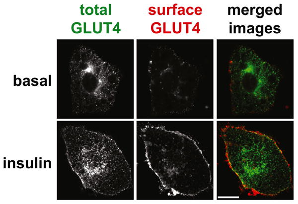
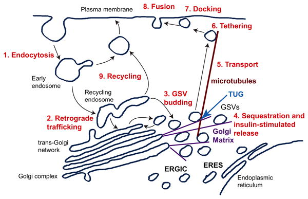
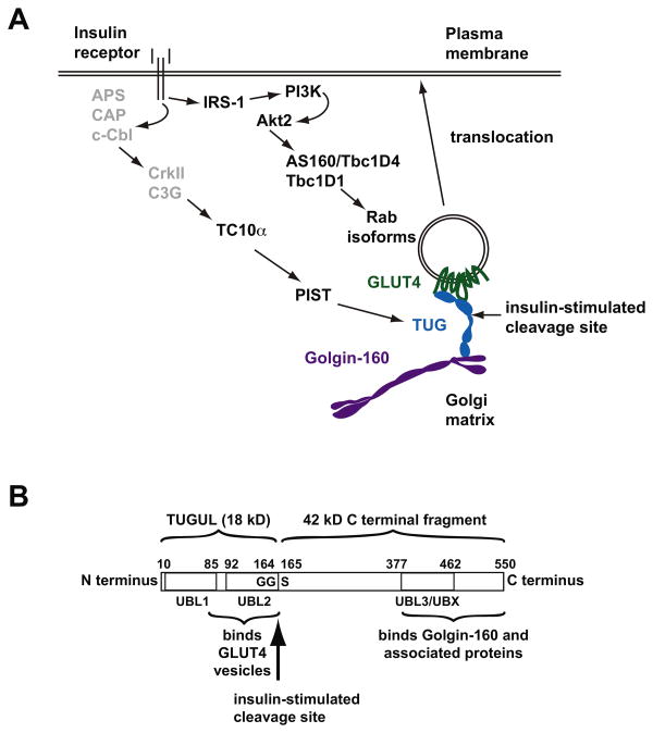
Insulin regulates glucose uptake by controlling the subcellular location of GLUT4 glucose transporters. GLUT4 is sequestered within fat and muscle cells during low-insulin states, and is translocated to the cell surface upon insulin stimulation. The TUG protein is a functional tether that sequesters GLUT4 at the Golgi matrix. To stimulate glucose uptake, insulin triggers TUG endoproteolytic cleavage. Cleavage accounts for a large proportion of the acute effect of insulin to mobilize GLUT4 to the cell surface. During ongoing insulin exposure, endocytosed GLUT4 recycles to the plasma membrane directly from endosomes, and bypasses a TUG-regulated trafficking step. Insulin acts through the TC10α GTPase and its effector protein, PIST, to stimulate TUG cleavage. This action is coordinated with insulin signals through AS160/Tbc1D4 and Tbc1D1 to modulate Rab GTPases, and with other signals to direct overall GLUT4 targeting. Data support the idea that the N-terminal TUG cleavage product, TUGUL, functions as a novel ubiquitin-like protein modifier to facilitate GLUT4 movement to the cell surface. The C-terminal TUG cleavage product is extracted from the Golgi matrix, which vacates an "anchoring" site to permit subsequent cycles of GLUT4 retention and release. Together, GLUT4 vesicle translocation and TUG cleavage may coordinate glucose uptake with physiologic effects of other proteins present in the GLUT4-containing vesicles, and with potential additional effects of the TUG C-terminal product. Understanding this TUG pathway for GLUT4 retention and release will shed light on the regulation of glucose uptake and the pathogenesis of type 2 diabetes.


Conflict of interest statement

Conflict of interest: The authors declare that they have no conflicts of interest relevant to the content of this review article.

Figures

Figure 1
Figure 1. Insulin stimulated GLUT4 glucose transporter translocation
The images show cultured 3T3-L1 adipocytes that express a GLUT4 reporter protein, which contains a 7Myc epitope tag in its first extracellular loop as well as GFP fused at the C-terminus. Cells were serum starved, treated with or without insulin as indicated, then stained to detect the externalized 7Myc epitope tag. Images were acquired by confocal microscopy of GFP (total GLUT4, shown in green in the merged images) and Myc epitope (surface GLUT4, shown in red in the merged images). Scale bar, 10 μM. Reproduced from Yu, C., et al., J Biol Chem (2007) 282, 7710-7722.
Figure 2
Figure 2. A model of GLUT4 trafficking pathways
GLUT4 undergoes endocytosis from the plasma membrane (1), and is targeted by retrograde trafficking to the recycling endosome and trans-Golgi network (2). Specialized, insulin-responsive GLUT4 Storage Vesicles (GSVs) likely bud from one or both of these locations (3), and become trapped at the Golgi matrix near the endoplasmic reticulum-Golgi intermediate compartment (ERGIC) and endoplasmic reticulum exit sites (ERES). The TUG protein is required for GSV sequestration at the Golgi matrix, and is cleaved upon insulin stimulation to mobilize the GSVs (4). The released GSVs are transported on microtubules to the cell periphery (5), and undergo insulin-stimulated tethering (6), docking (7), and fusion (8) at the plasma membrane. During the continued presence of insulin, endocytosed GLUT4 recycles directly to the plasma membrane from endosomes (9), and bypasses the TUG-regulated mechanism for GLUT4 sequestration and release. Adapted from Bogan, J.S. (2012) Annu Rev Biochem, 81, 507-532.
Figure 3
Figure 3. Insulin signaling and TUG endoproteolytic cleavage
A. Insulin signals through at least two pathways to mobilize GLUT4 to the plasma membrane. One pathway comprises IRS-1, phosphatidylinositol-3-kinase (PI3K), and Akt2, which phosphorylates and inactivates the Rab GTPase Activating Proteins AS160/Tbc1D1 and/or Tbc1D4 to modulate Rab GTPase isoforms and direct vesicle trafficking. A second pathway is less well studied, and is proposed to involve signaling through APS/CAP/c-Cbl to CrkII and the Rho-family GTP Exchange Factor C3G, which activates the TC10α GTPase. TC10α then signals through its effector, PIST, to trigger TUG endoproteolytic cleavage. Because conflicting data have been reported for the TC10α pathway, upstream components are shown in gray. Insulin-stimulated cleavage separates regions of TUG that bind the Golgi matrix, including Golgin-160 and possibly other proteins, from those that bind GSV proteins, including GLUT4 and likely other cargos. Cleavage thus liberates GLUT4 for exocytic translocation to the plasma membrane. B. The domain structure of the TUG protein is depicted, with residues numbered according to the sequence of the mouse protein. Tandem ubiquitin-like domains (UBL1 and UBL2) are present at the N-terminus, and interact with GLUT4 and possibly other proteins present in GLUT4 vesicles. A third ubiquitin-like region, specifically a UBX domain, is also indicated (UBL3/UBX), and C-terminal regions of TUG bind to Golgin-160 and associated proteins at the Golgi matrix. Insulin stimulates cleavage of the bond linking residues Gly164 and Ser165. This cleavage site follows a Gly-Gly sequence, which is typical of ubiquitin-like precursor proteins. Proteolysis produces TUGUL, an 18 kD N-terminal product thought to function as a ubiquitin-like protein modifier, as well as a 42 kD C-terminal product. Cleavage separates regions of TUG that bind GSVs from regions that bind the Golgi matrix, and thus can liberate GSVs that are sequestered intracellularly at the Golgi matrix. Adapted from Bogan, J.S. et al., (2012) J Biol Chem 287, 23932-23947.

References

    1. Cushman SW, Wardzala LJ. Potential mechanism of insulin action on glucose transport in the isolated rat adipose cell. Apparent translocation of intracellular transport systems to the plasma membrane. J Biol Chem. 1980;255(10):4758–4762. - PubMed
    1. Suzuki K, Kono T. Evidence that insulin causes translocation of glucose transport activity to the plasma membrane from an intracellular storage site. Proc Natl Acad Sci U S A. 1980;77(5):2542–2545. - PMC - PubMed
    1. Mueckler M, Caruso C, Baldwin SA, Panico M, Blench I, Morris HR, et al. Sequence and structure of a human glucose transporter. Science. 1985;229(4717):941–945. - PubMed
    1. Birnbaum MJ. Identification of a novel gene encoding an insulin-responsive glucose transporter protein. Cell. 1989;57(2):305–315. - PubMed
    1. James DE, Strube M, Mueckler M. Molecular cloning and characterization of an insulin-regulatable glucose transporter. Nature. 1989;338(6210):83–87. - PubMed

Publication types

LinkOut - more resources