Review
. 2014 Feb;16(1):43-65.
doi: 10.1007/s40272-013-0052-2.

Drug treatment of pulmonary hypertension in children

Affiliations
Review

Drug treatment of pulmonary hypertension in children

Erika E Vorhies et al. Paediatr Drugs. 2014 Feb.

Abstract

Pulmonary arterial hypertension (PAH) is a rare disease in infants and children that is associated with significant morbidity and mortality. The disease is characterized by progressive pulmonary vascular functional and structural changes resulting in increased pulmonary vascular resistance and eventual right heart failure and death. In the majority of pediatric patients, PAH is idiopathic or associated with congenital heart disease and rarely is associated with other conditions such as connective tissue or thromboembolic disease. Although treatment of the underlying disease and reversal of advanced structural changes has not yet been achieved with current therapy, quality of life and survival have been improved significantly. Targeted pulmonary vasodilator therapies, including endothelin receptor antagonists, prostacyclin analogs, and phosphodiesterase type 5 inhibitors, have demonstrated hemodynamic and functional improvement in children. The management of pediatric PAH remains challenging, as treatment decisions continue to depend largely on results from evidence-based adult studies and the clinical experience of pediatric experts. This article reviews the current drug therapies and their use in the management of PAH in children.


Figures

Fig. 1
Fig. 1
Pulmonary Vascular Research Institute Classification of Pediatric Pulmonary Hypertensive Vascular Disease (Panama 2011) (From Del Cerro MJ, et al. Pulm Circ. 2011, with permission)
Fig. 2
Fig. 2
Annual incidence rates for pediatric pulmonary hypertension based on epidemiological data from the Netherlands during a 15-year period. PH, pulmonary hypertension; PAH, pulmonary arterial hypertension; PAH-CHD, PAH associated with congenital heart disease; iPAH, idiopathic PAH. (From Van Loon RL, et al. Circulation. 2011, with permission)
Fig. 3
Fig. 3
Survival curves for idiopathic pulmonary arterial hypertension (IPAH) and associated pulmonary arterial hypertension (APAH). Cases were censored for time in the study and transplantation. (From Haworth SG, et al. Heart. 2009, with permission)
Fig. 4
Fig. 4
Survival curves for the subgroups within the APAH group. Shown is the number in each group (brackets), and the predicted survival out of a possible 5 years. APAH, associated pulmonary arterial hypertension; CT, controls. (From Haworth SG, et al. Heart. 2009, with permission)
Fig. 5
Fig. 5
Schematic diagram of endothelial vascular biology depicting the relevant vasoactive mediators that have led to targeted treatment of pulmonary hypertension, including the nitric oxide-cGMP system, the endothelin system and the prostacyclin system. (Reproduced with permission from Diller GP, et al. IJCP. 2010.)
Fig. 6
Fig. 6
A diagnostic algorithm for investigating pulmonary hypertension. CXR, chest x ray; PH, pulmonary hypertension; V/Q, ventilation/perfusion. (From Haworth SG, et al. Arch Dis Child. 2008, with permission)
Fig. 7
Fig. 7
Percentage of pediatric patients with pulmonary hypertension that demonstrated acute vasodilatory response to the evaluated agents. The greatest number of acute responders was seen with the use of iNO and oxygen. iNO, inhaled nitric oxide; 02, oxygen. (Reproduced with permission from Barst RJ, et al. Pediatr Cardiol. 2010.)
Fig. 8
Fig. 8
Treatment allocation in the STARTS-1 trial. Patients were randomized to placebo, low, medium or high dose oral sildenafil. (From Barst RJ, et al. Circulation. 2012, with permission)
Fig. 9
Fig. 9
Percent change in peak oxygen consumption (V02) from baseline to week 16 of sildenafil monotherapy in treatment-naïve children, aged 1–17 years, with pulmonary arterial hypertension (STARTS-1). Given the small number of developmentally able children in the 8- to 20-kg group, this group was combined with the 20- to 45-kg group. Greatest improvement in V02 was demonstrated with medium and high doses of sildenafil. (From Barst RJ, et al. Circulation. 2012, with permission)
Fig. 10
Fig. 10
Changes in systolic pulmonary artery pressures (sPAP) (A) and pulmonary/systemic systolic artery pressure (sPAP/ssBP) (B) as determined by echocardiogram in response to prolonged sildenafil therapy in infants with chronic lung disease. Median duration of treatment between studies was 58 days (range: 25 – 334). Individual data plotted together with mean ± SD. (Reproduced with permission from Mourani PM, et al. J Peds. 2009.)
Fig. 11
Fig. 11
Change in hemodynamic measures after transition from sildenafil to tadalafil for 14 pediatric patients with PAH. Hemodynamic data, including mPAP, PVRi and Rp/Rs, improved in comparison to the last catheterization data on sildenafil therapy during follow-up period (23.5 ± 8.3 months). mPAP, mean pulmonary arterial pressure; PVRi, pulmonary vascular resistance index; Rp/Rs (From Takatsuki S, et al. Pediatr Cardiol. 2012, with permission)
Fig. 12
Fig. 12
Change in indexed pulmonary vascular resistance (PVRi) from baseline to week 16 in placebo and bosentan groups (BREATHE-5). PVRi, the secondary endpoint, was significantly improved with bosentan therapy demonstrating a treatment effect of −472 (SE 221.9) dyn.sec.cm(−5). TE, treatment effect. (Reproduced with permission from Galie N, et al. Circulation. 2006)
Fig. 13
Fig. 13
Change in 6 minute walk distance (6MWD) from baseline to week 16 in placebo and bosentan groups (BREATHE-5). Exercise capacity measured by 6MWD was significantly improved with bosentan therapy demonstrating a treatment effect of 53.1 (SE 19.2) m. TE, treatment effect. (Reproduced with permission from Galie N, et al. Circulation. 2006)
Fig. 14
Fig. 14
Pharmacokinetics in FUTURE -1. Arithmetic mean (±SD) plasma concentration vs. time profiles of bosentan in patients with pediatric pulmonary arterial hypertension after multiple dose administration of bosentan at a dose of 2 and 4mg/kg twice daily. (n = 11). 2mg kg-1 (■); 4mg kg-1 (□)(Reproduced with permission from Beghetti M, et al. Br J Clin Pharmacol. 2009.)
Fig. 15
Fig. 15
Kaplan-Meier survival curve for a cohort of pediatric PAH patients receiving prostacyclin therapy, comprising patients on epoprostenol, treprostinil, and those who transitioned, with 95% confidence intervals (CI) depicted. Transplant-free 5-year survival was 70% (95% CI, 56% –80%). (Reproduced with permission from Siehr SL, et al. J Heart Lung Transplant. 2013.)
Fig. 16
Fig. 16
Change in pulmonary-to-systemic vascular resistance ratio (Rp/Rs) over time in pediatric PAH patients receiving intravenous epoprostenol or treprostinil. Initial improvement was seen in Rp/Rs at 1 to 2 years on therapy that was not sustained long-term. (Reproduced with permission from Siehr SL, et al. J Heart Lung Transplant. 2013.)
Fig. 17
Fig. 17
Hemodynamic change in the mean pulmonary artery pressure and the pulmonary vascular resistance index during acute pulmonary vasodilator testing with inhaled nitric oxide and inhaled treprostinil in children with PAH. Inhaled nitric oxide and inhaled treprostinil significantly decreased the mean pulmonary artery pressure and the pulmonary vascular resistance index. iNO, inhaled nitric oxide; NS, not significant. (From Takatsuki S, et al. Pediatr Cardiol. 2013, with permission)
Fig. 18
Fig. 18
Change in pulmonary-to-systemic vascular resistance ratio (Rp/Rs) in children with congenital heart disease and pulmonary hypertension in response to inhaled nitric oxide (iNO) and aerosolized iloprost. (Reproduced with permission from Rimensberger P, et al. Circulation 2001.)
Fig. 19
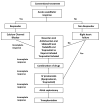
Fig. 19
Treatment algorithm in children with severe pulmonary arterial hypertension. (From Tissot C, et al. J Pediatr 2010, with permission)

References

    1. Rubin LJ. Primary pulmonary hypertension. N Engl J Med. 1997;336(2):111–7. - PubMed
    1. Allen KM, Haworth SG. Cytoskeletal features of immature pulmonary vascular smooth muscle cells: the influence of pulmonary hypertension on normal development. J Pathol. 1989 Aug;158(4):311–7. - PubMed
    1. Humbert M, Morrell NW, Archer SL, Stenmark KR, MacLean MR, Lang IM, et al. Cellular and molecular pathobiology of pulmonary arterial hypertension. J Am Coll Cardiol. 2004 Jun 16;43(12 Suppl S):13S–24S. - PubMed
    1. Mandegar M, Fung YC, Huang W, Remillard CV, Rubin LJ, Yuan JX. Cellular and molecular mechanisms of pulmonary vascular remodeling: role in the development of pulmonary hypertension. Microvasc Res. 2004 Sep;68(2):75–103. - PubMed
    1. Hassoun PM, Mouthon L, Barbera JA, Eddahibi S, Flores SC, Grimminger F, et al. Inflammation, growth factors, and pulmonary vascular remodeling. J Am Coll Cardiol. 2009 Jun 30;54(1 Suppl):S10–9. - PubMed

Publication types

MeSH terms