. 2013 Nov 22;288(47):33642-33653.
doi: 10.1074/jbc.M113.485490. Epub 2013 Oct 10.

Poxviral protein A52 stimulates p38 mitogen-activated protein kinase (MAPK) activation by causing tumor necrosis factor receptor-associated factor 6 (TRAF6) self-association leading to transforming growth factor β-activated kinase 1 (TAK1) recruitment

Affiliations

Poxviral protein A52 stimulates p38 mitogen-activated protein kinase (MAPK) activation by causing tumor necrosis factor receptor-associated factor 6 (TRAF6) self-association leading to transforming growth factor β-activated kinase 1 (TAK1) recruitment

Julianne Stack et al. J Biol Chem. .

Abstract

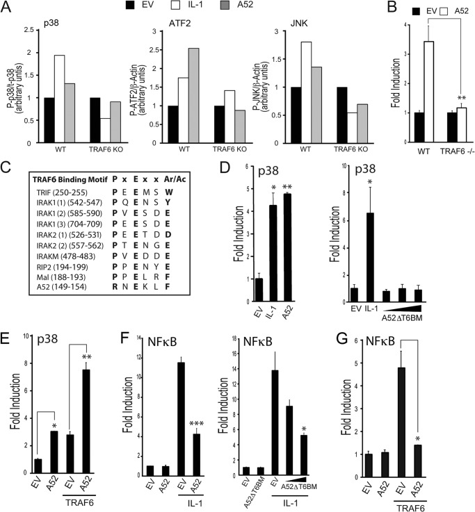
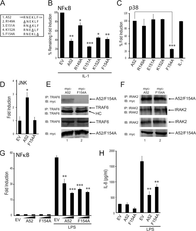
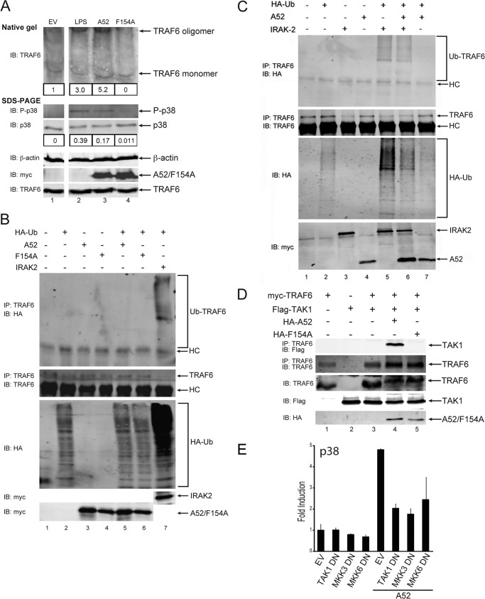
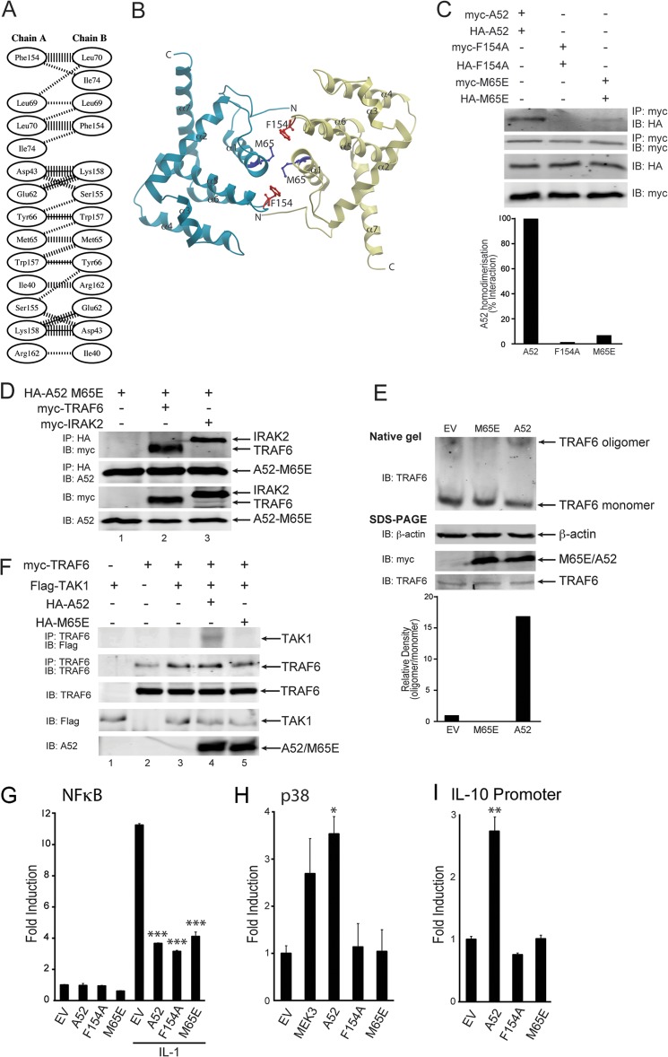
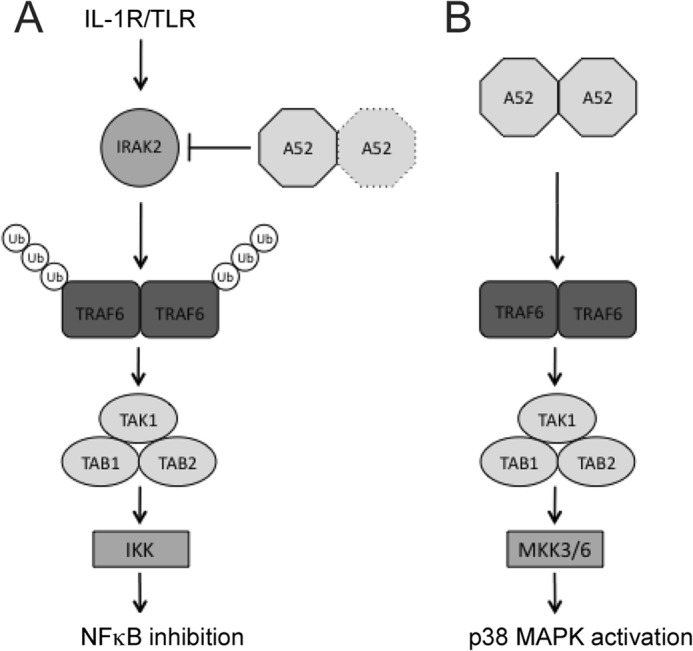
Vaccinia virus encodes a number of proteins that inhibit and manipulate innate immune signaling pathways that also have a role in virulence. These include A52, a protein shown to inhibit IL-1- and Toll-like receptor-stimulated NFκB activation, via interaction with interleukin-1 receptor-associated kinase 2 (IRAK2). Interestingly, A52 was also found to activate p38 MAPK and thus enhance Toll-like receptor-dependent IL-10 induction, which was TRAF6-dependent, but the manner in which A52 manipulates TRAF6 to stimulate p38 activation was unclear. Here, we show that A52 has a non-canonical TRAF6-binding motif that is essential for TRAF6 binding and p38 activation but dispensable for NFκB inhibition and IRAK2 interaction. Wild-type A52, but not a mutant defective in p38 activation and TRAF6 binding (F154A), caused TRAF6 oligomerization and subsequent TRAF6-TAK1 association. The crystal structure of A52 shows that it adopts a Bcl2-like fold and exists as a dimer in solution. Residue Met-65 was identified as being located in the A52 dimer interface, and consistent with that, A52-M65E was impaired in its ability to dimerize. A52-M65E although capable of interacting with TRAF6, was unable to cause either TRAF6 self-association, induce the TRAF6-TAK1 association, or activate p38 MAPK. The results suggest that an A52 dimer causes TRAF6 self-association, leading to TAK1 recruitment and p38 activation. This reveals a molecular mechanism whereby poxviruses manipulate TRAF6 to activate MAPKs (which can be proviral) without stimulating antiviral NFκB activation.

Keywords: NF-kappa B (NF-KB); Pox Viruses; Signal Transduction; TRAF6; Viral Protein; p38 MAPK.


Figures

FIGURE 1.
FIGURE 1.
A52 has a non-canonical TRAF6-binding domain required for p38 activation. A, MEFs from wild-type and TRAF6 knock-out mice were seeded into six-well plates (4 × 10⁵ cells) 24 h before transfection with 2 μg of empty vector or Myc-A52. Where indicated, cells were stimulated with 50 ng/ml IL-1α for 30 min 24 h after transfections. Lysates were analyzed by SDS-PAGE, and immunoblotting was performed. Immunoblots were subjected to densitometric analysis with levels of P-ATF2 and P-JNK normalized to β-actin and levels of p-p38 normalized to p38. B, wild-type or TRAF6−/− MEFs were transfected with 150 ng of Myc-A52 or pCMV-Myc empty vector (EV), along with the pFR luciferase reporter gene and CHOP-Gal4. Luciferase activity was measured 24 h after transfection. C, presence of PxExx(Ar/Ac) motifs in TRAF6-binding proteins compared with A52. D–G, HEK293-R1 cells were transfected for 24 h with either the pFR luciferase reporter gene and CHOP-Gal4 (D and E) or the NFκB luciferase reporter gene (F and G). Cells were co-transfected with either 150 ng of Myc-A52, A52ΔT6BM, or pCMV-Myc (EV) (D and F) or 100 ng of Myc-A52 with 50 ng of FLAG-TRAF6 (E and G). Cells were stimulated with 50 ng/ml IL-1α for 6 h where indicated, and luciferase reporter gene activity was measured. The data are mean ± S.D. of triplicate samples and are representative of at least three separate experiments. *, p < 0.05; **, p < 0.005; or ***, p < 0.0005 compared with EV alone.
FIGURE 2.
FIGURE 2.
A52 F154A cannot activate p38 MAPK activation nor bind TRAF6. A, four point mutations were made in the putative TRAF6-binding motif of A52. HEK293-R1 cells were transfected for 24 h with 150 ng of Myc-A52 or empty vector (EV), along with either the NFκB luciferase reporter gene (B) or the pFR luciferase reporter gene and CHOP-Gal4 (C). Cells were stimulated with 50 ng/ml IL-1α for 6 h, and luciferase reporter gene activity was measured. The data are presented as percentage remaining of fold induction (B) or percentage of fold induction (C) and are representative of at least three experiments. D, HEK293-R1 cells were transfected with the pFR luciferase reporter gene and c-Jun-Gal4 with either 150 ng of Myc-A52, A52-F154A, or pCMV-Myc (EV). Luciferase reporter gene activity was measured 24 h after transfection. E and F, HEK293T cells were transfected with Myc-A52 and Myc-A52 F154A. After 48 h, lysates were subject to immunoprecipitation, SDS-PAGE, and immunoblotting (IB) with the indicated antibodies. HC, heavy chain of anti-TRAF6 antibody. G, HEK293-TLR4 cells were transfected for 24 h with increasing amounts of Myc-A52 and Myc-A52-F154A, or empty vector (EV), along with the NFκB luciferase reporter gene. Cells were stimulated with 100 ng/ml LPS for 6 h, and luciferase reporter gene activity was measured. H, HEK293-TLR4 cells were transfected with Myc-A52, Myc-A52-F154A, or pCMV-Myc (EV). 24 h later, cells were stimulated with 50 ng/ml LPS for a further 24 h, after which the supernatants were assessed for IL-8 production by ELISA. The data are mean ± S.D. of triplicate samples and are representative of at least three separate experiments. *, p < 0.05; **, p < 0.005; or ***, p < 0.0005 compared with EV (B, D, G, and H) or A52 (C).
FIGURE 3.
FIGURE 3.
A52, but not A52-F154A, enhances TRAF6 self-association and causes TRAF6-TAK1 association. A, HEK293-TLR4 cells were transfected with empty vector (EV), Myc-A52, or Myc-A52-F154A. Cells were stimulated with 100 ng/ml LPS for 30 min 48 h after transfection. Lysates were analyzed by either native gel electrophoresis or SDS-PAGE. Immunoblotting was performed using standard conditions. Immunoblots (IB) were subjected to densitometric analysis with levels of the TRAF6 oligomer and phospho-p38 normalized to total levels of TRAF6 monomer and p38, respectively. B and C, HEK293T cells were transfected for 24 h with 4 μg of A52, A52-F154A, IRAK2, or HA-ubiquitin (Ub) as indicated. Endogenous TRAF6 was immunoprecipitated from cell lysates, and levels of ubiquitinated TRAF6 were assessed by immunoblotting with anti-HA antibody. HC, heavy chain of anti-TRAF6 antibody. D, HEK293T cells were transfected with Myc-TRAF6, FLAG-TAK1, HA-A52, and HA-A52 F154A. After 48 h, lysates were subject to immunoprecipitation, SDS-PAGE, and immunoblotting with the indicated antibodies. E, HEK293-Ts were transfected with 50 ng of Myc-A52 or pCMV-Myc (EV) and 100 ng of either TAK1 DN, MKK3 DN, or MKK6 DN, along with the pFR luciferase reporter gene and CHOP-Gal4. Luciferase reporter gene activity was measured 24 h after transfection. The data are mean ± S.D. of triplicate samples and are representative of at least three separate experiments.
FIGURE 4.
FIGURE 4.
The dimerization mutant, A52-M65E, can still interact with TRAF6 but cannot cause TRAF6-TAK1 association or TRAF6 self-association. A, schematic diagram of residue interactions across the A52 dimer interface generated by PDBsum (48). Parallel lines represent non-bonded contacts, and solid lines represent hydrogen bonds. For non-bonded contacts, the width of the parallel line is proportional to the number of atomic contacts. B, dimer interface of A52 homodimer generated using ICM Molsoft browser. The Phe-154 and Met-65 residues are represented as red and blue stick models, respectively. The dimer interface comprises of the N terminus (N), α1, and α6 of the Bcl-2 fold domain. C, HEK293T cells were transfected with 4 μg each of Myc-A52 and HA-A52 (lane 1), Myc-A52 F154A, and HA-A52 F154A (lane 2), or Myc-A52 M65E, and HA-A52 M65E (lane 3). After 48 h, lysates were subject to immunoprecipitation, SDS-PAGE, and immunoblotting (IB) with the indicated antibodies. Immunoblots were subjected to densitometric analysis with levels of co-immunoprecipitated HA-A52, HA-A52 F154A, and HA-A52 M65E normalized to total levels of immunoprecipitated Myc-A52, Myc-A52 F154A, and Myc-A52 M65E, respectively. D, HEK293T cells were transfected with 4 μg each of HA-A52 M65E, Myc-TRAF6, and Myc-IRAK2. After 48 h, lysates were subject to immunoprecipitation, SDS-PAGE, and immunoblotting with the indicated antibodies. E, HEK293T cells were seeded into 10-cm dishes (1.5 × 10⁶ cells) 24 h before transfection with 4 μg each of empty vector, Myc-A52, or Myc-A52-M65E. After 48 h, lysates were analyzed by either native gel electrophoresis or SDS-PAGE. Immunoblots were subjected to densitometric analysis with levels of TRAF6 oligomer normalized to total levels of TRAF6 monomer. F, HEK293T cells were transfected with the indicated amounts of Myc-TRAF6, FLAG-TAK1, HA-A52, and HA-A52 M65E. After 48 h, lysates were subject to immunoprecipitation, SDS-PAGE, and immunoblotting with the indicated antibodies. G–I, HEK293-R1 cells were transfected for 24 h with 150 ng of Myc-A52, Myc-A52 F154A, Myc-A52 M65E, or pCMV-Myc empty vector (EV), along with either the NFκB luciferase reporter gene (G), the pFR luciferase reporter gene and CHOP-Gal4 (H) or the IL-10 promoter luciferase reporter gene (I). MEK3 is the positive control for the PathdetectTM CHOP assay (H). Cells were stimulated with 50 ng/ml IL-1α for 6 h (G), and luciferase reporter gene activity was measured. The data are mean ± S.D. of triplicate samples and are representative of at least three separate experiments. *, p < 0.05; **, p < 0.005; or ***, p < 0.0005 compared with empty vector.
FIGURE 5.
FIGURE 5.
Model for manipulation of signaling by A52. A, upon TLR activation, IRAK2 associates with TRAF6. The interaction of IRAK2 with TRAF6 triggers the polyubiquitination of TRAF6 (19), which allows the subsequent recruitment of a complex containing TAB2, TAB1, and TAK1. This activates the kinase activity of TAK1 leading to the phosphorylation of IKKβ, culminating in the induction of NFκB. A52 inhibits IL-1R/TLR-dependent NFκB activation by interacting with IRAK2; its dimerization ability is not required for this function. B, as a dimer, A52 brings TRAF6 molecules together in the absence of TRAF6 polyubiquitination, inducing formation of the TRAF6-TAK1 complex. Activated TAK1 can then activate downstream kinases such as MKK3/6, leading to p38 phosphorylation. Ub, ubiquitin.

References

    1. Bowie A. G., Unterholzner L. (2008) Viral evasion and subversion of pattern-recognition receptor signalling. Nat. Rev. Immunol. 8, 911–922 - PMC - PubMed
    1. Stack J., Haga I. R., Schröder M., Bartlett N. W., Maloney G., Reading P. C., Fitzgerald K. A., Smith G. L., Bowie A. G. (2005) Vaccinia virus protein A46R targets multiple Toll-like-interleukin-1 receptor adaptors and contributes to virulence. J. Exp. Med. 201, 1007–1018 - PMC - PubMed
    1. Harte M. T., Haga I. R., Maloney G., Gray P., Reading P. C., Bartlett N. W., Smith G. L., Bowie A., O'Neill L. A. (2003) The poxvirus protein A52R targets Toll-like receptor signaling complexes to suppress host defense. J. Exp. Med. 197, 343–351 - PMC - PubMed
    1. DiPerna G., Stack J., Bowie A. G., Boyd A., Kotwal G., Zhang Z., Arvikar S., Latz E., Fitzgerald K. A., Marshall W. L. (2004) Poxvirus protein N1L targets the I-κB kinase complex, inhibits signaling to NF-κB by the tumor necrosis factor superfamily of receptors, and inhibits NF-κB and IRF3 signaling by toll-like receptors. J. Biol. Chem. 279, 36570–36578 - PubMed
    1. Chen R. A., Ryzhakov G., Cooray S., Randow F., Smith G. L. (2008) Inhibition of IκB kinase by vaccinia virus virulence factor B14. PLoS Pathog. 4, e22. - PMC - PubMed

Publication types

MeSH terms

Substances

LinkOut - more resources