Review
. 2013 Dec;22(12):1698-710.
doi: 10.1002/pro.2374. Epub 2013 Oct 19.

Molecular basis of MAP kinase regulation

Affiliations
Review

Molecular basis of MAP kinase regulation

Wolfgang Peti et al. Protein Sci. 2013 Dec.

Abstract

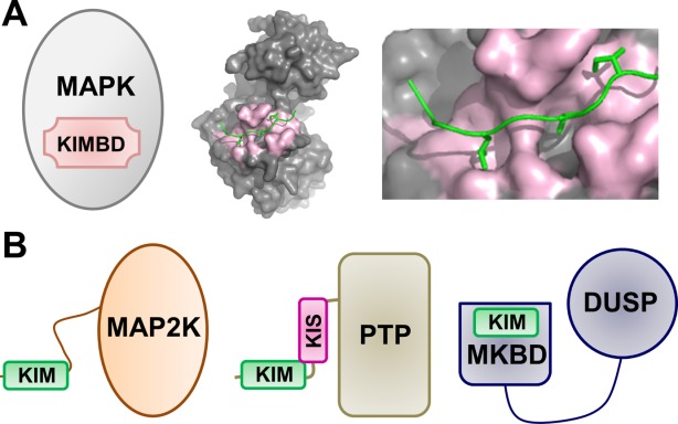
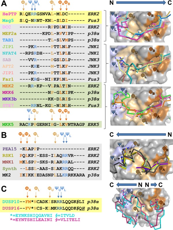
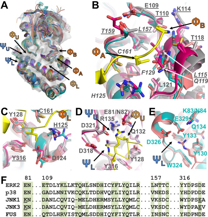
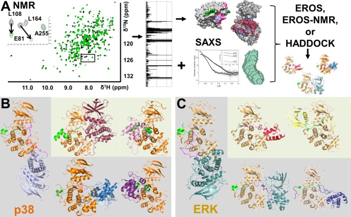
Mitogen-activated protein kinases (MAPKs; ERK1/2, p38, JNK, and ERK5) have evolved to transduce environmental and developmental signals (growth factors, stress) into adaptive and programmed responses (differentiation, inflammation, apoptosis). Almost 20 years ago, it was discovered that MAPKs contain a docking site in the C-terminal lobe that binds a conserved 13-16 amino acid sequence known as the D- or KIM-motif (kinase interaction motif). Recent crystal structures of MAPK:KIM-peptide complexes are leading to a precise understanding of how KIM sequences contribute to MAPK selectivity. In addition, new crystal and especially NMR studies are revealing how residues outside the canonical KIM motif interact with specific MAPKs and contribute further to MAPK selectivity and signaling pathway fidelity. In this review, we focus on these recent studies, with an emphasis on the use of NMR spectroscopy, isothermal titration calorimetry and small angle X-ray scattering to investigate these processes.

Keywords: D-motif; DUSP; ERK; JNK; KIM; MAP kinase; NMR; PTP; SAXS; kinase interaction motif; p38; structure.


Figures

Figure 1
Figure 1
MAPKs and MAPK regulatory proteins. (A) Cartoon of the MAPK KIM binding domain (KIMBD); p38 bound to the MEF2A KIM peptide (PDBID 1LEW). (B) Cartoons of MAPK regulatory proteins, including MAP2Ks (left), KIM-PTPs (middle) and DUSPs (right).
Figure 2
Figure 2
Structure-based sequence alignment of KIM peptides bound to MAPKs. (A) Left, structure-based sequence alignment of KIM peptides that bind the MAPK KIM binding pocket in the N→C direction; phosphatases on a yellow background, substrates/scaffolds on a grey background and kinases on a green background. Key interaction residues are colored. Positively charged amino acids that bind the electrostatic pocket are labeled ΨU and ΨL and colored blue; hydrophobic amino acids that bind the ΦA-X-ΦB binding groove are labeled ΦA and ΦB and colored orange; hydrophobic amino acids that bind the ΦL (also known as ΦA-2) and the ΦU (also known as ΦH) pockets are labeled and colored beige. The identities of the MAPKs present in the MAPK:KIMpeptide/protein complex structures are indicated to the right in italics. Right, MAPK:KIMpeptide/protein complexes superimposed using residues that optimally align the MAPK KIM docking grooves (Table II). MAPK surface is in grey, with the electrostatic binding pockets in light blue, the ΦA-X-ΦB binding groove in orange and the ΦL and ΦU binding pockets in beige. The bound KIM residues are shown as ribbons with the key interacting residues (ΨU, ΨL, ΦU, ΦL, ΦA, ΦB) shown as sticks. The ribbons are colored according to the MAPK interacting protein listed in A. B. Same as A, except KIM peptides bind in the C→N direction. C. Same as A, except bound KIM is part of a structured domain, the MAPK binding domain, and binds predominantly in the C→N direction. DUSP16 makes an additional interaction in a pocket below ΦL, which is indicated by H.
Figure 3
Figure 3
KIM binding pockets. (A) MAPKs listed in Table I aligned using the C-terminal cores described in Table II and displayed as cartoons; ERK2 shown as a transparent grey surface. Locations of KIM binding pockets labeled. (B) ΦA and ΦB binding pockets. ERK2 (grey; PDBID 3TEI), p38 (bright pink; PDBID 2Y8O), JNK1 (cyan, PDBID 2XRW), JNK3 (coral, PDBID 4H39), FUS3 (light pink, PDBID 2B9H); RSK1 KIM peptide in yellow (PDBID 3TEI). MAPK residues shown as sticks with ERK2 residues labeled; ΦA pocket residues underlined, ΦB pocket residues in italics. C. ΦL pocket, colored as in B. Residues that are also part of other KIM binding pockets underlined. D. ΦL, ΨU, ΨL pockets of ERK2, p38 and FUS3; colored as in B, labeled as in C. E. ΨL pocket of JNK1 and JNK3; colored as in B, labeled as in C, except JNK1 residues labeled. F. Sequence alignment of the MAPKs; residues that make up the KIM binding pockets highlighted in green (ERK2 residue numbers above alignment).
Figure 4
Figure 4
Interactions Outside the MAPK KIM Binding Groove. (A) Schematic illustration showing how NMR spectroscopy, using chemical shift perturbation (CSP) experiments, coupled with small angle X-ray scattering (SAXS) can be used to determine the structures of large (∼80 kDa) protein:protein complexes in solution. (B) p38:MAPK regulatory protein complexes. p38 shown in orange, the KIM residues of the MAPK regulatory proteins are shown in green and the KIS sequence of HePTP/STEP shown in pink. Structures determined using the procedures highlighted in A shown on a grey background while crystal structures are shown on a light green background (p38 in the same orientation in all figures). Left, p38:HePTP resting state complex (HePTP, light blue); upper middle, p38:MK2 (MK2, dark pink, PDBID 2OZA); upper right, p38:DUSP10 (DUSP10, pink, PDBID 3TG1); lower middle, p38:STEP (STEP, blue); lower right, p38:DUSP16 (DUSP16, purple). (C) ERK:MAPK regulatory protein complexes: ERK shown in beige while the KIM residues of the MAPK binding proteins are shown in green and the KIS sequence of HePTP shown in pink. ERK in the same orientation in all figures. Structures determined using the procedures highlighted in (A) shown on a grey background while crystal structures are shown in a light green background. Left, ERK:HePTP resting state (HePTP, light blue); upper middle, T185E-ERK:PEA-15 (PEA-15, red, PDBID 4IZ5); upper right, ERK5:MKK5 (MKK5, yellow; PDBID 4IC7); lower middle, pTpY-ERK:HePTPSTM (HePTPSTM, light blue); lower left, ERK:Ets-1 (Ets-1, dark blue).

References

    1. Hunter T, Sefton BM. Transforming gene product of Rous sarcoma virus phosphorylates tyrosine. Proc Natl Acad Sci U S A. 1980;77:1311–1315. - PMC - PubMed
    1. Kuo JF, Greengard P. Cyclic nucleotide-dependent protein kinases. IV. Widespread occurrence of adenosine 3′,5′-monophosphate-dependent protein kinase in various tissues and phyla of the animal kingdom. Proc Natl Acad Sci U S A. 1969;64:1349–1355. - PMC - PubMed
    1. Cuadrado A, Nebreda AR. Mechanisms and functions of p38 MAPK signalling. Biochem J. 2010;429:403–417. - PubMed
    1. Kim EK, Choi EJ. Pathological roles of MAPK signaling pathways in human diseases. Biochim Biophys Acta. 2010;1802:396–405. - PubMed
    1. Kim SC, Hahn JS, Min YH, Yoo NC, Ko YW, Lee WJ. Constitutive activation of extracellular signal-regulated kinase in human acute leukemias: combined role of activation of MEK, hyperexpression of extracellular signal-regulated kinase, and downregulation of a phosphatase, PAC1. Blood. 1999;93:3893–3899. - PubMed

Publication types

Substances