Review
. 2014 Jan;9(1):201-18.
doi: 10.2215/CJN.05900513. Epub 2013 Oct 10.

Cutaneous manifestations of ESRD

Affiliations
Review

Cutaneous manifestations of ESRD

Timur A Galperin et al. Clin J Am Soc Nephrol. 2014 Jan.

Abstract

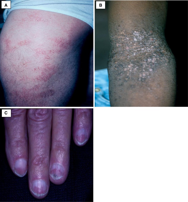
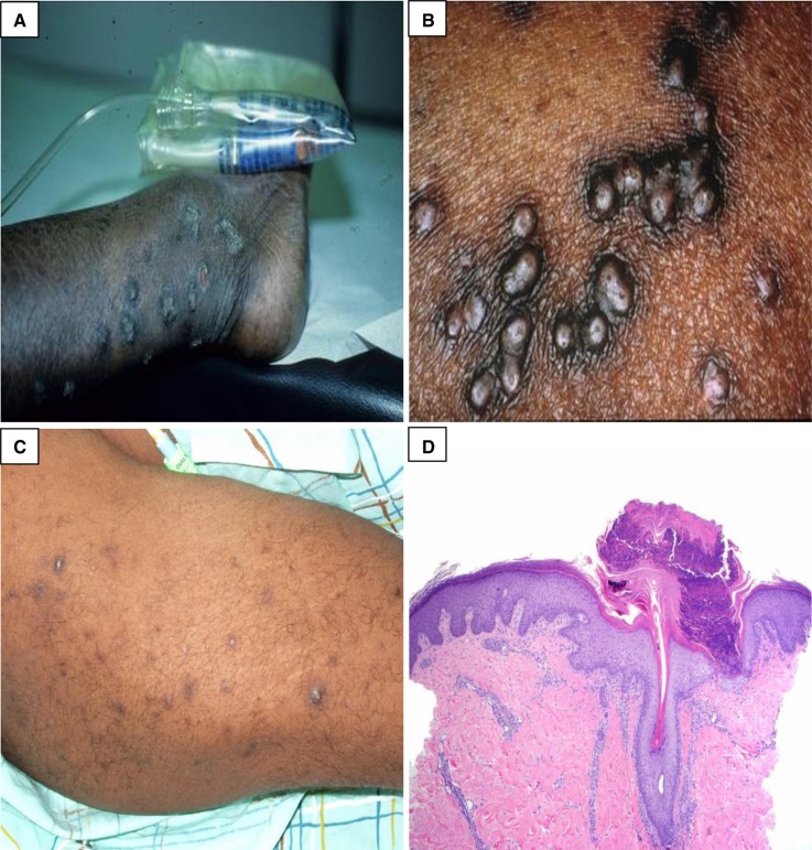
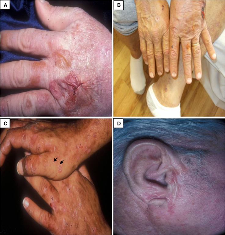
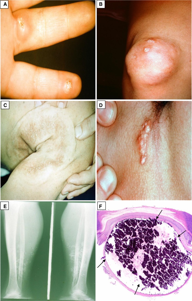
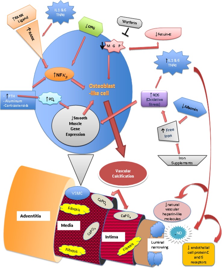
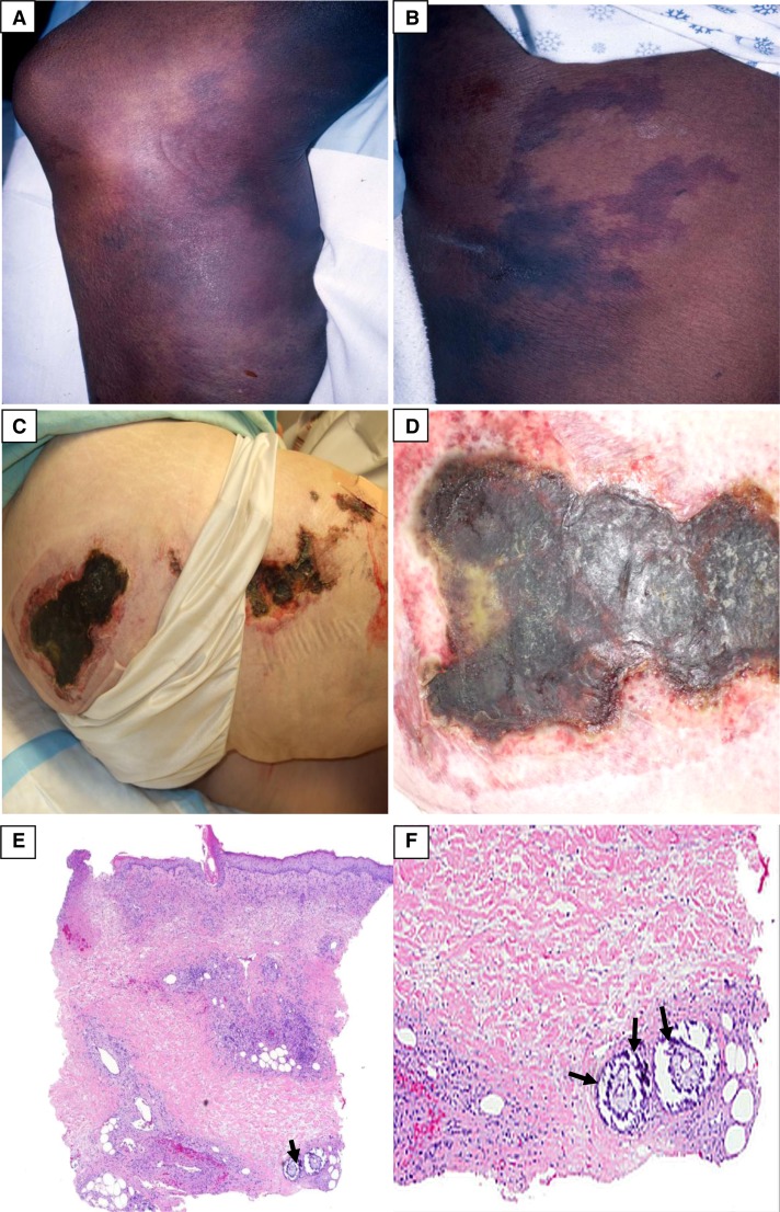
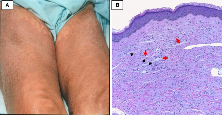
A broad range of skin diseases occurs in patients with ESRD: from the benign and asymptomatic to the physically disabling and life-threatening. Many of them negatively impact on quality of life. Their early recognition and treatment are essential in reducing morbidity and mortality. The cutaneous manifestations can be divided into two main categories: nonspecific and specific. The nonspecific manifestations are commonly seen and include skin color changes, xerosis, half-and-half nails, and pruritus. The specific disorders include acquired perforating dermatosis, bullous dermatoses, metastatic calcification, and nephrogenic systemic fibrosis. This review article describes these conditions and considers the underlying pathophysiology, clinical presentations, diagnosis, and treatment options.


Figures

Figure 1.
Figure 1.
The nonspecific cutaneous manifestations seen in ESRD. (A) Patches of xerotic eczema on the lateral thigh. (B) Lichenified plaque with few excoriations on the antecubital fossa from repeated scratching. (C) Half-and-half nails with the characteristic proximal white half of the nail and the distal red half of the nail. (Courtesy of Timothy G. Berger.)
Figure 2.
Figure 2.
Acquired perforating dermatosis. (A and B) Multiple clustered, hyperpigmented, dome-shaped nodules and coalescing plaques with a central keratotic plug. (C) Hyperpigmented macules and patches from lesions that have resolved. (D) There is a cup-shaped epidermal depression filled with parakeratosis and neutrophilic debris. At the base of the depression, the epidermis is thinned, and degenerated collagen fibers are noted protruding through this attenuated epidermis. (Courtesy of Timothy G. Berger, Anna K. Haemel, and Thaddeus W. Mully.)
Figure 3.
Figure 3.
Porphyria cutanea tarda. (A and B) Tense and ruptured bullae on the dorsal hand. (C) Erosions, crusts, atrophic scarring, and milia (at the tips of the black arrows) on the dorsal hands. (D) Sclerodermatous plaque on the right ear. (Courtesy of Timothy G. Berger and Anna K. Haemel.)
Figure 4.
Figure 4.
Calcinosis cutis. (A) Firm nodules on the palmar aspect of the fingers with central white calcium globules. (B) A calcified nodule around the knee. (C) A large, white, indurated calcified plaque on the right posterior shoulder and arm. (D) A linear, white calcified plaque in the axilla. (E) A plain radiograph of the lower extremity showing radio-opaque soft tissue calcium deposits. (F) There is a nodular deposit of basophilic refractile calcium in the dermis (black arrows). Minimal inflammation is present. (Courtesy of Timothy G. Berger and Thaddeus W. Mully.)
Figure 5.
Figure 5.
Pathophysiology of calcific uremic arteriolopathy. Nuclear factor κβ (NFκβ), a transcription factor involved in the production of cytokines and inflammatory mediators, the receptor activator of NFκβ (RANK), and its ligand are vital for bone mineral resorption and development (68,73,76). They can be activated by chronic inflammatory states, parathyroid hormone (PTH), aluminum, corticosteroids, free radicals, and infectious agents (73,76). Increased NFκβ activity leads to bone mineral loss and vascular calcification. Osteoprotegerin (OPG) is an antagonist of RANKL. RANK/RANKL and OPG are expressed on endothelial cells, osteoblasts/osteoclasts, and vascular smooth muscle cells (VSMCs) (73,76). Transformation of VSMCs into osteoblast-like cells is initiated by metabolic disturbances (particularly hyperphosphatemia), reactive oxygen species (ROS), and decrease of local vascular calcification inhibitors, such as matrix γ-carboxy-glutamate protein (MGP), a vitamin K-dependent protein (68,76). Fetuin-A, a hepatically synthesized protein that functions systemically to inhibit vascular calcification, is reduced in inflammatory states, renal failure, and calcific uremic arteriolopathy (68,76). Progressive subintimal fibrosis and medial-arteriolar calcification lead to endothelial injury and luminal narrowing. The low-flow rate within the cutaneous vessels combined with luminal narrowing leads to decreased blood flow and may lead to blood stasis (73). Systemic inflammation also causes endothelial dysfunction. It creates a procoagulant state in the narrowed vessels and increases the risk of thrombus formation, which can lead to localized tissue ischemia and necrosis (73,76,78).
Figure 6.
Figure 6.
Calcific uremic arteriolopathy. (A) Violaceous, indurated plaque with surrounding reticular purple discoloration. (B) Violaceous reticulation resembling livedo reticularis. (C) Necrotic ulcerations on the right buttock and thigh. (D) Close view of a necrotic ulcer with a violaceous border. (E and F) Low- and high-power magnification views showing basophilic calcium deposits (at the tips of the black arrows) in the deep dermal blood vessels. Minimal associated inflammation is noted. More superficially, congested blood vessels are identified. Focal epidermal necrosis is present. (Courtesy of Timothy G. Berger, Anna K. Haemel, and Thaddeus W. Mully.)
Figure 7.
Figure 7.
Nephrogenic systemic fibrosis. (A) Hyperpigmentation and skin thickening of the thighs bilaterally. (B) Increased number of small spindle cells (at the tips of the black arrowheads) and collagen bundles within the dermis. The spaces that are normally seen between collagen bundles in the reticular dermis are effaced by mucin deposition (red arrows) and fine collagen bundles. (Courtesy of Philip E. LeBoit.)

References

    1. National Institutes of Health; National Institute of Diabetes and Digestive and Kidney Diseases; US Renal Data System: Annual Data Report: Atlas of Chronic Kidney Disease and End-Stage Renal Disease in the United States, 2012. Available at: http://www.usrds.org/2012/pdf/v2_ch1_12.pdf Accessed April 10, 2013
    1. Goldman, L, Schafer AI : Chronic Kidney Disease. In: Goldman’s Cecil Medicine, Expert Consult Premium Edition, 24th Ed., Philadelphia, Elsevier Saunders, 2012
    1. Attia EA, Hassan SI, Youssef NM: Cutaneous disorders in uremic patients on hemodialysis: An Egyptian case-controlled study. Int J Dermatol 49: 1024–1030, 2010 - PubMed
    1. Markova A, Lester J, Wang J, Robinson-Bostom L: Diagnosis of common dermopathies in dialysis patients: A review and update. Semin Dial 25: 408–418, 2012 - PubMed
    1. Airaghi L, Garofalo L, Cutuli MG, Delgado R, Carlin A, Demitri MT, Badalamenti S, Graziani G, Lipton JM, Catania A: Plasma concentrations of alpha-melanocyte-stimulating hormone are elevated in patients on chronic haemodialysis. Nephrol Dial Transplant 15: 1212–1216, 2000 - PubMed