Review
. 2013 Jul;78(3):42-7.
doi: 10.12659/PJR.889403.

Variants of cerebral arteries - anterior circulation

Affiliations
Review

Variants of cerebral arteries - anterior circulation

Grzegorz Makowicz et al. Pol J Radiol. 2013 Jul.

Abstract

Advances in imaging techniques allow for in vivo identification of abnormalities and normal variants of cerebral arteries. These arterial variations can be asymptomatic and uncomplicated although, some of them increase the risk of aneurysm formation, acute intracranial hemorrhage, play a vital role in neurosurgical planning or can be misidentified as serious pathology and medical errors. The goal of this publication is to discuss arterial anomalies of anterior cerebral circulation, their prevalence and demonstrate radiological images of some of those variants. In this article we will discuss variants of internal carotid artery, anterior cerebral artery, anterior communicating artery, middle cerebral artery, persistent stapedial artery and fenestration.

Keywords: arterial abnormalities; congenital absence of the ICA; duplication; fenestration.


Figures
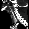
Figure 1.
Figure 1.
Congenital absence of the ICA. Lateral MIP projection of CT angiogram shows long narrowing of the cervical segment of the right internal carotid artery (ICA). The CCA and ECA indicate common carotid artery and external carotid artery respectively (own material).
Figure 2.
Figure 2.
Congenital absence of the ICA. Axial CT image (the same patient) of the skull base shows hypoplastic right carotid canal (RICA). The LICA indicates the normal left carotid canal (own material).
Figure 3.
Figure 3.
Fenestration. Posterior VR projection of CT angiogram shows dual fenestration of the anterior communicating artery (ACoA) (own material).
Figure 4.
Figure 4.
Azygos anterior cerebral artery. Posterior VR projection of CT angiogram shows an unpaired A2 segment of the anterior cerebral artery (Azygos-ACA) and saccular aneurysmal dilatation on the A2 and A3 junction (*). CT angiogram demonstrates a hypoplastic A1 segment of the right anterior cerebral artery (A1-ACA-R). The A1-ACA-L indicates a normal A1 segment of the left anterior cerebral artery (own material).
Figure 5.
Figure 5.
Triplication of the anterior cerebral artery. Postero-lateral VR projection of CT angiogram shows triplicated ACA. There are three A2 segments (<) that arise from the anterior communicating artery. Note saccular aneurysmal dilatation of the anterior communicating artery (*). The A1-ACA indicates normal A1 segments of the anterior cerebral arteries (own material).
Figure 6.
Figure 6.
Recurrent artery of Heubner. Superior VR projection of CT angiogram shows the anterior cerebral artery with a smaller-caliber accessory cerebral artery (RAH) with cortical branches that arises from the junction of the A1-A2 segment. This accessory artery can be named as a recurrent artery of Heubner or also as accessory middle cerebral artery (type-3 according to Manelfe). The < indicates cortical branches of the left middle cerebral artery. Note contralateral occlusion of the internal carotid artery and a proximal part of the M1 segment of the right middle cerebral artery (MCA-R). The A1-ACA indicates A1 segments of the anterior cerebral arteries (own material).

References

    1. Brenner DJ, Hall EJ. Computed tomography – an increasing source of radiation exposure. N Engl J Med. 2007;357:2277–84. - PubMed
    1. McKinney AM, Palmer CS, Truwit CL, et al. Detection of aneurysms by 64-section multidetector CT angiography in patients acutely suspected of having an intracranial aneurysm and comparison with digital subtraction and 3D rotational angiography. Am J Neuroradiol. 2008;29:594–602. - PMC - PubMed
    1. Villablanca JP, Rodriguez FJ, Stockman T, et al. MDCT angiography for detection and quantification of small intracranial arteries: comparison with conventional catheter angiography. Am J Roentgenol. 2007;188:593–602. - PubMed
    1. Skutta B, Fürst G, Eilers J, et al. Intracranial stenoocclusive disease: double-detector helical CT angiography versus digital subtraction angiography. Am J Neuroradiol. 1999;20:791–99. - PMC - PubMed
    1. Sanudo J, Vazquez R, Puerta J. Meaning and clinical interest of the anatomical variations in the 21st century. Eur J Anat. 2003;7(Suppl.1):1–3.

LinkOut - more resources