. 2013 Oct 7;8(10):e76037.
doi: 10.1371/journal.pone.0076037. eCollection 2013.

Molecular marker differences relate to developmental position and subsets of mesodiencephalic dopaminergic neurons

Affiliations

Molecular marker differences relate to developmental position and subsets of mesodiencephalic dopaminergic neurons

Simone M Smits et al. PLoS One. .

Abstract

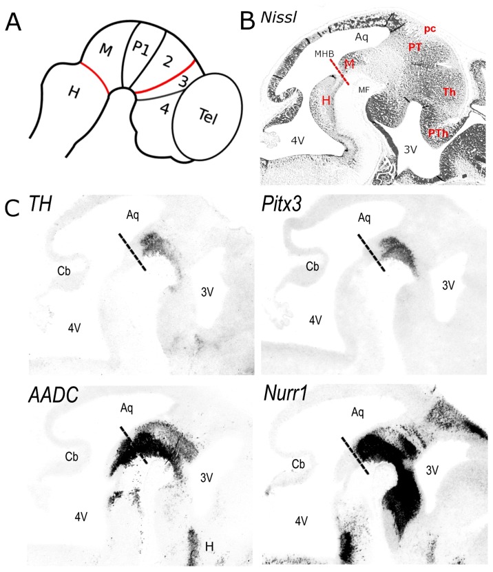
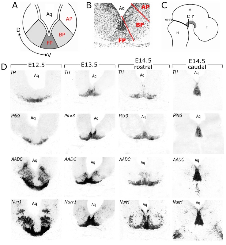
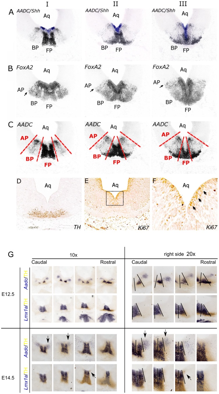
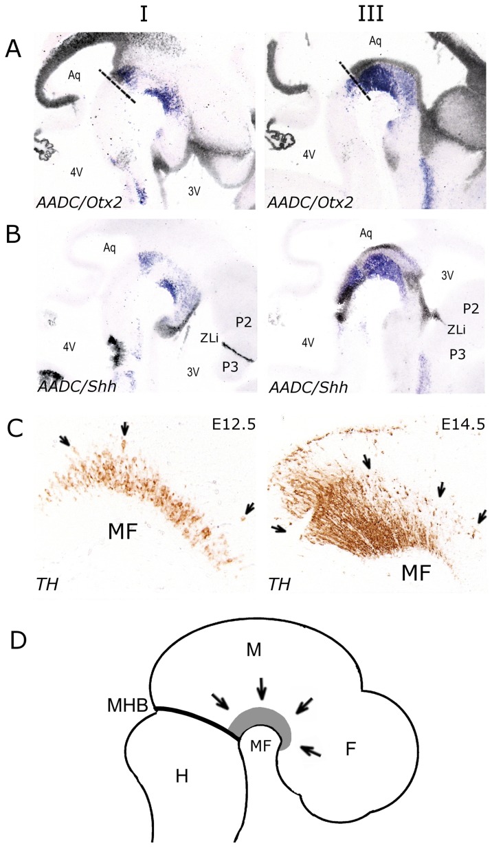
The development of mesodiencephalic dopaminergic (mdDA) neurons located in the substantia nigra compacta (SNc) and ventral tegmental area (VTA) follow a number of stages marked by distinct events. After preparation of the region by signals that provide induction and patterning, several transcription factors have been identified, which are involved in specifying the neuronal fate of these cells. The specific vulnerability of SNc neurons is thought to root in these specific developmental programs. The present study examines the positions of young postmitotic mdDA neurons to relate developmental position to mdDA subset specific markers. MdDA neurons were mapped relative to the neuromeric domains (prosomeres 1-3 (P1-3), midbrain, and hindbrain) as well as the longitudinal subdivisions (floor plate, basal plate, alar plate), as proposed by the prosomeric model. We found that postmitotic mdDA neurons are located mainly in the floorplate domain and very few in slightly more lateral domains. Moreover, mdDA neurons are present along a large proportion of the anterior/posterior axis extending from the midbrain to P3 in the diencephalon. The specific positions relate to some extent to the presence of specific subset markers as Ahd2. In the adult stage more of such subsets specific expressed genes are present and may represent a molecular map defining molecularly distinct groups of mdDA neurons.


Conflict of interest statement

Competing Interests: The authors have declared that no competing interests exist.

Figures

Figure 1
Figure 1. (A) Schematic sagittal representation of the prosomeric model as proposed by Puelles and Rubenstein (2003).
The transverse lines represent interneuromeric boundaries in which the mid/hindbrain border (MHB) and the zona limitans intrathalamica, a boundary between the thalamus (P2) and prethalamus (P3) are depicted in red. (B) Nissl stained E13.5 sagittal section for the anatomical orientation. (C) In situ hybridisation for Th, Pitx3, Aadc and Nurr1 on E13.5 sagittal sections. Abbreviations: 2, prosomere 2; 3, prosomere 3; 4, prosomere 4; 3V, third ventricle; 4V, fourth ventricle; Aq, aqueduct; Cb, cerebellum; H, hindbrain; M, midbrain; MHB, mid/hindbrain border (indicated by dashed lines); MF, mesencephalic flexure; P1, prosomere 1; pc, posterior commissure; PT, pretectum; PTh, prethalamus; Tel, telencephalon; Th, thalamus.
Figure 2
Figure 2. Identification of the transverse interneuromeric boundaries by region specific markers Gbx2 and Dlx2 on E13.5 sagittal sections at three different levels, from lateral to medial, I to III respectively.
(A) Overlays of mRNA expression patterns of Gbx2 (purple) and Dlx2 (black) on adjacent sections. Gbx2 is expressed in prosomere 2 (P2) creating a sharp boundary with the Dlx2 expression domain in P3. (B) Overlays of mRNA expression patterns of Aadc (purple) and Gbx2 (black) on adjacent sections. (C) Overlays of mRNA expression patterns of Aadc (purple) and Dlx2 (black) on adjacent sections. (D) The major transverse interneuromeric boundaries are illustrated by dashed lines in E13.5 sagittal sections. Note that Aadc positive neurons are located across multiple segments (M, P1, P2 and P3). Abbreviations: 3V, third ventricle; 4V, fourth ventricle; Aq, aqueduct; H, hindbrain; M, midbrain; MF, mesencephalic flexure; P1, prosomere 1; P2, prosomere 2; P3, prosomere 3.
Figure 3
Figure 3. Presentation dorsal-ventral boundaries at the rostral and caudal mesodiencephalon during development.
(A) Schematic coronal representation of the dorso-ventral organization of the floor plate (FP), basal plate (BP) and alar plate (AP). (B) Nissl stained E13.5 coronal section for the anatomical orientation. The floor-basal and basal-alar boundaries are shown in red dashed lines. (C) Schematic representation of a sagittal section showing the position of the coronal sections shown in G and F. The position of DA neurons are depicted in grey. (D–G) In situ hybridisation for Th, Pitx3, Aadc and Nurr1 on E12.5 (D), E13.5 (E), anterior E14.5 (F) and posterior E14.5 coronal sections (G). Abbreviations: Aq, aqueduct; F, forebrain; H, hindbrain; M, midbrain; MHB, mid/hindbrain border.
Figure 4
Figure 4. Identification of the longitudinal subdivisions by region specific markers Shh and FoxA2 on E13.5 sagital sections at three different levels, from posterior to anterior, I to III respectively.
(A) Overlays of mRNA expression patterns of Shh (purple) and Aadc (black) on adjacent sections. Shh is highly expressed in the ventricular zone of the basal plate, whereas the signal in the floor plate is less intense. (B) In situ hybridization for FoxA2 on adjacent sections marking the basal-alar plate boundary, as indicated arrows. (C) The major longitudinal subdivisions are illustrated by dashed red lines in E13.5 sagittal sections, in which in situ hybridization for Aadc identifies the putative DA neurons. Note that Aadc positive neurons are located in the floor and basal plate. (D) TH imunostaining on an E12.5 coronal section for the identification of fully differentiated mdDA neurons. (E) Ki67 immunostaining on adjacent E12.5 coronal sections identifies the proliferative cells in the ventricular zone. (F) Higher magnification of the boxed area in panel E. Note that the proliferating cells in the ventricular zone are orientated radially (see arrows). (G) Co-localization of the floorplate marker Lmx1a with Aadc and Th at E12.5 and E14.5. Note the expression of TH and Aadc outside the Lmx1a expression domain (arrows) Abbreviations: AP, alar plate; Aq, aqueduct; BP, basal plate; FP, floor plate.
Figure 5
Figure 5. Analysis of ventricular zone markers Otx2 (A) and Shh (B) in E13.5 sagittal sections at two different levels, lateral (I) and medial (III).
(A) Overlays of mRNA expression patterns of Otx2 (black) and Aadc (purple) on adjacent sections. (B) Overlays of mRNA expression patterns of Shh (black) and Aadc (purple) on adjacent sections. Note that young Aadc positive neurons in the posterior diencephalon are in proximity with the ventricular zone aligning the third ventricle. (C) High magnification of the mesencephalic flexure (MF) in sagittal E12.5 and E14.5 sections immunostained with TH. Anterior is to the right and posterior to the left. Note that TH-immunoreactive neurons are radially orientated along the A/P axis (see arrows). (D) Schematic representation of a sagittal E13.5 embryo, in which mdDA neurons are depicted in gray. The arrows indicate the radial migration of DA neurons. Abbreviations: 3V, third ventricle; 4V, fourth ventricle; Aq, aqueduct; F, forebrain; H, hindbrain; M, midbrain; MF, mesencephalic flexure; MHB, mid/hindbrain border; P1, prosomere 1; P2, prosomere 2; P3, prosomere 3; ZLi, zona limitans intrathalamica.
Figure 6
Figure 6. Th and Ahd2 mRNA expression in mdDA neurons of E14.5 wild-type (A) and E14.5 Pitx3-deficient embryos (B) at different levels along the A/P axis.
The boundary between floor and basal plate is illustrated by dashed red lines. Abbreviations: BP, basal plate; FP, floor plate.
Figure 7
Figure 7. Bitmap traces of expression patterns of adult mdDA subset markers (red) matched to Th (green).
(A) Rostral mdDA (SNc) neuronal enriched markers. (B) Caudal mdDA (VTA) enriched markers. (C) E13.5 sagital sections showing Th, Otx2, Cck and Adra expression.

References

    1. Hirsch E, Graybiel AM, Agid YA (1988) Melanized dopaminergic neurons are differentially susceptible to degeneration in Parkinson’s disease. Nature 334: 345–348 10.1038/334345a0 - DOI - PubMed
    1. Blum D, Torch S, Lambeng N, Nissou M, Benabid AL, et al. (2001) Molecular pathways involved in the neurotoxicity of 6-OHDA, dopamine and MPTP: contribution to the apoptotic theory in Parkinson’s disease. Prog Neurobiol 65: 135–172. - PubMed
    1. Greene JG, Dingledine R, Greenamyre JT (2005) Gene expression profiling of rat midbrain dopamine neurons: implications for selective vulnerability in parkinsonism. Neurobiol Dis 18: 19–31 10.1016/j.nbd.2004.10.003 - DOI - PubMed
    1. Grimm J, Mueller A, Hefti F, Rosenthal A (2004) Molecular basis for catecholaminergic neuron diversity. Proc Natl Acad Sci USA 101: 13891–13896 10.1073/pnas.0405340101 - DOI - PMC - PubMed
    1. Wolfart J, Neuhoff H, Franz O, Roeper J (2001) Differential expression of the small-conductance, calcium-activated potassium channel SK3 is critical for pacemaker control in dopaminergic midbrain neurons. J Neurosci 21: 3443–3456. - PMC - PubMed

Publication types

MeSH terms

LinkOut - more resources