

Effect of transcatheter intraarterial therapies on the distribution of Doxorubicin in liver cancer in a rabbit model
- PMID: 24116106
- PMCID: PMC3792963
- DOI: 10.1371/journal.pone.0076388
Effect of transcatheter intraarterial therapies on the distribution of Doxorubicin in liver cancer in a rabbit model
Abstract
Background and aims: Transcatheter intraarterial techniques can effectively deliver chemotherapeutic agents to tumor and improve the efficacy of chemotherapy. The present study is designed to evaluate the effect of transcatheter intraarterial techniques on the distribution of doxorubicin in relation to blood vessels in liver cancer.
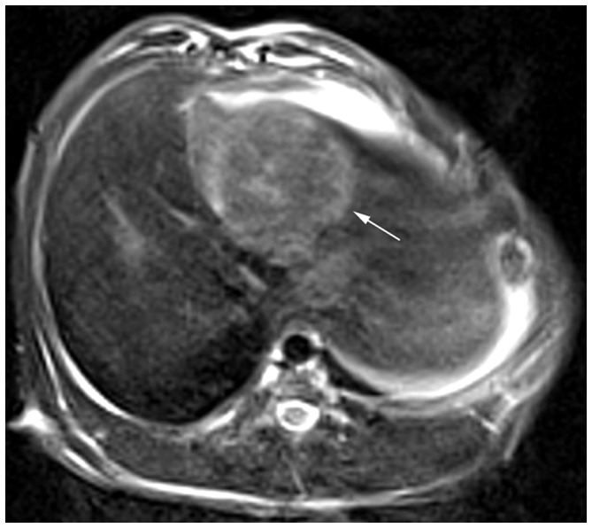
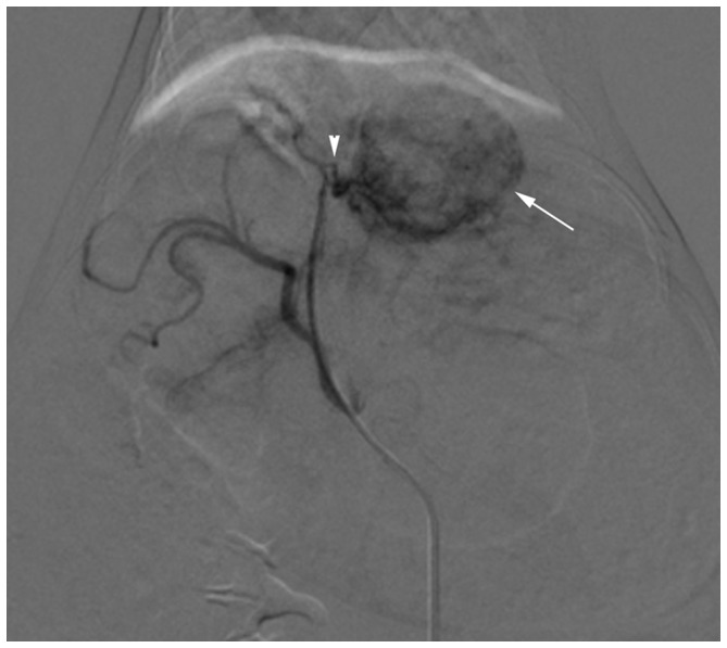
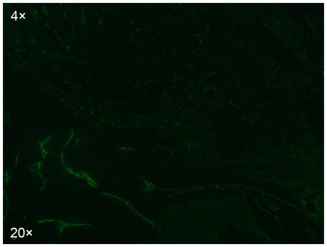
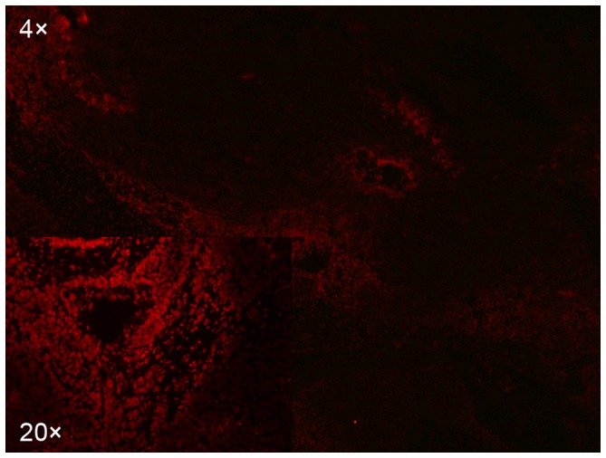
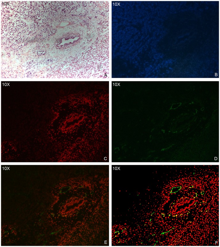
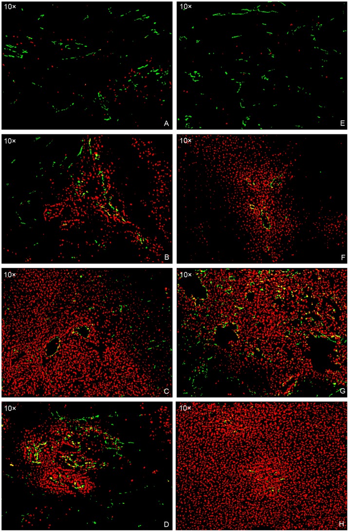
Methods: VX2 tumors were implanted in the livers of 32 rabbits. The animals were divided into 4 groups of 8 animals each. Group 1 (doxo iv) animals received doxorubicin intravenous injection; group 2 (doxo ia) received doxorubicin hepatic intraarterial infusion; group 3 (doxo ia + E) received doxorubicin hepatic intraarterial infusion followed by embolization; group 4 (doxo + L ia + E) received hepatic intraarterial infusion of doxorubicin mixed with Lipiodol followed by embolization. Ten minutes or 4 hours after treatment, the animals were sacrificed and tumors were sampled. Immunofluorescence techniques were used to evaluate the distribution of doxorubicin in relation to blood vessels.
Results: Doxorubicin fluorescence was distributed around tumor blood vessels and decreased with distance from the blood vessels. Tumor cells in avascular and adjacent regions were not exposed to detectable concentrations of doxorubicin. Tumors in the group 2, 3 and 4 had a significant increase in doxorubicin penetration compared with the group 1 tumors (P<0.05). Among the three groups of transcatheter therapies, doxorubicin penetration distance in group 3 was significantly larger than that in group 2 and 4 (P<0.05), and no significant difference was found between group 2 and 4 tumors (P>0.05) at 10 minutes. In contrast, at 4 hours and in total, both group 3 and 4 tumors had significant increases in drug penetration compared with group 2 (P<0.05), and no significant difference was noted between group 3 and 4 tumors (P>0.05).
Conclusion: Transcatheter intraarterial therapies improve doxorubicin penetration in liver cancer; nevertheless their effect on drug distribution is somewhat limited.
Conflict of interest statement
Figures
Similar articles
-
Effect of Transcatheter Intra-Arterial Therapies on Tumor Interstitial Fluid Pressure and Its Relation to Drug Penetration in a Rabbit Liver Tumor Model.J Vasc Interv Radiol. 2015 Dec;26(12):1879-86. doi: 10.1016/j.jvir.2015.05.031. Epub 2015 Aug 4. J Vasc Interv Radiol. 2015. PMID: 26254117
-
Transarterial Infusion of iRGD-Modified ZrO2 Nanoparticles with Lipiodol Improves the Tissue Distribution of Doxorubicin and Its Antitumor Efficacy.J Vasc Interv Radiol. 2019 Dec;30(12):2026-2035.e2. doi: 10.1016/j.jvir.2019.04.014. Epub 2019 Oct 4. J Vasc Interv Radiol. 2019. PMID: 31590966
-
The antitumor effect and hepatotoxicity of a hexokinase II inhibitor 3-bromopyruvate: in vivo investigation of intraarterial administration in a rabbit VX2 hepatoma model.Korean J Radiol. 2009 Nov-Dec;10(6):596-603. doi: 10.3348/kjr.2009.10.6.596. Korean J Radiol. 2009. PMID: 19885316 Free PMC article.
-
Chemoembolization of liver cancer with doxorubicin-loaded CalliSpheres microspheres: plasma pharmacokinetics, intratumoral drug concentration, and tumor necrosis in a rabbit model.Drug Deliv Transl Res. 2020 Feb;10(1):185-191. doi: 10.1007/s13346-019-00672-9. Drug Deliv Transl Res. 2020. PMID: 31482517
-
Comparison of transcatheter intraarterial perfusion MR imaging and fluorescent microsphere perfusion measurements during transcatheter arterial embolization of rabbit liver tumors.J Vasc Interv Radiol. 2007 Oct;18(10):1280-6. doi: 10.1016/j.jvir.2007.07.008. J Vasc Interv Radiol. 2007. PMID: 17911519
Cited by
-
Antitumor Effects of Intra-Arterial Delivery of Albumin-Doxorubicin Nanoparticle Conjugated Microbubbles Combined with Ultrasound-Targeted Microbubble Activation on VX2 Rabbit Liver Tumors.Cancers (Basel). 2019 Apr 24;11(4):581. doi: 10.3390/cancers11040581. Cancers (Basel). 2019. PMID: 31022951 Free PMC article.
-
In vitro cytotoxicity of Indonesian stingless bee products against human cancer cell lines.Asian Pac J Trop Biomed. 2014 Jul;4(7):549-56. doi: 10.12980/APJTB.4.2014APJTB-2013-0039. Asian Pac J Trop Biomed. 2014. PMID: 25183275 Free PMC article.
-
Liver-Directed Therapies for Hepatocellular Carcinoma and Intrahepatic Cholangiocarcinoma.Cancer Control. 2017 Jul-Sep;24(3):1073274817729244. doi: 10.1177/1073274817729244. Cancer Control. 2017. PMID: 28975829 Free PMC article. Review.
-
Endovascular Embolization by Transcatheter Delivery of Particles: Past, Present, and Future.J Funct Biomater. 2017 Apr 3;8(2):12. doi: 10.3390/jfb8020012. J Funct Biomater. 2017. PMID: 28368345 Free PMC article. Review.
-
Hepatic Artery Infusion Chemotherapy for Hepatocellular Carcinoma: Clinical Advancements.Curr Oncol. 2025 May 28;32(6):313. doi: 10.3390/curroncol32060313. Curr Oncol. 2025. PMID: 40558256 Free PMC article. Review.
References
-
- Forner A, Llovet JM, Bruix J (2012) Hepatocellular carcinoma. Lancet 379: 1245–1255. - PubMed
-
- Brown DB, Gould JE, Gervais DA, Goldberg SN, Murthy R, et al. (2007) Transcatheter therapy for hepatic malignancy: standardization of terminology and reporting criteria. J Vasc Interv Radiol 18: 1469–1478. - PubMed
-
- Llovet JM, Real MI, Montaña X, Planas R, Coll S, et al. (2002) Arterial embolisation or chemoembolisation versus symptomatic treatment in patients with unresectable hepatocellular carcinoma: a randomised controlled trial. 359: 1734–1739. - PubMed
-
- Lo CM, Ngan H, Tso WK, Liu CL, Lam CM, et al. (2002) Randomized controlled trial of transarterial lipiodol chemoembolization for unresectable hepatocellular carcinoma. Hepatology 35: 1164–1171. - PubMed
Publication types
MeSH terms
Substances
LinkOut - more resources
Full Text Sources
Other Literature Sources
