

The Sequencing Bead Array (SBA), a next-generation digital suspension array
- PMID: 24116138
- PMCID: PMC3792038
- DOI: 10.1371/journal.pone.0076696
The Sequencing Bead Array (SBA), a next-generation digital suspension array
Abstract
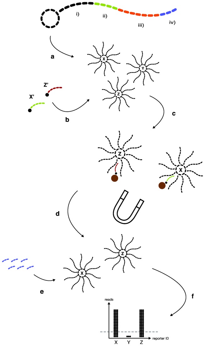
Here we describe the novel Sequencing Bead Array (SBA), a complete assay for molecular diagnostics and typing applications. SBA is a digital suspension array using Next-Generation Sequencing (NGS), to replace conventional optical readout platforms. The technology allows for reducing the number of instruments required in a laboratory setting, where the same NGS instrument could be employed from whole-genome and targeted sequencing to SBA broad-range biomarker detection and genotyping. As proof-of-concept, a model assay was designed that could distinguish ten Human Papillomavirus (HPV) genotypes associated with cervical cancer progression. SBA was used to genotype 20 cervical tumor samples and, when compared with amplicon pyrosequencing, was able to detect two additional co-infections due to increased sensitivity. We also introduce in-house software Sphix, enabling easy accessibility and interpretation of results. The technology offers a multi-parallel, rapid, robust, and scalable system that is readily adaptable for a multitude of microarray diagnostic and typing applications, e.g. genetic signatures, single nucleotide polymorphisms (SNPs), structural variations, and immunoassays. SBA has the potential to dramatically change the way we perform probe-based applications, and allow for a smooth transition towards the technology offered by genomic sequencing.
Conflict of interest statement
Figures
Similar articles
-
Targeted next generation sequencing panel for HPV genotyping in cervical cancer.Exp Mol Pathol. 2021 Feb;118:104568. doi: 10.1016/j.yexmp.2020.104568. Epub 2020 Nov 7. Exp Mol Pathol. 2021. PMID: 33171155
-
Sequencing-based genotyping of mixed human papillomavirus infections by use of RipSeq software.J Clin Microbiol. 2013 Apr;51(4):1278-80. doi: 10.1128/JCM.00091-13. Epub 2013 Jan 30. J Clin Microbiol. 2013. PMID: 23363820 Free PMC article.
-
Human papillomavirus genotyping by Linear Array and Next-Generation Sequencing in cervical samples from Western Mexico.Virol J. 2015 Oct 6;12:161. doi: 10.1186/s12985-015-0391-4. Virol J. 2015. PMID: 26444975 Free PMC article.
-
Profile of MeltPro® HPV test for human papillomavirus genotyping and cervical precancer screening.Expert Rev Mol Diagn. 2019 Oct;19(10):857-862. doi: 10.1080/14737159.2019.1662299. Epub 2019 Sep 5. Expert Rev Mol Diagn. 2019. PMID: 31466483 Review.
-
Clinical significance of human papillomavirus genotyping.J Gynecol Oncol. 2016 Mar;27(2):e21. doi: 10.3802/jgo.2016.27.e21. J Gynecol Oncol. 2016. PMID: 26768784 Free PMC article. Review.
Cited by
-
Suspension arrays based on nanoparticle-encoded microspheres for high-throughput multiplexed detection.Chem Soc Rev. 2015 Aug 7;44(15):5552-95. doi: 10.1039/c4cs00382a. Epub 2015 May 29. Chem Soc Rev. 2015. PMID: 26021602 Free PMC article. Review.
-
Particle-Based Microarrays of Oligonucleotides and Oligopeptides.Microarrays (Basel). 2014 Oct 28;3(4):245-62. doi: 10.3390/microarrays3040245. Microarrays (Basel). 2014. PMID: 27600347 Free PMC article. Review.
-
Predicting the outcomes for out-of-hospital cardiac arrest patients using multiple biomarkers and suspension microarray assays.Sci Rep. 2016 Jun 3;6:27187. doi: 10.1038/srep27187. Sci Rep. 2016. PMID: 27256246 Free PMC article.
-
Deconvolution of nucleic-acid length distributions: a gel electrophoresis analysis tool and applications.Nucleic Acids Res. 2019 Sep 19;47(16):e92. doi: 10.1093/nar/gkz534. Nucleic Acids Res. 2019. PMID: 31226202 Free PMC article.
References
Publication types
MeSH terms
Substances
Grants and funding
LinkOut - more resources
Full Text Sources
Other Literature Sources
Medical
Research Materials
