

Temporal changes of CB1 cannabinoid receptor in the basal ganglia as a possible structure-specific plasticity process in 6-OHDA lesioned rats
- PMID: 24116178
- PMCID: PMC3792868
- DOI: 10.1371/journal.pone.0076874
Temporal changes of CB1 cannabinoid receptor in the basal ganglia as a possible structure-specific plasticity process in 6-OHDA lesioned rats
Abstract
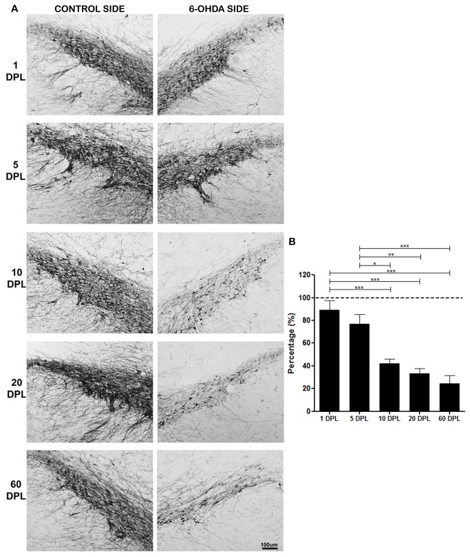
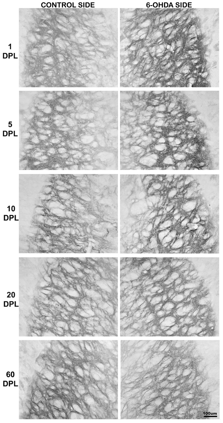
The endocannabinoid system has been implicated in several neurobiological processes, including neurodegeneration, neuroprotection and neuronal plasticity. The CB1 cannabinoid receptors are abundantly expressed in the basal ganglia, the circuitry that is mostly affected in Parkinson's Disease (PD). Some studies show variation of CB1 expression in basal ganglia in different animal models of PD, however the results are quite controversial, due to the differences in the procedures employed to induce the parkinsonism and the periods analyzed after the lesion. The present study evaluated the CB1 expression in four basal ganglia structures, namely striatum, external globus pallidus (EGP), internal globus pallidus (IGP) and substantia nigra pars reticulata (SNpr) of rats 1, 5, 10, 20, and 60 days after unilateral intrastriatal 6-hydroxydopamine injections, that causes retrograde dopaminergic degeneration. We also investigated tyrosine hydroxylase (TH), parvalbumin, calbindin and glutamic acid decarboxylase (GAD) expression to verify the status of dopaminergic and GABAergic systems. We observed a structure-specific modulation of CB1 expression at different periods after lesions. In general, there were no changes in the striatum, decreased CB1 in IGP and SNpr and increased CB1 in EGP, but this increase was not sustained over time. No changes in GAD and parvalbumin expression were observed in basal ganglia, whereas TH levels were decreased and the calbindin increased in striatum in short periods after lesion. We believe that the structure-specific variation of CB1 in basal ganglia in the 6-hydroxydopamine PD model could be related to a compensatory process involving the GABAergic transmission, which is impaired due to the lack of dopamine. Our data, therefore, suggest that the changes of CB1 and calbindin expression may represent a plasticity process in this PD model.
Conflict of interest statement
Figures


Similar articles
-
Morphological and biochemical adaptations to unilateral dopamine denervation of the neostriatum in newborn rats.Neuroscience. 1997 Apr;77(3):753-66. doi: 10.1016/s0306-4522(96)00500-3. Neuroscience. 1997. PMID: 9070750
-
Adenosine A(2A) receptor-mediated modulation of GABA and glutamate release in the output regions of the basal ganglia in a rodent model of Parkinson's disease.Neuroscience. 2004;127(1):223-31. doi: 10.1016/j.neuroscience.2004.04.050. Neuroscience. 2004. PMID: 15219684
-
Loss of cannabinoid CB1 receptor expression in the 6-hydroxydopamine-induced nigrostriatal terminal lesion model of Parkinson's disease in the rat.Brain Res Bull. 2010 Apr 5;81(6):543-8. doi: 10.1016/j.brainresbull.2010.01.009. Epub 2010 Jan 25. Brain Res Bull. 2010. PMID: 20097273 Free PMC article.
-
Parkinson's disease related alterations in cannabinoid transmission.Brain Res Bull. 2022 Jan;178:82-96. doi: 10.1016/j.brainresbull.2021.11.009. Epub 2021 Nov 20. Brain Res Bull. 2022. PMID: 34808322 Review.
-
Roles of the Cannabinoid System in the Basal Ganglia in Parkinson's Disease.Front Cell Neurosci. 2022 Feb 21;16:832854. doi: 10.3389/fncel.2022.832854. eCollection 2022. Front Cell Neurosci. 2022. PMID: 35264932 Free PMC article. Review.
Cited by
-
Cannabinoid Receptors: An Update on Cell Signaling, Pathophysiological Roles and Therapeutic Opportunities in Neurological, Cardiovascular, and Inflammatory Diseases.Int J Mol Sci. 2020 Oct 17;21(20):7693. doi: 10.3390/ijms21207693. Int J Mol Sci. 2020. PMID: 33080916 Free PMC article.
-
The Role of Lipids in Parkinson's Disease.Cells. 2019 Jan 7;8(1):27. doi: 10.3390/cells8010027. Cells. 2019. PMID: 30621069 Free PMC article. Review.
-
M3 Receptor Pathway Stimulates Rapid Transcription of the CB1 Receptor Activation through Calcium Signalling and the CNR1 Gene Promoter.Int J Mol Sci. 2023 Jan 9;24(2):1308. doi: 10.3390/ijms24021308. Int J Mol Sci. 2023. PMID: 36674826 Free PMC article.
-
Design and validation of recombinant protein standards for quantitative Western blot analysis of cannabinoid CB1 receptor density in cell membranes: an alternative to radioligand binding methods.Microb Cell Fact. 2022 Sep 15;21(1):192. doi: 10.1186/s12934-022-01914-1. Microb Cell Fact. 2022. PMID: 36109736 Free PMC article.
-
Cannabidiol as a Therapeutic Target: Evidence of its Neuroprotective and Neuromodulatory Function in Parkinson's Disease.Front Pharmacol. 2020 Dec 15;11:595635. doi: 10.3389/fphar.2020.595635. eCollection 2020. Front Pharmacol. 2020. PMID: 33384602 Free PMC article. Review.
References
Publication types
MeSH terms
Substances
LinkOut - more resources
Full Text Sources
Other Literature Sources
Miscellaneous
