. 2013 Oct 8;8(10):e77112.
doi: 10.1371/journal.pone.0077112. eCollection 2013.

An anti-CD34 antibody-functionalized clinical-grade POSS-PCU nanocomposite polymer for cardiovascular stent coating applications: a preliminary assessment of endothelial progenitor cell capture and hemocompatibility

Affiliations

An anti-CD34 antibody-functionalized clinical-grade POSS-PCU nanocomposite polymer for cardiovascular stent coating applications: a preliminary assessment of endothelial progenitor cell capture and hemocompatibility

Aaron Tan et al. PLoS One. .

Abstract

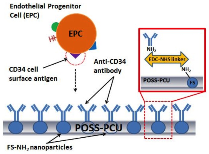
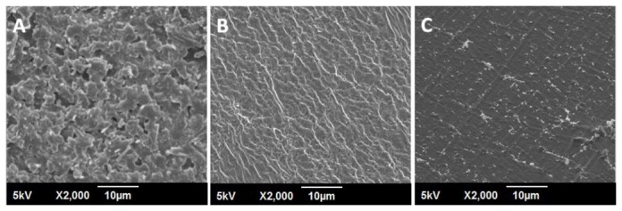
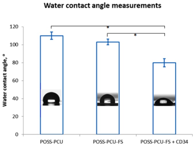
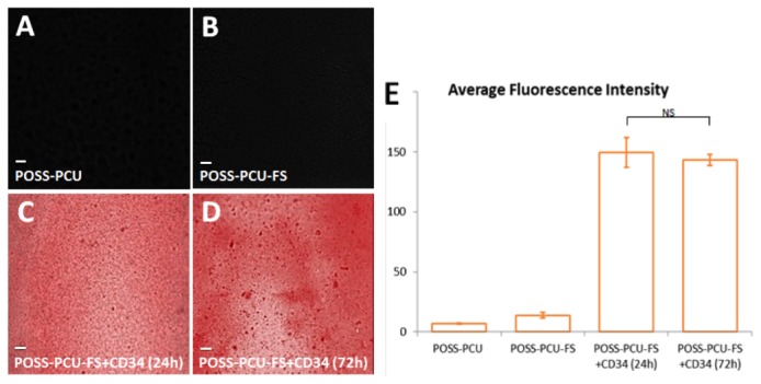
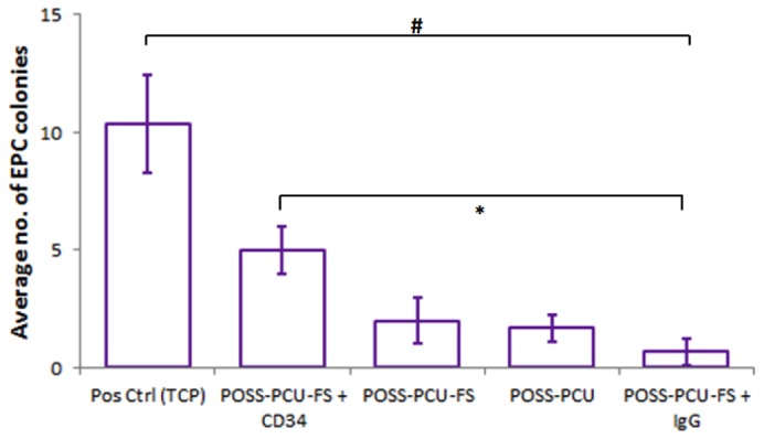
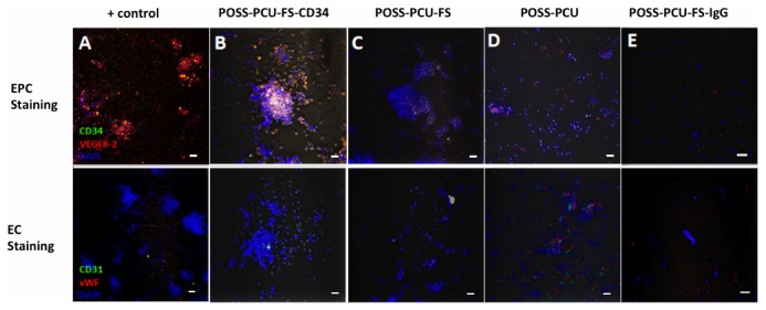
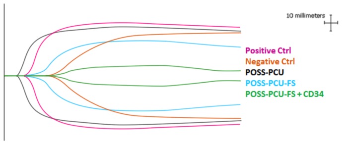
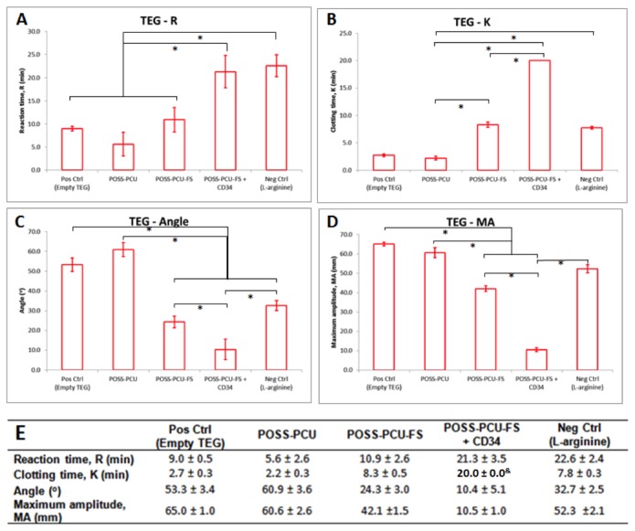
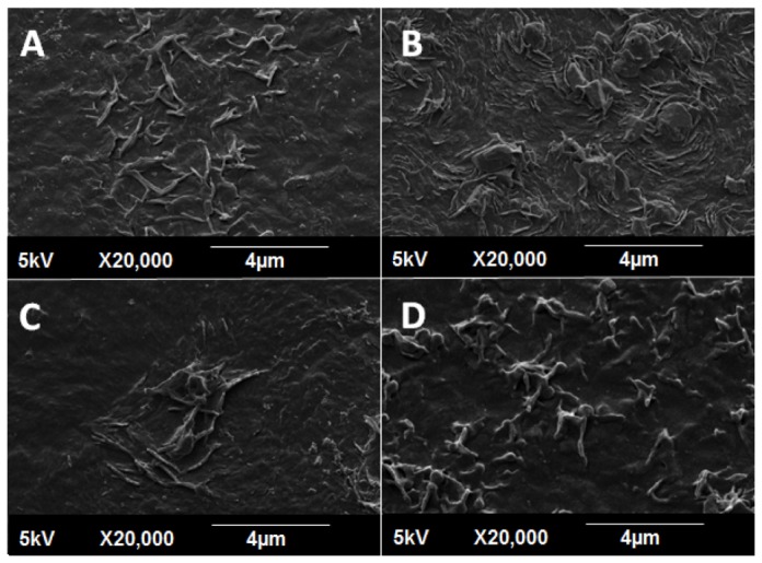
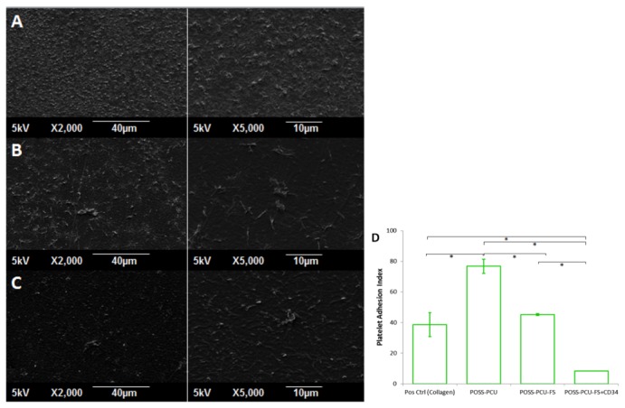
In situ endothelialization of cardiovascular implants has emerged in recent years as an attractive means of targeting the persistent problems of thrombosis and intimal hyperplasia. This study aimed to investigate the efficacy of immobilizing anti-CD34 antibodies onto a POSS-PCU nanocomposite polymer surface to sequester endothelial progenitor cells (EPCs) from human blood, and to characterize the surface properties and hemocompatibility of this surface. Amine-functionalized fumed silica was used to covalently conjugate anti-CD34 to the polymer surface. Water contact angle, fluorescence microscopy, and scanning electron microscopy were used for surface characterization. Peripheral blood mononuclear cells (PBMCs) were seeded on modified and pristine POSS-PCU polymer films. After 7 days, adhered cells were immunostained for the expression of EPC and endothelial cell markers, and assessed for the formation of EPC colonies. Hemocompatibility was assessed by thromboelastography, and platelet activation and adhesion assays. The number of EPC colonies formed on anti-CD34-coated POSS-PCU surfaces was not significantly higher than that of POSS-PCU (5.0±1.0 vs. 1.7±0.6, p>0.05). However, antibody conjugation significantly improved hemocompatibility, as seen from the prolonged reaction and clotting times, decreased angle and maximum amplitude (p<0.05), as well as decreased platelet adhesion (76.8±7.8 vs. 8.4±0.7, p<0.05) and activation. Here, we demonstrate that POSS-PCU surface immobilized anti-CD34 antibodies selectively captured CD34+ cells from peripheral blood, although only a minority of these were EPCs. Nevertheless, antibody conjugation significantly improves the hemocompatibility of POSS-PCU, and should therefore continue to be explored in combination with other strategies to improve the specificity of EPC capture to promote in situ endothelialization.


Conflict of interest statement

Competing Interests: MSA is employed by a commercial company, Pharmidex Pharmaceutical Services Ltd. However, we can confirm that this does not alter the authors' adherence to all PLOS ONE policies on data sharing and materials

Figures

Figure 1
Figure 1. Anti-CD34 antibody immobilization on POSS-PCU polymer surface via amine-functionalized fumed silica nanoparticles and EDC-NHS linker.
This method could represent a novel way of attracting circulating endothelial progenitor cells for accelerated endothelialization.
Figure 2
Figure 2. Surface characterization of unmodified and modified POSS-PCU surfaces by scanning electron microscopy at 2000x magnification.
A POSS-PCU B POSS-PCU-FS C POSS-PCU-FS+CD34. POSS-PCU-FS: POSS-PCU with fumed silica anchors, POSS-PCU-FS+CD34: POSS-PCU biofunctionalized with anti-CD34 antibodies.
Figure 3
Figure 3. Water contact angle measurements of unmodified and modified POSS-PCU surfaces, using a sessile drop method.
Biofunctionalization with anti-CD34 antibodies significantly reduces the mean water contact angle value compared to POSS-PCU and POSS-PCU-FS. Error bars: ± SD; * denotes p < 0.05. POSS-PCU-FS: POSS-PCU with fumed silica anchors, POSS-PCU-FS+CD34: POSS-PCU biofunctionalized with anti-CD34 antibodies.
Figure 4
Figure 4. Detection of surface-immobilized anti-CD34 antibodies using quantum dots.
When films were immunostained with red QD-IgG fluorescent labels, (A) control POSS-PCU and (B) POSS-PCU-FS did not exhibit fluorescence, as compared to (C-D) POSS-PCU-FS+CD34 films, which showed uniform immobilization of bound antibodies on the film even after washing by mechanical shaking for 24 and 72 hrs (10x magnification). (E) Measurements of samples’ residual fluorescent intensities after 24 and 72 hrs of washing demonstrates that antibodies remain stably bound. Error bars: ± SD; NS denotes no significant difference (p > 0.05). POSS-PCU-FS: POSS-PCU with fumed silica anchors, POSS-PCU-FS+CD34: POSS-PCU biofunctionalized with anti-CD34 antibodies.
Figure 5
Figure 5. EPC extraction from peripheral blood.
The buffy coat layer contains EPCs, which were cultured on test samples.
Figure 6
Figure 6. Morphological changes of human endothelial progenitor cells (EPCs) isolated from peripheral blood, and cultured for 7 days.
(A) Peripheral blood mononuclear cells (PBMCs) containing a heterogenous population of EPCs, monocytes, and granulophages, plated on normal uncoated tissue culture plate on day 1 (40x magnification). (B) White arrows point towards EPC colonies observed at day 7 (10x magnification). (C) An EPC colony at day 7, defined morphologically as central cluster of rounded cells surrounded by multiple spindle-shaped cells (20x magnification). Inset: Black arrows point to multipotent stem cells; white arrows point to EPCs (40x magnification). Scale bar represents 20 µm.
Figure 7
Figure 7. EPCs under light microscopy.
(A) An EPC colony, defined morphologically as a central cluster of rounded cells surrounded by multiple spindle-shaped cells (20x magnification). (B-D) Expression of VEGFR-2 (red) and CD34 (green) was assessed under laser scanning confocal microscopy (10x magnification). Double-positive colonies (yellow) were identified as EPC colonies. Scale bar represents 20 µm.
Figure 8
Figure 8. EPC colony counts.
Number of double-positive CD34+ / VEGFR-2+ EPC colonies formed per 106 cells plated was enumerated after 7 days of culture on the different surfaces. Error bars: ± SD; # denotes a significant difference (p < 0.05) between positive control and all other groups; * denotes a significant difference (p < 0.05) between POSS-PCU-FS+CD34 and POSS-PCU-FS+IgG. Scale bar represents 20 µm. POSS-PCU-FS: POSS-PCU with fumed silica anchors, POSS-PCU-FS+CD34: POSS-PCU biofunctionalized with anti-CD34 antibodies.
Figure 9
Figure 9. EPC capture assay.
Cells were double-stained at day 7 for either EPC (CD34/VEGFR-2) or EC (CD31/vWF) markers, and counterstained with DAPI for nuclei. (A) Positive control, (B) POSS-PCU-FS+CD34, (C) POSS-PCU-FS, (D) POSS-PCU, (E) POSS-PCU-FS+IgG isotype negative control (20x magnification). Scale bar represents 20 µm. POSS-PCU-FS: POSS-PCU with fumed silica anchors, POSS-PCU-FS+CD34: POSS-PCU biofunctionalized with anti-CD34 antibodies, POSS-PCU-FS+IgG: POSS-PCU biofunctionalized with goat anti-mouse antibodies.
Figure 10
Figure 10. Representative TEG profiles of blood in contact with the modified and unmodified POSS-PCU surfaces.
Standard unmodified TEG cups with citrated whole blood were used as positive controls, whereas standard cups containing citrated blood mixed with the anti-coagulant L-arginine were used as negative controls. Profiles demonstrate a similar cigar-shape, indicating functional hemostasis. POSS-PCU-FS: POSS-PCU with fumed silica anchors, POSS-PCU-FS+CD34: POSS-PCU biofunctionalized with anti-CD34 antibodies.
Figure 11
Figure 11. TEG parameters.
Samples differed in (A) reaction time, (B and C) clotting rate, and (D) maximum clot strength. The mean of each clotting parameter was plotted (n=3). Error bars: ± SD; *denotes p < 0.05. (E) Table representing mean TEG parameters. &Anti-CD34-coated samples never reached 20 mm clot size after 1.5 hrs of testing, hence k value was set as 20 min for ease of statistical testing. Standard unmodified TEG cups with citrated whole blood were used as positive controls, whereas standard-coated cups containing citrated blood mixed with the anti-coagulant L-arginine were used as negative controls. POSS-PCU-FS: POSS-PCU with fumed silica anchors, POSS-PCU-FS+CD34: POSS-PCU biofunctionalized with anti-CD34 antibodies.
Figure 12
Figure 12. Platelet activation assay.
SEM images (2000x magnification) of adhered platelets show distinctly different morphological appearances. (A) Collagen, (B) POSS-PCU, (C) POSS-PCU-FS, (D) POSS-PCU-FS+CD34. Adhered platelets found on collagen-coated surfaces (positive control, A), showed the highest degree of activation, with formation of distinct pseudopodia and hyaloplasm spreading. Platelets adhering on POSS-PCU (B) and POSS-PCU-FS (C) were mostly dendritic-spread, with prominent pseudopodia as well as some flattening. Those adhered to POSS-PCU-FS+CD34 (D) remained dendritic with a clear spherical body and without any evident flattening. POSS-PCU-FS: POSS-PCU with fumed silica anchors, POSS-PCU-FS+CD34: POSS-PCU biofunctionalized with anti-CD34 antibodies.
Figure 13
Figure 13. Platelet adhesion assay.
(A-C) SEM images (2000x and 5000x magnification) show the greatest number of platelets adhering to POSS-PCU surfaces. (A) POSS-PCU, (B) POSS-PCU-FS, (C) POSS-PCU-FS+CD34. (D) The degree of platelet adhesion, expressed as the Platelet Adhesion Index, was significantly reduced by incorporation of FS and subsequent conjugation of anti-CD34 antibodies. *denotes a significant difference (p < 0.05). POSS-PCU-FS: POSS-PCU with fumed silica anchors, POSS-PCU-FS+CD34: POSS-PCU biofunctionalized with anti-CD34 antibodies.

References

    1. Boudriot E, Thiele H, Walther T, Liebetrau C, Boeckstegers P et al. (2011) Randomized comparison of percutaneous coronary intervention with sirolimus-eluting stents versus coronary artery bypass grafting in unprotected left main stem stenosis. J Am Coll Cardiol 57: 538-545. doi:10.1016/S0735-1097(11)60538-X. PubMed: 21272743. - DOI - PubMed
    1. Mohr FW, Morice M-C, Kappetein AP, Feldman TE, Ståhle E et al. (2013) Coronary artery bypass graft surgery versus percutaneous coronary intervention in patients with three-vessel disease and left main coronary disease: 5-year follow-up of the randomised, clinical SYNTAX trial. Lancet 381: 629-638. doi:10.1016/S0140-6736(13)60141-5. PubMed: 23439102. - DOI - PubMed
    1. Goh D, Tan A, Farhatnia Y, Rajadas J, Alavijeh MS et al. (2013) Nanotechnology-Based Gene-Eluting Stents. Mol Pharm 10(4): 1279-1298. doi:10.1021/mp3006616. PubMed: 23394068. - DOI - PubMed
    1. Tan A, Rafiei Y, de Mel A, Rajadas J, Alavijeh MS et al. (2013) Inception to Actualization: Next Generation Coronary Stent Coatings Incorporating Nanotechnology. J Biotechnol 164(1): 151-170. doi:10.1016/j.jbiotec.2013.01.020. PubMed: 23376617. - DOI - PubMed
    1. Padfield GJ, Newby DE, Mills NL (2010) Understanding the role of endothelial progenitor cells in percutaneous coronary intervention. J Am Coll Cardiol 55: 1553-1565. doi:10.1016/j.jacc.2009.10.070. PubMed: 20378071. - DOI - PubMed

Publication types