. 2013 Oct 8;8(10):e77316.
doi: 10.1371/journal.pone.0077316. eCollection 2013.

Regional fibronectin and collagen fibril co-assembly directs cell proliferation and microtissue morphology

Affiliations

Regional fibronectin and collagen fibril co-assembly directs cell proliferation and microtissue morphology

Carlos A Sevilla et al. PLoS One. .

Abstract

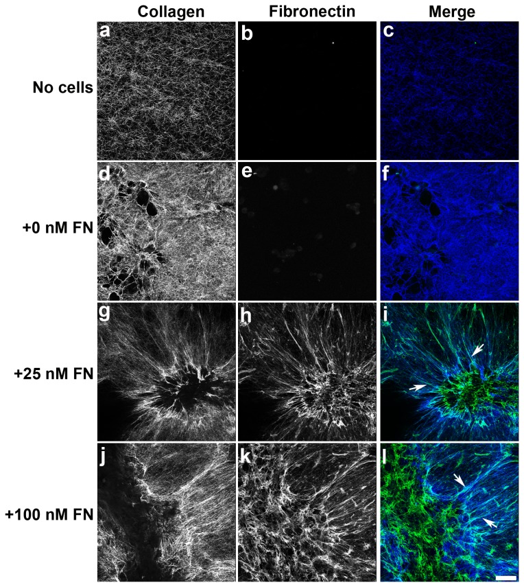
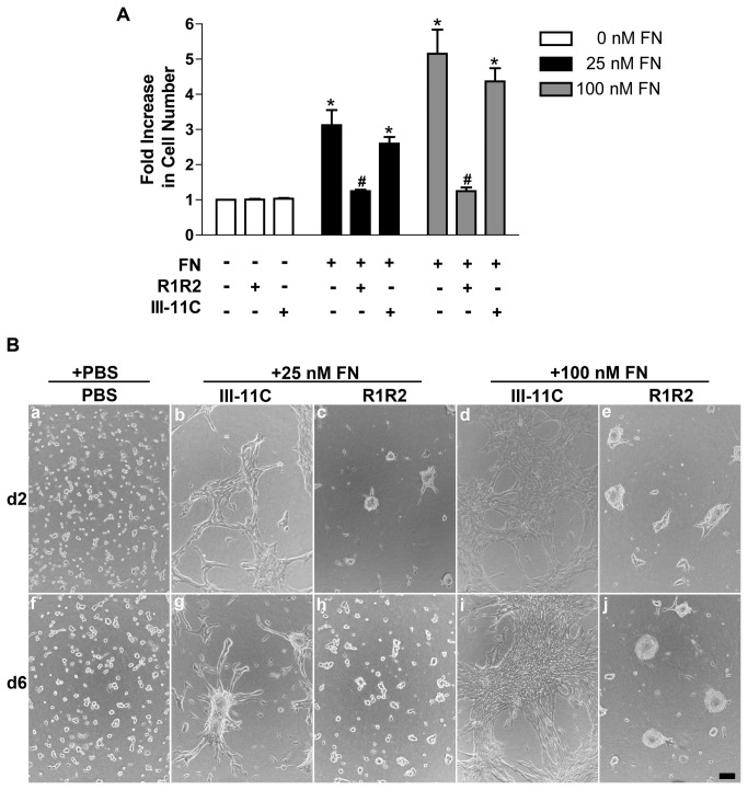
The extracellular matrix protein, fibronectin stimulates cells to self-assemble into three-dimensional multicellular structures by a mechanism that requires the cell-dependent conversion of soluble fibronectin molecules into insoluble fibrils. Fibronectin also binds to collagen type I and mediates the co-assembly of collagen fibrils into the extracellular matrix. Here, the role of collagen-fibronectin binding in fibronectin-induced cellular self-assembly was investigated using fibronectin-null fibroblasts in an in vitro model of tissue formation. High resolution, two-photon immunofluorescence microscopy was combined with second harmonic generation imaging to examine spatial and temporal relationships among fibronectin and collagen fibrils, actin organization, cell proliferation, and microtissue morphology. Time course studies coupled with simultaneous 4-channel multiphoton imaging identified regional differences in fibronectin fibril conformation, collagen fibril remodeling, actin organization, and cell proliferation during three-dimensional cellular self-assembly. Regional differences in cell proliferation and fibronectin structure were dependent on both soluble fibronectin concentration and fibronectin-collagen interactions. Fibronectin-collagen binding was not necessary for either fibronectin matrix formation or intercellular cohesion. However, inhibiting fibronectin binding to collagen reduced collagen fibril remodeling, decreased fibronectin fibril extension, blocked fibronectin-induced cell proliferation, and altered microtissue morphology. Furthermore, continual fibronectin-collagen binding was necessary to maintain both cell proliferation and microtissue morphology. Collectively, these data suggest that the complex changes in extracellular matrix and cytoskeletal remodeling that mediate tissue assembly are driven, in part, by regional variations in cell-mediated fibronectin-collagen co-assembly.


Conflict of interest statement

Competing Interests: The authors have declared that no competing interests exist.

Figures

Figure 1
Figure 1. Spatial and temporal changes in cell proliferation, actin organization, and fibronectin matrix.
FN-null MEFs adherent to native collagen type I gels were treated with 25 or 100 nM fibronectin (FN). (A) After a 6-day incubation, samples were immunostained for fibronectin. Z-slices (1-μm step size) were reconstructed in 3D and projected along the x-y plane. Images represent 1 of 3 experiments performed in triplicate. (B) After either a 2- or 4-day incubation, cells were processed for immunofluorescence microscopy. DAPI, BrdU, actin, and FN images were collected simultaneously along the z-axis at 1-μm intervals using two-photon microscopy. Z-slices corresponding to the microtissue-collagen interface were projected along the z-plane using ImageJ. Scale bar, 50 µm. Images represent 1 of 3 experiments performed in triplicate.
Figure 2
Figure 2. Merged 4-channel images of Day 2 and Day 4 microtissues.
Collagen-adherent FN-null MEFs were treated with 25 or 100 nM fibronectin (FN). After a 2- or 4-day incubation, cells were processed for immunofluorescence microscopy. (A) DAPI (blue), BrdU (red), actin (green), and FN (white) images were collected simultaneously as described in the legend to Figure 1. Z-slices corresponding to the microtissue-collagen interface were projected along the z-plane, merged, and aligned using ImageJ. Scale bar, 50 µm. Images represent 1 of 3 experiments performed in triplicate. (B) Representative FN staining obtained from the core (a) and periphery (b) of day 4 microtissues formed in response to 25 nM FN. Note the different organizational patterns of pericellular (a) and extended (b) fibronectin fibrils. Scale bar, 100 μm.
Figure 3
Figure 3. Spatial and temporal analysis of fibronectin-induced cell proliferation.
Collagen-adherent FN-null MEFs were treated with 25 nM or 100 nM fibronectin (FN) for either 2 or 4 days, and then processed for immunofluorescence microscopy. Four channel images of fibronectin, actin, BrdU, and DAPI staining were obtained and processed to quantify the spatial distribution of proliferating cells in microtissues. (A) The relative distance of all BrdU-positive cells from the centroid (r a/rb) was calculated. Data are grouped to display the frequency of occurrence of proliferating cells from the centroid (r a/rb = 0) to the periphery (ra/rb = 1). Data are presented as the mean percent of BrdU-positive cells ± SEM of 3 independent experiments. (B) Representative x-y projections showing BrdU (red) and DAPI (blue) staining on day 4. (C) Total numbers of proliferating and non-proliferating cells were determined from BrdU and DAPI staining. Data are presented as mean percent BrdU-positive cells + SEM of 3 experiments performed in triplicate.
Figure 4
Figure 4. Collagen and fibronectin fibril co-localization within cell networks.
Collagen-adherent FN-null cells were incubated for 2 days in the absence (0 nM FN; d-f) or presence of either 25 nM (g-i) or 100 nM (j-l) fibronectin (FN). Also included are images of polymerized collagen gels incubated for 2 days in the absence of both cells and FN (a-c). Collagen (a,d,g,j) was visualized using second harmonic generation microscopy. FN (b,e,h,k) was visualized using an anti-FN pAb followed by Alexa488-conjugated anti-rabbit IgG. FN and collagen were simultaneously visualized along the z-axis at 1-μm intervals using two-photon microscopy and then projected onto the z-plane using ImageJ. Note that images were collected at and above the collagen surface, and do not include images collected below the surface. Merged images of collagen (blue) and FN (green) staining are shown (c,f,i,l). Co-localized signals produce cyan (arrows). Scale bar, 50 μm.
Figure 5
Figure 5. Collagen-fibronectin co-localization at the microtissue-substrate interface.
Representative x-y projections of 2-day microtissues formed in response to either 25 nM or 100 nM fibronectin (FN). FN (green) and collagen (blue) were visualized as described in the legend to Figure 4. Z-slices (1-μm step size) were collected beginning below the collagen surface and extending above the microtissue. Images were reconstructed in 3D and then projected along the x-y plane. Arrows denote areas of co-localization (cyan). Note the non-overlapping FN and collagen signals on the apical and basal surfaces, respectively.
Figure 6
Figure 6. Cell proliferation and microtissue morphology require fibronectin-collagen binding.
Collagen-adherent FN-null MEFs were treated with PBS (0 FN) or fibronectin (25 or 100 nM) in the absence or presence of R1R2, the control peptide III-11C, or an equal volume of the vehicle, PBS. (A) Following a 6-day incubation, cell number was determined using MTT. Data are presented as fold increase in cell number versus non-treated (+PBS/PBS) controls + SEM of 3 experiments performed in triplicate. *Significant vs. +PBS/PBS, p<0.001 (ANOVA). #Significant vs. respective +FN/PBS and +/III-11C, p<0.001 (ANOVA). (B) Representative phase contrast images collected on days 2 and 6 of culture. Images represent 1 of 3 experiments performed in triplicate. Scale bar, 50 μm.
Figure 7
Figure 7. Collagen-fibronectin binding is required for ECM fibril organization.
FN-null MEFs adherent to polymerized collagen were treated with fibronectin (25 or 100 nM) in the presence of either R1R2 or the control peptide III-11C. Following a 4-day incubation, cells were processed for immunofluorescence microscopy. Fibronectin (green) and collagen (blue) were visualized using an anti-FN pAb and second harmonic generation imaging, respectively. Images were collected at and above the collagen substrate and then projected onto the z-plane using ImageJ. Dashed red lines denote spheroid location. Arrowheads indicate colocalizing fibronectin and collagen fibrils. Arrows show fibronectin fibrils that do not colocalize with collagen fibrils. Scale bar, 50 μm.
Figure 8
Figure 8. Microtissue morphology depends on fibronectin-collagen binding.
Collected images of 4-day microtissues formed in response to either 25 nM or 100 nM fibronectin (FN), pretreated with either R1R2 or III-11C, were reconstructed in 3D and projected onto the x-y plane. Fibronectin (green) and collagen (blue) are shown. Scale bar, 50 μm.
Figure 9
Figure 9. Disrupting collagen-fibronectin binding alters microtissue shape and reduces cell proliferation.
Collagen-adherent FN-null MEFs were treated with 25 or 100 nM fibronectin. Following a 3-day incubation, media were removed and replaced with media containing fibronectin (25 or 100 nM) pretreated with R1R2 or III-11C. Cells were then incubated an additional 3 days. (A) Phase contrast images were collected on day 3 prior to treatment with R1R2 (a,b) and again on day 6 (c,d). Images represent 1 of 3 experiments performed in triplicate. Scale bar, 100 μm. (B) Cell number was determined on day 6 using MTT. Data are presented as fold increase in cell number versus non-treated (0 nM FN) controls + SEM of 3 experiments performed in triplicate. *Significant vs. ‘-FN’, p<0.001 (ANOVA). #Significant vs. respective ‘+FN’ and ‘+FN/III-11C’, p<0.001 (ANOVA).
Figure 10
Figure 10. Proposed role of fibronectin-collagen fibrils in tissue self-assembly.
Schematic representation of collagen-fibronectin co-assembly in the development of regional differences in cell proliferation and intercellular cohesion. Fibronectin matrix assembly by cells adherent to native collagen type I substrates stimulates cell network formation and cell contraction. Co-assembled collagen-fibronectin bundles provide anchors for cell networks to develop isometric tension and extended fibronectin fibrils that promote cell proliferation.

Similar articles

Cited by

References

    1. Ingber DE, Mow VC, Butler D, Niklason L, Huard J et al. (2006) Tissue engineering and developmental biology: going biomimetic. Tissue Eng 12: 3265-3283. doi:10.1089/ten.2006.12.3265. PubMed: 17518669. - DOI - PubMed
    1. Mironov V, Visconti RP, Kasyanov V, Forgacs G, Drake CJ et al. (2009) Organ printing: tissue spheroids as building blocks. Biomaterials 30: 2164-2174. doi:10.1016/j.biomaterials.2008.12.084. PubMed: 19176247. - DOI - PMC - PubMed
    1. Lubarsky B, Krasnow MA (2003) Tube morphogenesis: making and shaping biological tubes. Cell 112: 19-28. doi:10.1016/S0092-8674(02)01283-7. PubMed: 12526790. - DOI - PubMed
    1. Nogawa H, Morita K, Cardoso WV (1998) Bud formation precedes the appearance of differential cell proliferation during branching morphogenesis of mouse lung epithelium in vitro. Dev Dyn 213: 228-235. doi:10.1002/(SICI)1097-0177(199810)213:2. PubMed: 9786423. - DOI - PubMed
    1. Patel VN, Rebustini IT, Hoffman MP (2006) Salivary gland branching morphogenesis. Differentiation 74: 349-364. doi:10.1111/j.1432-0436.2006.00088.x. PubMed: 16916374. - DOI - PubMed

Publication types

LinkOut - more resources